Unrecognized Gastrostomy Tube Migration Eventually Causing Bowel Obstruction
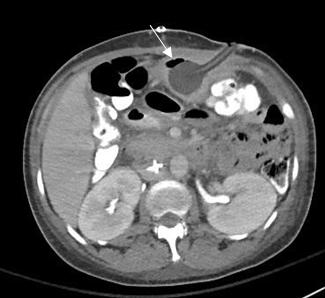
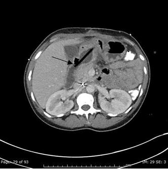
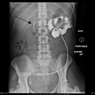
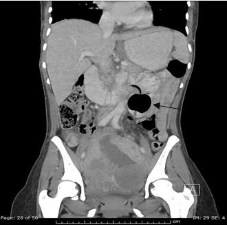
A 45 year old female initially presented with traumatic evisceration of bowel requiring gastrostomy tube (G-tube) placement. An initial CT showed the G-tube to be in appropriate position. An additional inpatient CT scan performed 2 weeks later for abdominal pain showed unrecognized, and therefore not reported, distal migration of the G-tube to the proximal duodenum without signs of obstruction at that time. Fluoroscopy performed prior to discharge demonstrated the G-tube to be intraluminal; however, the G-tube did not appear to terminate within the stomach, which was not reported. Additional oblique views may have demonstrated the G-tube balloon to be deep within the abdomen rather than opposed to the skin surface. The patient was then discharged. The patient presented 2 weeks later with symptoms of nausea and bilious emesis and CT scan obtained at that time showed the G-tube balloon to be in the proximal jejunum causing obstruction of the more proximal bowel. This case illustrates a likely preventable G-tube malposition related complication due to failure to recognize and report the G-tube’s migration on CT and failure to obtain oblique images during the fluoroscopic tube placement verification study.
Introduction
Percutaneous gastrostomy tubes are routinely placed in patients with an inability to feed by mouth and requiring long term (>4-6 weeks) enteral nutrition. They are considered safe and have been commonly placed since 1980, with an estimated 160,000-200,000 placement procedures performed annually. Procedure-related morbidity has been reported at 9.4% and a mortality of 0.53%, with 1-3% of procedures associated with major
References
-
Hawatmeh A, Alkhateeb A, Arqoub AA, Jumean, K, Shaaban H (2016) Gastrostomy tube migration complicated with acute pancreatitis: Two case reports with review of literature. Int J Crit Illn Inj Sci 6(1): 48-50.
-
Milanchi S, Wilson MT (2008) Malposition of percutaneous endoscopic-guided gastrostomy: Guideline and management. J Minim Access Surg 4(1): 1-4.
-
Potack JZ, Chokhavatia S (2008) Complications of and Controversies Associated With Percutaneous Endoscopic Gastrostomy: Report of a Case and Literature Review. Medscape J Med 10(6): 142.
- Ultrasound Guided Therapeutic Nerve Blocks
- Cyclops Lesion Without ACL Reconstruction: A Rare Case in a Patient with Intact Anterior Cruciate Ligament and Tibial Plateau Fracture
- Dosimetric Comparison between Two Dose Calculation Algorithms in SBRT Treatment of Lung Cancer in Ring-based and C-arm Radiation Therapy Equipment
- Adolescent Testicular Adrenal Rest Tumors: A Case Report and Review of the Literature
- Giant Intrathoracic Lipoma: A Rare Presentation
- Image of a Right Renal Angiomyolipoma Complicated by Hemorrhage