Chromoendoscopy Features of Melanosis Coli
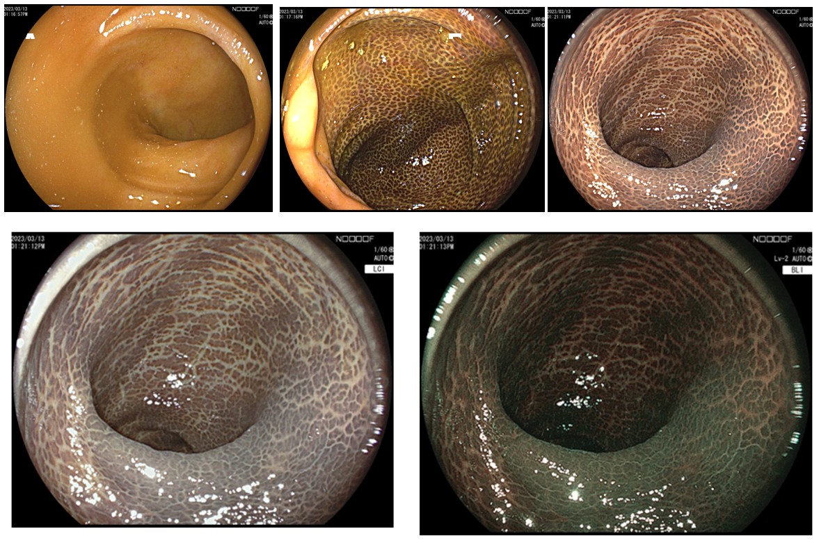
A 64-year-old woman with life-long constipation and extensive use of laxatives containing anthraquinone received colonoscopy examination during a health checkup. Lower gastrointestinal endoscopy showed diffuse dark pigmentation in the colonic mucosa that contrasted dramatically with the normal pink ileal mucosa of the ileocecal valve (Figure 1A, 1B).
Image Article
A 64-year-old woman with life-long constipation and extensive use of laxatives containing anthraquinone received colonoscopy examination during a health check- up. Lower gastrointestinal endoscopy showed diffuse dark pigmentation in the colonic mucosa that contrasted dramatically with the normal pink ileal mucosa of the ileo- cecal valve (Figure 1A, 1B). Colorful features of melanosis coli were observed by chromoendoscopy. Linked color imaging (LCI) and blue laser imaging (BLI) reviews of melanosis Image Article coli in the rectal mucosa recognized amazing differences in mucosal color (Figure 1C-E). Histopathology of the specimen revealed pigmented macrophages in the lamina propria, consistent with melanosis coli. Melanosis coli is considered to be associated with chronic use of laxatives containing anthraquinone [1]. The production of lipofuscin by epithelial cells causes the dark pigmentation in patients with chronic use of laxatives containing anthraquinone [2, 3]. Gastroenterologists may use chromoendoscopy to evaluate the benign aspects of melanosis ilei.
Conflicts of Interest: The authors have no conflicts of interest to declare.
Ethical Statement: The authors are accountable for all aspects of the work in ensuring that questions related to the accuracy or integrity of any part of the work are appropriately investigated and resolved. Written informed consent was obtained from the patient for publication of this “GI Image”.
References
-
Li D, Browne LW, Ladabaum U (2009) Melanosis coli. Clin Gastroenterol Hepatol 7(9): A20.
-
Nishikawa J, Tanaka T, Sugiyama T (2012) Melanosis ilei. Clin Gastroenterol Hepatol 10(11): A24.
-
Hung CY, Shyung LR, Chen MJ (2012) Pigmentation sparing on melanosis coli. Gastroenterology 142(3): e10-11.
- Management of Gallbladder Perforations: A Review
- From The Mouth to the Gut: The Oral Microbiome's Role in Promoting Gastrointestinal Disease
- Case Report: Intraductal Papillary Mucinous Neoplasm (IPMN) Complicated by Portal Vein Plaquing and Biliary Obstruction Mimicking Pancreatic Metastatic Malignancy
- Management of Non-Cirrhotic Portal Hypertension during Pregnancy: A Review
- Effectiveness of Omeprazole versus Pantoprazole for Symptomatic Relief of Gastro-Esophageal Reflux Disease (GERD)/ Acid Peptic Disease (APD): A Real-World Evidence (RWE) Study
- Case of Splenic Infarction; A Rare Presentation of Complicated Enteric Fever in a Pediatric Patient