Hypoglycaemic Mechanism of Manosrin from Anisopus Mannii N. E. Br.
<p>Medicinal Plants from various parts of the world have been tested for their hypoglycaemic properties and other related ailments. These have resulted into the isolation and purification of novel compounds which have been made into drugs and commercialized. The MeOH sub-fraction of Anisopus mannii N. E. Br.yielded ‘Manosrin’ (3, 23, 28 Trihydroxy-12-oleanen-3-O-(β-D-glucopyranosyl-(1,6)-β-D-glucopyranosyl(1,6)-β-D-xylopyranosyl)-28-O-β-D-glucopyranosyl-(1,6)-β-D-glucopyranoside) as a novel compound. To understand the mechanism of the drug action using ICR mice and other possible effect of the drug as hypoglycaemic compound. Manosrin was subjected to evaluations, such as oral glucose tolerance test (OGTT), standard toxicity studies, pathological investigations and route of efficiency, by intraperitoneal injection and oral administration using ICR mice, and the drug mechanism of action by co-treatment with isosorbide dinitrate and nifedipine as Ca+ and K+ ion channel regulators. Oral administration of the methanol sub-fraction (250 mg/kg bw) inhibited blood glucose increase, but stimulated blood glucose lowering in post prandial ICR mice. Significant body weight gains were observed both in the 2,000 and 5,000 mg/kg bw treated groups at day 3 to 14.All of the treated groups showed significant (P < 0.05) decrease in organ weights compared to the control group (kidney and liver). The BUN and creatinine levels decreased in Manosrin treated groups in the 2,000 and 5,000 mg/kg bw treated groups. However, a dose dependent elevation was observed for AST, ALT and Total bilirubin levels. The oral and intraperitoneal administration of Manosrin showed higher efficiency than Glibenclamide but lower than insulin. The co-treatment of ICR mice with ion channel regulators, showed that Manosrin was not dependent on the closure of K+ and opening of the Ca2+ channels. It could therefore be speculated that Manosrin may have enhanced insulin activity as a way of FBG reduction in the diabetic mice. Manosrin has exhibited a Thiazolidinedione-like action as its modus operandi. The hypoglycaemic effect observed prompts further investigation on other possible potentials of this compound in the management of Diabetes mellitus and related diseases.</p>
Introduction
Diabetes mellitus (DM) is a complex disease linked with a condition of high glucose level in the blood, or hyperglycaemia, resulting from deficiencies in the amount of insulin secretion, inaction, or both. Diabetic patients thus suffer from complications of macro- and micro vascular tissues, which if not properly managed, lead to frequent hospitalization and complications, together with increased risk of cardiovascular diseases [1]. Diabetes mellitus is a condition which afflicts approximately 387 million people across the globe [2]. The Centre for Disease Control and Prevention about 86 million people had prediabetes condition and 15–30% of them developed into full-blown diabetes [3]. The African story of Diabetes mellitus is that of ignorance and poverty. As the ailment spreads due to these factors, several researchers have subjected alternative therapies linked to different communities in and around Africa, South America and Asia [4]. Amidst must of these researches on traditional Medicinal plants or remedies, little is talked of concerning the mechanism of action for the potent hypoglycaemic remedies. Where these are discussed, the conclusions are usually postulations based on plausible assumptions. This research is aimed at investigating one of such results of a potent hypoglycaemic compound from an African traditional medicinal plants Anisopus mannii N. E. Br. [5]. The new compound of interest called ‘Manosrin’ (3, 23, 28–Trihydroxy-12-oleanen-3-O-(β-D- glucopyranosyl-(1,6)-β-D-glucopyranosyl-(1,6)-β-D- xylopyranosyl)-28-O-β-D-glucopyranosyl-(1,6)-β-D- glucopyranoside). The new compound though still at the infancy stage showed highly potent hypoglycaemic effect on mice [6]. This research strives to evaluate certain parameters like free radical scavenging activity of the crude extract, Oral glucose tolerance test (OGTT), acute oral toxicity studies and other methods towards the understanding of the compound’s mechanism of action, and the pathological studies of the hypoglycaemic compound.
Materials and Methods
Experimental Animals
Adult male and female ICR mice weighing between 24 – 27 g purchased from the Faculty of Medicine, Chiang Mai University and the National Laboratory Animal Center- Mahidol University, Thailand, were used. The mice were fed with the standard diet, water ad libitum and were maintained under the standard conditions of temperature, humidity and light (23 ± 1oC; 70% RH and 12 h light/dark). The experiments was complied with the Organization for Economic Co-operation and Development (OECD/OCDE: 425) Guidelines for Testing of Chemicals [7].
Production of the Diabetic Mice
Diabetic mice were produced as previously described by Zhou, et al. [8]. Briefly, the mice were injected at the tail vein with alloxan monohydrate in sterile normal saline solution at the dose of 75 mg/kg bw. Diabetes was confirmed on the third day after alloxan administration. The mice having blood glucose levels greater than 200 mg/dl were considered diabetic and selected for further study [9]. Fasting Blood Glucose (FBG) was assayed from the tail vein blood of the mice using Finetest Glucometer (Infopia Co., Ltd. Korea). Three doses of the plants extract (100, 200 and 400 mg kg-1 bw) [10] were orally administered to the 18 h fasted normal/diabetic mice (n = 5) using feeding tube [11]. Noticeable irritation or restlessness should not be observed after administration of the extracts. Blood glucose was measured hourly for 4 hrs. Control mice were fed with distilled water (oral), whereas insulin 0.5 IU/kg (injection/ip) and glibenclamide 1 mg/kg (oral) were used as reference hypoglycemic drugs.
Hypoglycaemic Compound (Manosrin)
The compound Manosrin was obtained from Anisopus mannii N. E. Br. according to the method described by Zaruwa, et al. [6].
Oral Glucose Tolerance Test (OGTT)
OGTT was performed by using the modified methods as described by Wu, et al. [12]. Briefly, normoglycemic mice were divided into five groups of 5 mice each. The mice were fasted for 18 h, and then treated with the agent orally using a feeding tube. After two hours, DW or different carbohydrates were administered as indicated; glucose (2.5 g/kg, o.p.), Sucrose (2.5 g/kg, o.p.), Corn starch (6 g/kg, o.p.) and lactose (6 g/kg, o.p). The blood glucose were tested at 0, 1, 2, 3 and 4 h. Glibenclamide (Glb - 1.0 mg/kg bw) and Insulin (Ins - 0.5 iu/kg bw) served as control (1 mg/kg bw, o.p.).
Co-treatment with Ca+ and K+ ion Channel Regulators
Normoglycemic mice were divided into four groups (n=5) with three other groups treated with Ca+
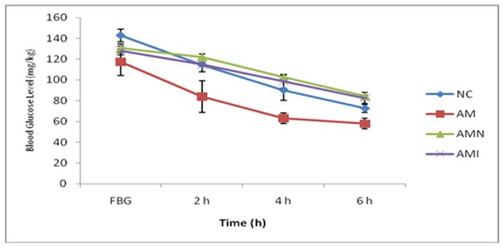
(Nifedipine) or K+ (Isosorbate dinitrate) channel regulators (Wu et al., 2010). The NC group was treated with normal saline (i.p.), AMS (250 mg/kg bw + normal saline, i.p.), AMN (250 mg/kg bw + normal saline + Nifedipine 13.6 mg/kg, o.p.) and AMI (250 mg/kg bw + normal saline + Isosorbide dinitrate 6.8 mg/kg bw) respectively. Blood glucose was tested at 0, 2, 4 and 6 h post treatment.
Intraperitoneal Injection and Oral Administration Studies
Mice were kept under standard conditions of temperature and humidity. The animals were fasted for 18 hours and 75 mg/kg bw alloxan monohydrate administered via the tail vein. Diabetic mice (< 200 mg/kg bw) were selected and grouped into three (n =5). To the control group was administered distilled water (DW), group 1 received AM (62.5 mg/kg bw, i.p.) and group 2 received AM (250 mg/kg bw, o.p.). Blood glucose was monitored at 0, 1, 2, 3 and 4 h post treatment.
Acute Oral Toxicity Studies
The acute oral toxicity study was done with modified protocol as described by Adeneye, et al. [13]. Manosrin was prepared using 2 ml normal saline solution and administered sequentially over a 12 h period using a feeding tube. Studies were conducted using the limit test of up and down procedure according to the OECD/OCDE Test Guidelines [7]. A total of five female mice were selected at random. The mice were fasted for 18 h. Body weight of each mouse was determined and dosed with equivalent of 2000 mg/kg bw (because no lethal effect was expected since the medicine has been in use among humans) of the MeOH sub-fraction dissolved in distilled water using a feeding tube. Mortalities, clinical signs, body weight changes and gross findings were monitored for 14 days (1 – 15) [14]. Each animal was observed each time for the first 10 min post dosing for signs of regurgitation and thereafter for every 30 min for the successive 6 h and daily for 14 days for possible lethal outcome. Behavioral manifestations of acute oral toxicity were also noted for each mouse. The same procedure was repeated with 5000 mg/kg bw of the Manosrin on different groups of mice.
Experimental Execution/Histopathological Examination
All mice were observed for 14 days and their body weights taken every 72 h. On the 14th day, each mouse was decapitated with a shape surgical blade and blood collected in clean appendorf tubes and centrifuged at 3000 rpm for 10 minutes. Sera were collected for each mouse in an appendorf tube, labelled and stored at 4OC, to be used for blood urea nitrogen (BUN), Creatinine, AST, ALT and total bilirubin analyses. The kidney and liver were removed, washed in normal saline, wiped and weighed on a digital balance and preserved in 10% formalin for histopathology examination. In the histopathological examination, the kidney and liver of mice from the three groups were embedded in paraffin, cut into 6 and 12 μm thick, respectively. They were later stained with haematoxylin and eosin and examined under a light microscope [15].
Ethical Clearance
The animal experimental methods were approved by Chiang Mai University Animal Ethics Committee, Protocol Number: 40/2552.
Statistical Analysis
The data are expressed as mean ± SEM or mean ± SD calculated from Microsoft excel 2003. Differences between 2 means were compared using student’s t- test. Values were considered statistically significant at p < 0.05.
Results and Discussion
Modified Oral Glucose Tolerance Test
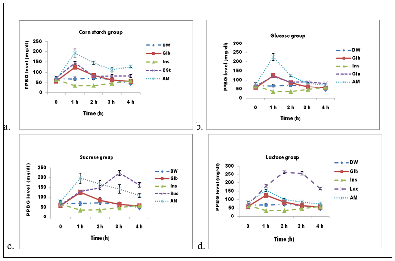
The hypoglycaemic effects of Manosrin in post-prandial hyperglycaemia in normal mice treated with various carbohydrates are shown in Figure 1. DW: distilled water, Cst: corn starch, Glu: glucose, Suc: sucrose, Lac: lactose, AM: Manosrin, Glib: Glibenclamide, Ins: insulin, PPBG: postprandial blood glucose. The test was performed by feeding the mice with various carbohydrates. The carbohydrates were given 2h after being fed with Manosrin (250 mg/kg bw) or Glibenclamide (1.00 mg/kg bw). a) Manosrin was ineffective on the PPBG levels in corn starch fed group.
b) Manosrin effectively reduced the PPBG levels in glucose fed group.
c)Manosrin was ineffective on the PPBG level in the sucrose fed group.
d) Manosrin effectively reduced the PPBG levels in the lactose fed group (*p < 0.05).
Effect of Oral and Intraperitoneal Administration of Manosrin
Both oral and intraperitoneal administration of the Manosrin showed similar effect.
Effects of Calcium (Nifedipine) and Potassium (Isosorbide) Channel Regulators on Hypoglycaemic Activity of Manosrin
Mean Body Weights (g) of Mice (n=5) at 14 days after Treated with Manosrin
The body and organ weights of mice treated with Manosrin at the doses of 2,000 and 5,000 mg/kg bw are shown in Tables 1 and 2. There were no animal deaths in the former group. Significant body weight gains were observed both in the 2,000 and 5,000 mg/kg bw treated groups at day 3 to 14. The relative body weights in the
2,000 mg/kg bw group were surprisingly higher than those in the 5,000 mg/kg bw group. The body weight gains were attributed to the effect of Manosrin. The mean ± SEM of kidney and liver weights with their relative organ weights compared to the body weights are shown in Table 2. Relative weight gains were observed for all the organs when compared to the untreated group.
| Mean body Weight (me: ±SEM | |||||||||||
|---|---|---|---|---|---|---|---|---|---|---|---|
| Day | Control | ||||||||||
| 2,000 mg/kg | 5,000 mg/kg | ||||||||||
| 0 | 24.20±0.86 | 23.40±0.52 | 24.40±0.66 | ||||||||
| 3 | 23.40±0.88 | 24.80±0.26*a | 24.80±0.26 | ||||||||
| 6 | 24.20±0.63 | 24.60±0.32*a | 25.20±0.63*a | ||||||||
| 9 | 24.60±0.52 | 25.20±0.26*a | 25.60±0.52*a | ||||||||
| 12 | 24.20±0.48 | 24.80±0.26*a | 25.80±0.63*a | ||||||||
| 14 | 25.20±0.26 | 25.40±0.32a | 26.00±0.58*a |
Table 1: Mean body weights (g) of mice (n=5) at 14 days after being treated with Manosrin at the doses of 2,000 and
| Control | Fraction of A. mannii (M) | |||||||||||||||||||
|---|---|---|---|---|---|---|---|---|---|---|---|---|---|---|---|---|---|---|---|---|
| Organ | ||||||||||||||||||||
| Entreated | RW (%) | 2000 (mg) | RW (%) | 5000 (mg) | RW (%) | |||||||||||||||
| Kidney | 0.3925±0.02 | 1.5699±0.09 | 0.3306±0.04* | 1.3223±0.17* | 0.3484±0.01* | 1.3937±0.03 | ||||||||||||||
| Liver | 1.4778±0.06 | 5.9113±0.24 | 1.3379±0.06* | 5.32517±0.23* | 1.3750±0.01* | 5.50±0.02* |
Table 2: Mean organ weights (g) of mice (n=5) at 14 days after being treated with the Manosrinat doses of 2,000 and Table 2: Mean
Table 2: Mean organ weights (g) of mice (n=5) at 14 days after being treated with the Manosrinat doses of 2,000 and Table 2: Mean organ weights (g) of mice (n=5) at 14 days after being treated with the Manosrinat doses of 2,000 and 5,000 mg/kg bw. *Significant difference from the control, p<0.05. RW: Relative weight = organ weight/body weight x 100
Liver and Kidney Function Tests of the ICR Mice treated with 2000 and 5000 mg/kg bw of Manosrin
All of the treated groups showed significant (P<0.05) decrease in organ weights in compared to the control group.
The BUN and creatinine levels decreased in Manosrin treated groups in the 2,000 and 5,000 mg/kg bw treated (Figures 4a and b). However, a dose dependent elevation was observed for AST, ALT and Total bilirubin levels.
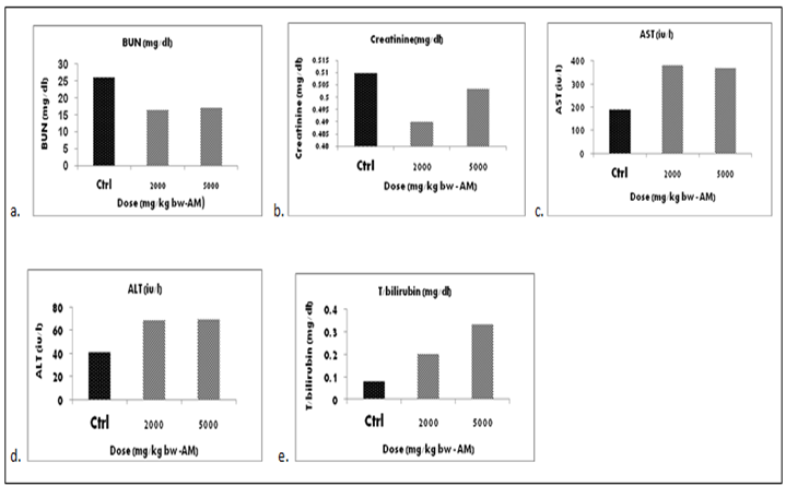
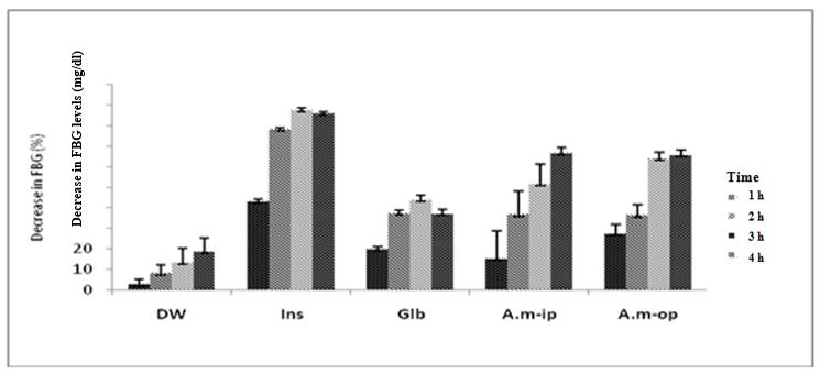
group which implied that, Manosrin may possess glucose oxidase and lactase (β-galactosidase) inhibition activity. A single dose treatment of Manosrin (ip or po) was able to decrease FBG levels at similar rates, thus, Manosrin showed efficiency in both fasting and postprandial states, which lead to the conclusion that Manosrin may have acted on the beta cells of the pancreas to stimulate insulin secretion and also enhanced insulin sensitivity by the cells [4]. Both oral and intraperitoneal administration of Manosrin showed similar outcome when administered orally or by intraperitoneal routes (Figure 2). The effect of a single dose of Manosrin (62.5 mg/kg bw and 120 mg/kg bw) were comparable with the standard drugs insulin (0.5 iu/kg bw- iv) and Glibenclamide (1.0 mg/kg bw- po). Manosrin treated mice showed higher efficiency in blood sugar reduction, than Glibenclamide, but lower than insulin (Figure 3). It was previously reported that glucose stimulates insulin secretion via K+ ATP channel pathway [17, 18] by closure of the cell surface ATP- sensitive K+ channels and the resulting opening of cell-surface voltage-dependent Ca2+ which facilitates the extracellular Ca2+ influx into beta cells and triggers the exocytosis of insulin [12]. This phenomenon was supported by the hypoglycaemic effect of Manosrin, which was observed in mice co-treated with isosorbide dinitrate and nifedipine. It indicated that, the hypoglycaemic activity shown by Manosrin was not dependent on the closure of K+ and opening of the Ca2+ channels. It could therefore be speculated that Manosrin may have enhanced insulin activity as a way of FBG reduction in the diabetic mice (Figure 3). In the acute toxicity studies with Manosrin_, no sign of behavioural changes, toxic signs as shown by the normal appearance of respiration pattern, colour of body surface, frequency of movement both voluntary and involuntary were observed. However, both body and organ weights were significantly (p <0.05) altered (Tables 1&2). Surprisingly, weight gains were higher in the mice treated with Manosrin of 2,000 mg/kg bw (6.7%) dosage than in 5,000 mg/kg bw (4.4%) group. A similar characteristic was also reported by Sani, et al., [19]. Evidences of likely impaired glomerular function were observed by the alteration of BUN and creatinine in the serum (Figure 4a and 4b). However, a reduction in serum level of blood urea could indicate that Manosrin may be tolerable to the body as observed with other medicinal plant extracts such as _Bridelia ferruginea [20]. It was assumed therefore that, the kidney was able to clear the waste products from the blood. The elevation of AST and ALT in Manosrin treated mice may be an indication of temporary liver dysfunction. The increased levels were possibly due to the leakage of these enzymes from the liver cytosol or other organs into the blood stream. The results concurs with the observation of Singh, et al. [21], that the administration of some medicinal plant extracts resulted in the elevated levels of AST and ALT in the animal serum. On the other hand, the severe elevation of bilirubin by 50% or 75% in the treated groups was probably as a result of the excess destruction of haemoglobin or that the liver was not actively treating the haemoglobin it received [22]. The histopathological results however did not show any damage to the cells (not shown), which lead to the conclusion that the abnormality observed in AST, ALT and total bilirubin may be from some undiscovered factors, probably some other phytochemical substances [23]. These were characterized by a reduction in the animal’s body and organ weights [15, 24]. The significant (p<0.05) increase in body weight, however was probably due to the effect of Manosrin on anti-diuretic hormone resulting in fluid retention similar to other medicinal plants [25]. It was possible that, since the traditional herbalists understood polydipsia and polyuria as symptoms of Diabetes Mellitus [1], this characteristic (fluid retention) was exploited for reducing the craving for water (taste) and polyuria, while it enhanced insulin secretion for the metabolic utilization of the blood glucose.
Conclusion
The emergence of Manosrin (3, 23, 28–Trihydroxy-12- oleanen-3-O-(β-D-glucopyranosyl-(1,6)-β-D- glucopyranosyl-(1,6)-β-D-xylopyranosyl)-28-O-β-D- glucopyranosyl-(1,6)-β-D-glucopyranoside) from Anisopus mannii N. E. Br. as apotent hypoglycaemic compound give credence to the assumption that, hypoglycaemic medicinal plants and remedies which serve as folk medicine in North-Eastern Nigeria, are indeed credible for the management of Diabetes mellitus. Though the safety studies were good, the mechanistic studies being preliminary showed a Thiazolidinedione- like action. The hypoglycaemic effect observed from Manosrin prompts further investigation on the possible potentials of this compound in the management of Diabetes mellitus and related diseases.
References
-
Kharroubi AT, Darwish HM (2015) Diabetes mellitus: The epidemic of the century. World J Diabetes 6(6): 850-867.
-
Fasanmade OA, Dagogo-Jack S (2015) Diabetes Care in Nigeria. Ann Glob Health 81(6): 821-829.
-
McCain J (2016) Prediabetes: Pre- Does Not Mean Preordained. Review. Managed Care, pp: 35-41.
-
Manosroi J, Zaruwa MZ, Manosroi A (2011) Potent hypoglycemic effect of Nigerian anti-diabetic medicinal plants. J Complement Integr Med 8(1): 1- 16.
-
Zaruwa MZ, Manosroi A, Akihisa T, Manosroi W, Rangdaeng S, et al. (2013) Hypoglycemic activity of _Anisopus mannii_ methanolic leaf extract in normal and alloxan induced diabetic mice. J Complement Integr Med 10(1): 1-10.
-
Zaruwa MZ (2011) Hypoglycaemic activity of Thai and Nigerian Medicinal Plants. A Doctoral Thesis submitted to the Faculty of Pharmacy, Chiang Mai University, Chiang Mai, Thailand.
-
OECD (2000) Guidance Document on Acute Oral Toxicity. Environmental Health and Safety Monograph Series on Testing and Assessment No 24.
-
Zhou T, Luo D, Li X, Luo Y (2009) Hypoglycemic and hypolipidaemic effects of flavonoids from lotus (_Nulumbo nuficera_ Gaertn) leaf in diabetic mice. Journal of Medicinal Plant Research 3(4): 290-293.
-
Cunha WR, Arantes GM, Ferreira DS, Lucarini R, Silva ML. et al. (2008) Hypoglycemic effect of _Leandra_ _lacunosa_ in Normal and alloxan- induced diabetic rats. Fitoterapia 79(5): 356-360.
-
Tanko Y, Yerima M, Mahdi MA, Yaro AH, Musa KY, et al. (2008) Hypoglycemic activity of methanolic stem bark of _Adansonnia digitata_ extract on blood glucose levels of streptozotocin induced diabetic Wistar rats. International Journal of Applied Research in National Product 1(2): 33-36.
-
Moufid A (2009) Mechanistic study of antidiabetic effect of _Chamaemelum nobile_ in diabetic mice. Advances in Phytotherapy Research (Res. Signpost) 37/661(2).
-
Wu C, Li Y, Chen Y, Lao X, Sheng L, et al. (2011) Hypoglycemic effect of _Belacanda chinensis_ leaf extract in normal and STZ-induced diabetic rats and its potential active fraction. Phytomedicine 18(4): 292- 297.
-
Adeneye AA, Ajagbonna OP, Adeleke TI, Bello SO (2006) Preliminary toxicity and phytochemical studies of the stem bark of _Musanga cecropiodes_ in rats. J Ethnopharmacol 105(3): 374-379.
-
Ha YW, Na YC, Seo JJ, Kim SN, Linhardt RJ, et al. (2006) Quanlitative and quantitative determination of ten major saponins in _Platycodi Radix_ by HPLC with ELSD and MS J Chromatogr A 1135(1): 27-35.
-
Huo Y, Winters WD, Yao DL (2003) Prevention of diet-induced type 2 diabetes in C57BL/6J mouse model by an anti-diabetic herbal formulation_._ Phytother Res17(1): 48-55.
-
World Health Organization (1999) Definition, Diagnosis and Classification of Diabetes Mellitus and its Complications. Report of WHO consultation. Department of Non-communicable Disease Surveillance. Geneva, Switzerland.
-
Yang G, Wu L, Jiang B, Yang W, Qi J, et al. (2008) H2S as a physiologic vasorelaxant: hypertension in Smice with deletion of cystathionine gamma-lyase. Science 322(5901): 587-590.
-
Seghers V, Nakazaki M, DeMayo F, Aguilar-Bryan L, Bryan J (2000) Sur 1 knockout mice. A model for K (ATP) channel-independent regulation of insulin secretion. J Biol Chem 275(13): 9270-9277.
-
Sani D, Sani S, Ngulde SI (2009) Phytochemical and microbial screening of the stem aqueous extract of _Anisopus mannii_. Journal of Medicinal Plant Research 3(3): 112-115.
-
Kolawole OM, Sunmonu TO (2010) Effect of wastewater treated with methanolic bark extract of _Bridelia ferrugine_a Benth on rat kidney and liver. Journal of Applied Sciences and Environmental Sanitation 5: 55-64.
-
Singh A, Duggal S, Suttee A (2009) Acanthus ilicifolius Linn. - Lesser Known Medicinal Plants with Significant Pharmacological Activities. International Journal of Phytomedicine 1(1): 1-3.
-
Tietz NW, Burtis CA (2000) Fundamentals of Clinical Chemistry. 6th (Edn.), Weekly 49(32): 729-731. 11.
-
Pereira PS, Franca SC, Olivera PVA, Breves CMS, Pereira SIV, et al. (2008) Chemical constituents from Tabernaemontana catharinensis root bark: a brief NMR review of indole alkaloids and _in vitro_ cytotoxicity_._ Quim Nova 31(1): 20-24.
-
Jahn AI, Gunzel PK (1997) The value of spermatology in male reproductive toxicology: Do spermatologic examinations in fertility studies provide new and additional information relevant for safety assessment? Reprod Toxicol 11(2-3): 171-178.
-
Chandrasekar R, Sivagami B (2017) Indian medicinal plant with diuretic activity. Indo American Journal of Pharmaceutical Research 7(1): 7359-7380.
- An Efficient and Affordable Method for Isolating Bone Marrow- Derived Mesenchymal Stem Cells from Swiss Albino Mice
- Superposition of Cryo-EM and AlphaFold Predictions of Dengue Antigen-Antibody Complexes
- Jugular-Applied Coherent Low-Level Laser Therapy Enhances Systemic Mitochondrial Metabolic Function and Antioxidant Response
- Role of OMC32 Polypeptide in Acrosin-Mediated Exocytosis during the Bovine Sperm Acrosome Reaction
- Association of Galectin-3 but not Laminin in Tamoxifen-Induced Growth Suppression in Breast Cancer MCF-7 Cells
- Effect of Different Wavelengths of Light on the Rate of Photosynthesis