Orally Administered Bisphenol A Impairs Normal Thyroid Functions Altering Thyroid Hormones Homeostasis in Female Wistar Rats
Background Objective: Bisphenol A (BPA) is an endocrine disrupting chemical used on a wide range in the industry. Studies on BPA suggests that both low and high doses of BPA affects plasma hormone levels There is increasing evidence that exposure to BPA, impair normal thyroid function, reduced bound circulating and tissue thyroid hormones but the effect of BPA varies at different levels of the thyroid system. The present study aims to investigate the effect of BPA on the thyroid gland of female rats. Materials and Methods: The rats received a daily oral administration of BPA (0.05 – 1.0 mg/kg for 13 weeks). Thyroid stimulating hormone (TSH), Thyroxine (T4) and Triiodithyroxine (T3) were assayed using Autochemical analyser and data obtained were subjected to statistical analysis with the SPSS software. Results: It was found that BPA at the eleven studied doses induced a significant increase in the thyroxin stimulating hormone (TSH), free thyroxin (FT4), total thyroxin (TT4) level, while the bound thyroxin is low compared to the control. The free triiodothyroxine (FT3) and total triiodothyroxine (TT3) were initially low at group 1 but at the other doses it were on the increase. The bound triidothyroxine are lower when compared to that of the control throughout the studied time intervals. Conclusion: These results suggest that BPA has thyroid toxic effects which are mediated by the oxidative stress resulting from the overproduction of free radicals, BPA may also participate in the thyroid gland function disturbances.
Introduction
Bisphenol A, is an organic compound that has two phenol functional groups. It is prepared by the condensation reaction of acetone and two equivalents of phenol Liu J, et al. [1] with hydrochloric acid Silver MK, et al. [2] (Figure 1).
$$ \mathrm {E} = \frac {1}{2} \mathrm {A} ^ {2} + \frac {1}{2} \mathrm {B} ^ {2} + \frac {1}{2} \mathrm {C} ^ {2} $$ (CH3)2CO + 2C6H5OH (CH3)2C(C6H4OH)2 + H2O

Bisphenol A has a phenolic odour with melting point of 155˚C and specific gravity of 1.060-1.195g/cm [3, 4]. It is soluble in nonpolar solvent [5]. Aerobic biodegration and biodegradation half-life for BPA in river water and soil is about 4.5 days Teppala S, et al. [3] and the photo-oxidation half-life for BPA in air is about 4 hours [6]. It is known that the global population is subject to repeated exposure to BPA is via the diet LaKind JS, et al. [7] with detectable metabolites in the urine [8]. Daily dietary intake of about 0.2μg/kg body weight in breast-fed babies and 1.5μg/kg body weight in adults Xiaoqian G, et al. [9] relevant doses of BPA causes increases in weight and size of the prostate gland, decreases in sperm efficiency, earlier puberty Shelby MD [10], and abnormalities in the oocytes [11]. Invernizzi P [12] showed that BPA trigers ductal and alveolar structures proliferations, development of ductal hyperplasia Murray TJ, et al. [13], modifications of the mammary gland architecture Moral R, et al. [14], mammary carcinogenesis Jenkins S, et al. [15], inflammatory cytokine dysregulation Jonathan ERB, et al. [16], and mitochondrial mediated apoptosis [17]. Health implications associated with BPA exposure include diabetes Lang IA, et al. [18], cardiovascular disease Meeker JD, et al. [19], altered liver enzymes activities Oguazu CE, et al. [20] and obesity-promoting effects [21]. BPA alters glucose homeostasis, increased pancreatic insulin and induced insulin resistance Magdalena PA, et al. [22], BPA induces oxidative stress Mourad IM, et al. [23], coronary artery disease Melzer D, et al. [24], activates Maxi-K ion channels Asano S, et al. [25], increased BP and decreased heart rate (HR) Bae S, et al. [26], increased risk of hypertension Erickson B [27], decreased efficiency of sperm production Mourad IM, et al. [23] and increased ovarian cancer cell proliferation [28]. According to this, Miyagawa et al. [29] reported impaired memory, increased aggressiveness Jones BA, et al. [30], alters anxiety Donald GS, et al. [31], loss or reduction of sexual dimorphisms Christensen KL, et al. [32], loss of sex difference in corticotrophin-releasing hormones Funabashi T, et al. [33], reduced the number of tyrosine by dioxylase-immunoreactive [34]. Studies on BPA suggests that both low and high doses of BPA affects plasma hormone levels There is increasing evidence that exposure to BPA, impair normal thyroid function, reduced bound circulating and tissue thyroid hormones but the effect of BPA varies at different levels of the thyroid system. Epidemiologic studies have revealed an association between BPA exposure and altered thyroid hormones Wang T, et al. [35], increased thyroid function [35]. The NHANES study also reported a suggestive inverse relationship between urinary BPA and total T4 and TSH [36]. Another survey observed a significantly negative correlation between serum BPA and FT4 level, but BPA was associated with TSH Sriphrapradang C, et al. [37] and reduced bound T4 women and decreased TSH in male [38]. BPA have several in vitro effects on the thyroid receptor β (TRβ), such as repressing the transcription of the thyroid receptor β Sheng ZG, et al. [39], and having an antagonistic role on the thyroid receptor β. There is an interference on the negative feed-back that the thyroid hormones carry out on TSH release Zoeller RT, et al. [40], accelerated embryonic development and advanced hatching through its effect on the thyroid receptor Ramakrishnan S, et al. [41], and interfered with T3 action during metamorphosis processes [42]. BPA exposure up-regulate genes involved in the synthesis of thyroid hormones in the thyroid follicle Gentilcore D, et al. [43], decrease in serum bound T4 level Du Y, et al. [44] and increase in free T4 levels [40]. The aim of this study is to establish the possible effects and physiological disposition of Bisphenol A on sex and thyroid hormones, in female wistar albino rats.
Materials and Methods
Study area
The study was carried out at Applied Biochemistry Lab, Nnamdi Azikiwe University, Awka, Nigeria and Biochemistry Lab, Gregory University Uturu, Abia state, Nigeria from June- September, 2021.
Methodology
Total 60 non-pregnant female rats of 5 weeks age were acclimatized in the laboratory for 7 days and randomly divided into 11 experimental groups of 5 rats each and respectively administered; 0.05, 0.1, 0.2, 0.3, 0.4, 0.5, 0.6, 0.7, 0.8, 0.9, and 1 mg of BPA/kg bw/day. The first group which served as control did not receive any treatment, but distilled water instead. The graded doses of BPA were dissolved in distilled water and administered by oral gavage using an intubation cannula (Lars Medicare Pvt. Ltd, New Delhi, India). Blood was obtained from the tail of the various groups by capillary action, weekly, after BPA administration for 13 weeks. Blood samples were processed for clinical assay.
Animals were housed in aluminum wire-mesh cages in a well-ventilated animal house with a 12 h dark/light cycle and at room temperature and were provided commercial rat pellets (Vital feed from Vital group of Company, Nigeria) and water ad libitum.
At the end of the experiments serum TSH, Thyroxine (T4) and Triiodithyroxine (T3) were assayed using an Autochemical analyser (Lx 20 pro Autoanalyser, Beckman Coulter, Woerden, Netherland and Chemwell chemical Analyzer, Manufacturer: Roche Hitachi, GMI.). All reagents were commercially obtained as already prepared kits. The kits for TSH, Thyroxine (T4) and Triiodithyroxine (T3) were purchased from Phoenix Pharmaceuticals, Burligame, CA; Enzo Life Sciences Inc, Boulevard Farmingale, NY; Diagnostic automation/Cortez Diagnostics Inc, Calabasas, CA. Individual tests were carried out according to the kit specifications as follow:
Thyroid Stimulating Hormone (TSH) Assay
Principle: The TSH ELISA test is based on the Principle of a solid phase enzyme linked immunosorbent assay. The assay system utilizes a unique monoclonal antibody directed against a distinct antigenic determinant on the intact TSH molecule. Mouse monoclonal anti-TSH antibody is used for solid phase immobilization. A goat anti-TSH antibody is in the antibody-enzyme (horseradish peroxidase) conjugate solution. The test sample is allowed to react simultaneously with the two antibodies, resulting in the TSH molecules being sandwiched between the solid phase and enzyme-linked antibodies. After 60-minute incubation at room temperature, the wells are washed with water to remove unbound labeled antibodies. A solution of TMB reagent is added and incubated for 20 minutes, resulting in the development of a blue color. The color development is stopped with the addition of stop solution, changing the color to yellow. The concentration of TSH is directly proportional to the color intensity of the test sample. Concentration is measured spectrophotometrically at 450 nm.
Kit Reagent
- Murine Monoclonal Anti-TSH-coated microtiter wells.
- Set of Reference Reagent.
- Enzyme Conjugate Reagent.
- TMB Reagent (One-Step).
- Stop Solution (1N HCl).
Procedure 1. The desired number of coated wells was placed in the holder. 2. 100 μl of blank, specimens, and controls were dispensed into appropriate wells. After which 100 μl of enzyme conjugate reagent was added into each well. The mixtures were thoroughly mixed for 30 seconds. And incubated at 25°C for 60 minutes.
3. The incubation mixture was removed by flicking plate contents. The microtiter wells were rinsed and flicked 5 times with distilled water and sharply striked onto absorbent paper towels to remove all residual water droplets. 4. 100 μl of TMB reagent was dispensed into each well. The mixture was gently mixed for 10 seconds and incubated at 25°C for 20 minutes. 5. The reaction was stopped by adding 100 μl of stop solution to each well and mixed for 30 seconds. 6. The concentrations were read at 450 nm within 15 minutes.
Thyroxine (T4) Assay
Principle: To measure T4 by competitive immunoassay techniques, a sample of serum or plasma containing the T4 to be quantified is mixed with labeled T4 and T4 antibody. In this T4 EIA, antibody to T4 is coated on a solid phase (microtiter well). A measured amount of patient serum and a constant amount of T4 labeled with horseradish peroxidase are added. During incubation, T4 in the patient sample and enzyme-labeled T4 compete for the limited binding sites on the T4 antibody. After 60-minute incubation at room temperature, the solid phase is washed with water to remove unbound-labeled T4. A solution of tetramethylbenzidine (TMB) is added and incubated for 20 minutes, resulting in the development of a blue color. The color development is stopped with the addition of 1N HCI, and the resulting yellow color is measured spectrophotometrically at 450 nm. The intensity of the color formed is proportional to the amount of enzyme present and is inversely related to the amount of T4 in the sample.
Kit Reagent
- Antibody-Coated Wells.
- Enzyme Conjugate Concentrate.
- Enzyme Conjugate Diluent.
- Reference Set.
- TMB Reagent.
- Stop Solution.
Procedure
- The desired numbers of coated wells were placed in the holder.
- 25μL of blank, specimens, and controls was pipetted into appropriate wells. Then 100 μL of working conjugate reagent was added into each well and was thoroughly mixed for 30 seconds. The mixture was incubated at 25°C for 60 minutes.
- The incubation mixture was removed by flicking plate contents into a waste container. The microtiter wells rinsed and flicked 5 times with distilled water and the wells were striked sharply onto absorbent paper towels to remove all residual water droplets.
- Then 100μL of TMB reagent was dispenced into each well and was mixed gently for 5 seconds. Again, the mixture was incubated at 25°C, in the dark, for 20 minutes.
- Stop solution (100μL) was added to each well to stop the reaction. The content of the wells were gently mixed for 30 seconds.
- The concentration was measured at 450nm within 15 minutes.
Triiodothyroxine (T3) Assay
Principle Competitive Enzyme Immunoassay – Analog Method for Free T3 The essential reagents required for a solid phase enzyme immunoassay include immobilized T3 antibody, enzyme-T3 conjugate and native free T3 antigen. The enzyme-T3 conjugate should have no measurable binding to serum proteins especially TBG and albumin. The method achieves this goal. Upon mixing immobilized antibody, enzyme-T3 conjugate and a serum containing the native free T3 antigen, a competition reaction results between the native free T3 and the enzyme-T3 conjugate for a limited number of insolubulized binding sites. The interaction is illustrated by the followed equation:
- T3 Antibody Coated Plate
- Wash Solution
- Substrate A
- Substrate B
- Stop Solution Procedure 1. The desired numbers of coated wells were placed in the holder. 2. 50 μl of the appropriate serum reference, control and specimen was pipetted into the assigned well. 100μl of fT3-enzyme reagent solution was added to all wells. The microplate was swirled gently for 30 seconds to mix; it was then covered and incubated 60 minutes at 25°C. 3. The content of the microplate was discarded by aspiration. Then 300μl of wash buffer was added to each wells and aspirated. The wash process was repeated two additional times for a total of three washes. 4. 100μl of working substrate solution was added to all wells and incubated for 15 minutes at 25°C. 5. 50 μl of stop solution was added to each well and mixed for 20 seconds. 6. The concentrations in each well were measured at 450nm.
Statistical Analysis
Differences between obtained values (mean±SD) were carried out by one-way analysis of variance (ANOVA) using SPSS software version 20.0 followed by the Tukey-Kramer multiple comparison test. At p≤0.05 was taken as a criterion for a statistically significant difference.
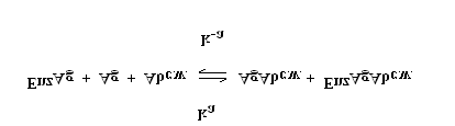
Ab c.w. = Monospecific Immobilized Antibody (Constant Quantity) Ag = Native Free Antigen (Variable Quantity) EnzAg = Enzyme-antigen Conjugate (Constant Quantity) AgAb c.w. = Antigen-Antibody Complex EnzAg Ab c.w. = Enzyme-antigen Conjugate -Antibody Complex Ka = Rate Constant of Association k-a = Rate Constant of Disassociation K = ka / k-a = Equilibrium Constant After equilibrium is attained, the antibody-bound fraction is separated from unbound antigen by decantation or aspiration. The enzyme concentration in the antibody-bound fraction is inversely proportional to the native free antigen concentration.
Kit Reagent
- Serum References
- fT3 –Enzyme Reagent
Thyroid Stimulating Hormone
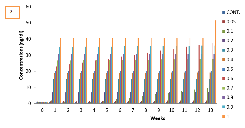
The experimental groups that were administered with 0.2-1 mg/kg bpa showed dose dependent significant increase in the concentration of thyroid stimulating hormones when compared with the control (Figure 2), the increase over time appeared to be relatively constant except 0.6mg/kg bpa test group that showed a constant increase in TSH with time and 1mg/kg bpa test group that showed high levels of thyroid stimulating hormone at week 13. The test exposed 0.05mg/kg bpa showed a nonsignificant increase in TSH that remained relatively constant with time. The group that received 0.1mg/kg bpa responded different during the study, it was observed that at the first week of exposure there was in increase in TSH, which gradually decreased through week 3, but suprisingly a consistant rise in the concentration of TSH was record from week 4 to 13. All the weeks of exposure showed a characteristic dose dependent effect of BPA on TSH, as the administered dose increases, the effect of BPA on TSH increases. The weeks of exposure showed sensitivity at two point (0.1 and 0.6) but was constant at other points of exposure.
Total Thyroxine (TT4)
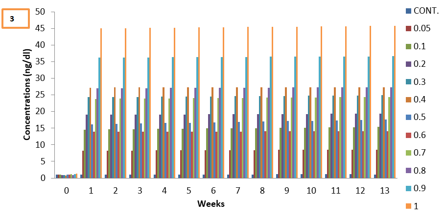
All the experimental groups that were administered with (0.05–1mg/kg) BPA showed significant increase in the concentration of TT4 when compared with the control (Figure 3), amidst the increase TT4, the test groups exposed to 0.05-0.4, and 0.6-1mgkg showed a dose dependent effect, while the TT4 level of the test groups exposed to 0.5mg/ kgbpa decreased relative to those of 0.4mg/kg, again those of 0.6mg/kg test group decreased further relative to those of 0.5mg/kgbpa test group. All the weeks of exposure showed a characteristic high but not dose dependent effect of BPA on TT4. The weeks of exposure showed nonsensitivity response with the lest observed effect at point 0.9.
Free Thyroxine (FT4)
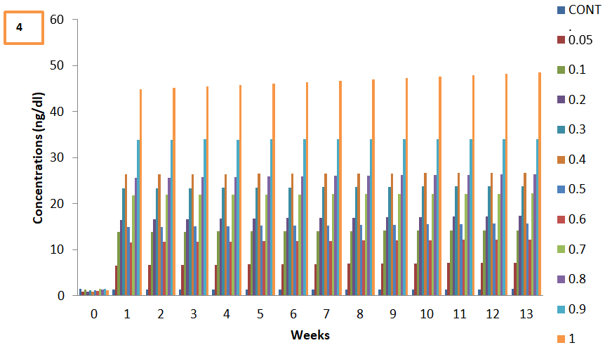
All the experimental groups that were administered with (0.05–1mg/kg) BPA showed significant increase in the concentration of FT4 when compared with the control (Figure 4), that appears to be relatively constant with time of exposure. Amidst the increase FT4, the test groups exposed to 0.05-0.4, and 0.6-1mgkg showed a dose dependent effect, while the FT4 level of the test groups exposed to 0.5mg/ kgbpa decreased relative to those of 0.4mg/kg, again those of 0.6mg/kg test group decreased further relative to those of 0.5mg/kgbpa test group. All the weeks of exposure showed a characteristic high and dose dependent effect of BPA on FT4 on the 0.05 – 0.4 doses of BPA. The weeks of exposure showed decrease response at piont 0.5 -06. mg/kgbpa and tend to rise from 0.7–1mg.kg.
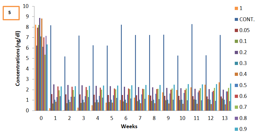
Conjugated Thyroxine (BT4)
All the experimental groups that were administered with (0.05–1mg/kg) BPA showed significant noncharacteristic decrease in the concentration of conjugated thyroxine (BT4) when compared with the control (Figure 5). All the weeks of exposure showed a noncharacteristic low but not dose dependent effect of BPA on BT4. All The weeks of exposure showed slight sensitivity response with the highest observed effect at point 1mg/kg.
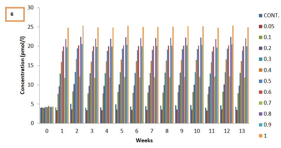
Total Triiodothyroxine (TT3)
The experimental groups that were administered with 0.1–1mg/kg bpa showed dose dependent significant increase in the concentration of total triiodothyroxine (TT3) when compared with the control (Figure 6), the increase over time appeared to be relatively constant except 0.7 and 0.9 mg/ kg bpa test group that showed a decrease in TT3 relative to 0.6 and 0.8mg/kg bpa test group respectively. The test exposed 0.05mg/kg bpa showed a nonsignificant decrease in TT3 that remained relatively constant with time (Figure 6). All the weeks of exposure showed a characteristic relative dose dependent effect of BPA on TT3, as the administered dose increases, the effect effect of BPA on TT3 increases. The weeks of exposure showed low level of TT3 at 0.7 point, falling below the control at point 0.05 .
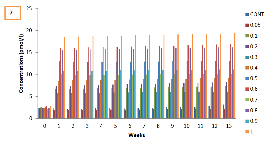
Free Triiodothyroxine (FT3)
The experimental groups that were administered with 0.1–1mg/kg bpa showed non dose dependent significant increase in the concentration of free triiodothyroxine (FT3) when compared with the control (Figure 7). The test exposed
0.05mg/kg bpa showed a nonsignificant decrease in FT3 that remained relatively constant with time (Figure 7). All the weeks of exposure showed a characteristic relative effect of BPA on FT3. The weeks of exposure showed low level of FT3 at 0.7 point, falling below the control at point 0.05 .
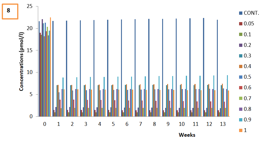
Conjugated Triiodothyroxine (BT3)
The experimental groups that were administered with 0.1–1mg/kg bpa showed significant decrease in the concentration of conjugated triiodothyroxine (BT3) when compared with the control (Figure 8). All the weeks of exposure showed a characteristic relative nondose dependent effect of BPA on BT3.
Discussion
In this research, it was observed that the thyroxin stimulating hormone (TSH), free thyroxin (FT4), total thyroxin (TT4) level were higher than that of the control, while the bound thyroxin is low compared to the control. The free triiodothyroxine (FT3) and total triiodothyroxine (TT3) were initially low at group 1 but at the other doses it were on the increase. The bound triidothyroxine are lower when compared to that of the control. There is increasing evidence that exposure to BPA, can impair normal thyroid function. From this experiment, BPA alters bound circulating and thyroid hormone concentrations and BPA were observed to alter thyroid hormone status.
Previous studies have revealed that BPA exposure altered thyroid hormones by increasing free triiodothyronine concentrations (FT3) Wang T, et al. [35], increased thyroid function Wang T, et al. [35], altered total T4 and TSH Meeker JD, et al. [36], altered FT4 level, and TSH Sriphrapradang C, et al. [37], reduced bound T4 [38]. Almudena VL, et al. [45] showed an increase in free T3 levels. Du, et al. [44] revealed decrease in serum bound T4 level. Zoeller, et al. [40] reported plasma free T4 increase.
BPA elicits its effect on the T4 hormone of the thyroid system as proposed: BPA inhibits the activity on the thyroid peroxidase (TPO) enzyme of the thyroid follicles, a key enzyme involved in the synthesis of T3 and T4 [39]. BPA affects the thyroid receptor β physiology (TRβ), by repressing the transcription of TRβ and having an antagonistic effect on the TRβ, thereby interfering with the negative feed-back that the thyroid hormones have on TSH release. Again, BPA alteration of thyroid hormones could be due to its ability to bind competitively with thyroid hormone transport proteins, and induce UDP-glucuronosyltransferase activity which amplified biliary excretion of thyroxine [46]. Also, BPA causes direct damage to the thyroid gland44. Increase estrogen trigers decreases serum bound T4, as proposed by Zhai, et al. [47].
For triiodothyroxine (T3) the effect of exposure to BPA could be explained by an increase of free T3 synthesis in the thyroid follicles and a higher T4 deiodination in the liver. BPA up-regulate genes involved in the synthesis of thyroid hormones in the thyroid follicle Gentilcore D, et al. [43] that gives support that BPA could trigger an increase of T3 synthesis. Also, considering the similar structure of BPA and T3, BPA could impair thyroid hormone action by inhibiting T3 binding to the TR and by suppressing its transcriptional activity. The BPA may silence or inhibiting the expression of a number of genes that govern normal thhyroid development.
BPA have a direct effect on thyroid follicular cell and leads to an altered expression of the genes involved in thyroid hormones synthesis. Other potential mechanisms for the effect of BPA on thyroid hormones include inhibiting thyroid hormone pathways Heimeier RA, et al. [48] and thyroid hormone receptor (TR) transcription suppression Sheng ZG, et al. [39], competitive binding with thyroid hormone for the thyroid plasma transporter [49]. Also, BPA act as an antagonist of thyroid hormone receptor because of its structural similarity to thyroid hormone. BPA inhibits thyroperoxidase activity, and accordingly block thyroid- induced metamorphism [42]. At receptor level BPA bind to thyroid hormone receptor as a ligand and act as antagonist, Inhibiting TR-mediated transcriptional activity [50, 51]. BPA displace TH from serum binding proteins and because it can displace TH from these binding proteins, it causes a decline in the bound serum hormone levels [5, 44]. Zhai, et al. [47] suggested another explanation to these results, is the enhanced T4 glucoronization in the liver and excretion of T4 into the bile, decreasing the amount of bound T4 in plasma. Another explanation could be an increase of T4 synthesis in the thyroid gland, driven by higher TSH levels induced after BPA exposure as observed in the study.
BPA have effects on the thyroid receptor β (TRβ), by repressing the transcription of the thyroid receptor β Sheng ZG, et al. [39], and having an antagonistic role on the thyroid receptor β. There is an interference on the negative feed-back that the thyroid hormones carry out on TSH release Zoeller RT, et al. [40], accelerated embryonic development and advanced hatching Ramakrishnan S, et al. [41], and interfered with T3 action during metamorphosis processes [52].There is increasing evidence that exposure to BPA, impair normal thyroid function Hatch EE, et al. [53], reduced bound circulating and tissue thyroid hormone concentrations [54, 55]. Bisphenol A have shown high affinity for TTR and prealbumin, competing with thyroid hormones for these plasma transporters and decreasing plasma bound thyroid levels [5, 56]. BPA alter thyroid hormone status Esseboom CK, et al. [57], increased thyroid function [35]. An inverse relationship between urinary BPA and total T4 and TSH have been reported [36]. There is a significantly negative correlation between serum BPA and FT4 level [37]. BPA exposure up-regulate genes involved in the synthesis of thyroid hormones in the thyroid follicle Gentilcore D, et al. [43], reduced bound T4 and decreased TSH Chevrier J, et al. [38], decrease in serum bound T4 level Du Y, et al. [44] and increase in free T4 levels [40]. Studies have shown that BPA can compete with T3 to bind with the thyroid plasma transporter, which could lead to a decrease in plasma bound T3 [49]. BPA impair thyroid hormone action by inhibiting T3 binding to the TR and by suppressing its transcriptional activity. Heimeier, et al. [48], showed that doses of BPA affected the gene expression that is controlled by T3 hormone. BPA altered the expression of many genes known to be turned on by thyroid hormone. BPA has the potential to affect genes that are regulated by thyroid hormone during human development. BPA also altered T3 gene expression. Also, according to Heimeier, et al. [48], BPA represents a serious risk to human development through disruption of T3 signaling pathways.
Conclusion
In conclusion, BPA is a potent inhibitor of thyroid hormone, which directs development. BPA alters a subset of important genes controlled by T3 that contribute to proper development, represents a serious risk to human development through disruption of TH signaling pathways. The study confirms past research showing BPA interferes with thyroid hormone activity. This research provides additional evidence into the fact that BPA interferes primarily with thyroid hormone level in addition to estrogen signals.
Significance Statement
BPA perturb thyroid hormone action throughout the body and interferes with thyroid hormone functions and homeostasis by inhibiting hormone synthesis, altering serum transport proteins, or increasing catabolism of thyroid hormones. Suggestively, the altered thyroid functions can lead to thyroid abnormalities, the risk for developing thyroid nodules and obesity. This study will help the researcher to uncover the critical areas of Bisphenol A toxicity on thyroid activity and thyroid hormone receptor that many researchers were not able to explore. Thus a new theory on the association between BPA exposure and thyroid function may be arrived at.
Conflict of Interest
The author hereby declares no conflict of interest.
Author’s Contribution
Chinenye E. Oguazu –analysis of thyroid hormones and result. Francis C. Ezeonu – supervisor, Enamali M.O and Charles C. Dike - Animal experiment which includes feeding, administration of graded doses of BPA. Ikechukwu K. Ubaoji and Chinenye E. Oguazu – statistical analysis and result presentation Charles C. Dike and Oguazu Chinenye E - blood sample collection and processing.
References
-
Liu J, Yu P, Qian W, Li Y, Zhao J, et al. (2013) Perinatal bisphenol A exposure and adult glucose homeostasis: identifying critical windows of exposure. PLoS One 8(5): e64143.
-
Silver MK, O’Neill MS, Sowers MR, Park SK (2011) Urinary bisphenol A and type-2 diabetes in U.S. adults: data from NHANES 2003–2008. PLoS One 6(10): e26868.
-
Teppala S, Madhavan S, Shankar A (2012) Bisphenol A and metabolic syndrome: results from NHANES. International Journal of Endocrinology 5: 98180.
-
Wang T, Li M, Chen B, Xu M, Xu Y, et al. (2012) Urinary bisphenol A (BPA) concentration associates with obesity and insulin resistance. J Clin Endocrinol Metab 97(2): E223–E227.
-
Vandenberg LN, Ehrlich S, Belcher SM, Jonathan NB, Dolinoy DC, et al. (2013) Low dose effects of bisphenol A: An integrated review of in vitro, laboratory animal, and epidemiology studies. Journal of Endocrine Disruptors 1(1): E25078.
-
Vandenberg LN, Chahoud I, Padmanabhan V, Paumgartten FJ, Schoenfelder G (2010) Biomonitoring studies should be used by regulatory agencies to assess human exposure levels and safety of bisphenol A. Environ Health Perspect 118: 1051-1054.
-
LaKind JS, Goodman M, Naiman DQ (2012) Use of NHANES data to link chemical exposures to chronic diseases: a cautionary tale. PLoS One 7(12): e51086.
-
Ye X, Tao LJ, Needham LL, Calafat AM (2008) Automated on-line column-switch HPLC-MS/MS method for measuring environmental phenols and prarbens in serum. Talanta 76(4): 865-871.
-
Xiaoqian G, Sheng WH (2014) Impact of bisphenol A on the cardiovascular system - epidemiological and experimental evidence and molecular mechanisms. Int J Environ Res Public Health 11(8): 8399-8413.
-
Shelby MD (2008) NTP-CERHR MonoGraph on the Potential Human reproductive and Developmental Effect of Bisphenol A. NTP CERHR MON.
-
Durando M, Kass L, Piva J, Sonnenschein C, Soto AM, et al. (2007) Prenatal bisphenol A exposure induces preneoplastic lesions in the mammary gland in Wistar rats. Environ Health Perspect 11(1): 580-586.
-
Invernizzi P (2013) Liver auto-immunology: the paradox of autoimmunity in a tolerogenic organ. J Autoimmun 46: 1-6.
-
Murray TJ, Maffini MV, Ucci AA (2007) Induction of mammary gland ductal hyperplasias and carcinoma in situ following fetal bisphenol A exposure. Reprod Toxicol 23(3): 383-390.
-
Moral R, Wang R, Russo IH, Lamartiniere CA, Pereira J, et al. (2008) Effect of prenatal exposure to the endocrine disruptor bisphenol A on mammary gland morphology and gene expression signature. J Endocrinol 196(1): 101-112.
-
Jenkins S, Raghuraman N, Eltoum I, Carpenter M, Russo J, et al. (2009) Oral exposure to bisphenol A increases dimethylbenzanthracene-induced mammary cancer in rats. Environ Health Perspect 117(6): 910-915.
-
Jonathan ERB, Hugo TD, Brandebourg AA (2009) Effects of bisphenol A on adipokine release from human adipose tissue: Implications for the metabolic syndrome. Journal of Molecular Cellular Endocrinology 304(1-2): 49-54.
-
Xia W, Jiang Y, Li Y, Wan Y, Liu J (2014) Early-life exposure to bisphenol A induces liver injury in rats involvement of mitochondria-mediated apoptosis. PLoS One 9(2): 234- 256.
-
Lang IA, Galloway TS, Scarlett A, Henley WE, Depledge M, et al. (2008) Association of urinary bisphenol Aconcentration with medical disorders and laboratory abnormalities in adults. JAMA 300(11): 1303-1313.
-
Meeker JD, Calafat AM, Hauser R (2010) Urinary bisphenol A concentrations in relation to serum thyroid and reproductive hormone levels in men from an infertility clinic. Environ Sci Technol 44(4): 1458-1463.
-
Oguazu CE, Ezeonu FC, Ubaoji KI, Anajekwu B (2015) Bisphol A Exerts a Transient Perturbation of Liver Function in Wistar Albino Rats at Acute and Sub-chronic Exposure Doses. Journal of Pharmacological Science and Bioscientific Research 5(3): 274-278.
-
Oguazu CE, Ezeonu FC (2017) Bisphenol A (BPA) Increases Blood Triglycerides and Low Density Lipoproteins in Albino Wistar Rats. Journal of Experimental Research 5(1): 24-27.
-
Magdalena PA, Ropero AB, Soriano S, Arevalo MG, Ripoll C, et al. (2012) Bisphenol-A acts as a potent estrogen via non-classical estrogen triggered pathways. Mol Cell Endocrinol 355(2): 201-207.
-
Mourad IM, Khadrawy YA (2012) The sensitivity of liver, kidney and testis of rats to oxidative stress induced by different doses of bisphenol A. International Journal of Life Science and Pharmacology Research 2(2): L19-L28.
-
Melzer D, Osborne NJ, Henley WE, Cipelli R., Young A, et al. (2012) Urinary bisphenol A concentration and risk of future coronary artery disease in apparently healthy men and women. Circulation 125(12): 1482-1490.
-
Asano S, Tune JD, Dick GM (2010) Bisphenol A activates Maxi-K (KCa1.1) channels in coronary smooth muscle. British Journal Pharmacology 160(1): 160-170.
-
Bae S, Kim JH, Lim YH, Park HY, Hong YC (2012) Associations of bisphenol A exposure with heart rate variability and blood pressure. Hypertension 60(3): 786-793.
-
Erickson B (2010) FDA raises flag on bisphenol A. Chem Eng News 88(4): 8.
-
Park YJ, Kwon WS, Oh SA, Pang MG (2012) Fertility- related proteomic profiling bull spermatozoa separated by percoll. J Proteome Res 11(8): 4162-4168.
-
Miyagawa K, Narita M, Narita M, Akama H, Suzuki T (2007) Memory impairment associated with a dysfunction of the hippocampal cholinergic system induced by prenatal and neonatal exposures to bisphenol-A. Neurosci Lett 418(3): 236-241.
-
Jones BA, Watson NV (2012) Perinatal BPA exposure demasculinizes males in measures of affect but has no effect on water maze learning in adulthood. Hormonal Behaviour 61(4): 605-610.
-
Donald GS, Beck MJ, Radovsky A, Garman HG, Freshwater LL, et al. (2010) Developmental neurotoxicity study of dietary bisphenol A in Sprague-Dawley rats. Toxicol Sci 115(1): 167-182.
-
Christensen KL, Lorber M, Koslitz S, Bruning T, Koch HM (2012) The contribution of diet to total Bisphenol A body burden in humans: results of a 48 hour fasting study. Journal of Environment International 50(1): 7-14.
-
Funabashi T, Kawaguchi M, Furuta M, Fukushima A, Kimura F (2004) Exposure to bisphenol A during gestation and lactation causes loss of sex difference in corticotropin-releasing hormone-immunoreactive neurons in the bed nucleus of the stria terminalis of rats. Psychoneuroendocrinology 29(4): 475-485.
-
Rubin BS, Murray MK, Damassa DA, King JC, Soto AM (2016) Perinatal exposure to low doses of bisphenol A affects body weight, patterns of estrous cyclicity, and plasma LH levels. Journal of Environmental Health Perspective 109(7): 675-680.
-
Wang T, Lu J, Xu M, Xu Y, Li M, et al. (2013) Urinary bisphenol a concentration and thyroid function in Chinese adults. Epidemiology 24(2): 295-302.
-
Meeker JD, Ferguson KK (2011) Relationship between urinary phthalate and bisphenol A concentrations and serum thyroid measures in U.S. adults and adolescents from the National Health and Nutrition Examination Survey (NHANES) 2007–2008. Environ Health Perspect 119(10): 1396-1402.
-
Sriphrapradang C, Chailurkit LO, Aekplakorn W, Ongphiphadhanakul B (2013) Association between bisphenol A and abnormal free thyroxine level in men. Endocrine 44(2): 441-447.
-
Chevrier J, Gunier RB, Bradman A, Holland NT, Calafat AM, et al. (2013) Maternal urinary bisphenol a during pregnancy and maternal and neonatal thyroid function in the CHAMACOS study. Environ Health Perspect 121(1): 138-144.
-
Sheng ZG, Tang Y, Liu YX, Yuan Y, Zhao BQ, et al. (2012) Low concentrations of bisphenol a suppress thyroid hormone receptor transcription through a nongenomic mechanism. Toxicol Appl Pharmacol 259(1): 133-142.
-
Zoeller RT, Bansal R, Parris C (2005) Bisphenol-A an environmental contaminant that act as a thyroid hormone receptor antagonist in vitro increase serum thyroxin and alters RC3/neurogranin expression in the developing rat brain. Endocrinology 146(2): 607-612.
-
Ramakrishnan S, Wayne NL (2008) Impact of bisphenol-A on early embryonic development and reproductive maturation. Journal of Reproductive Toxicology 25(2): 177-183.
-
Iwamuro S, Sakakibara M, Terao M, Ozawa A, Kurobe C, et al. (2006) Teratogenic and antimetamorphic effects of bisphenol A on embryonic and larval Xenopus laevis. General and comparative endocrinology 133(2): 189- 198.
-
Gentilcore D, Porreca I, Rizzo F, Ganbaatar E, Carchia E, et al. (2013) Bisphenol A interferes with thyroid specific gene expression. Toxicology 304: 21-31.
-
Du Y, Gao Y, Meng F, Liu S, Fan Z, et al. (2014) Iodine deficiency and excess coexist in china and induce thyroid dysfunction and disease: a cross-sectional study. PLoS One 9(11): e111937.
-
Almudena VL, Subramaniam P, Kurunthachalam K, Heather B, Patisaul DC, et al. (2015) Impact of Gestational Bisphenol A on Oxidative Stress and Free Fatty Acids: Human Association and Interspecies Animal Testing Studies. Endocrinology 156(3): 911-922.
-
Schmutzler C, Bacinski A, Gotthardt I, Huhne K, Ambrugger P, et al. (2007) The ultaviolet filter benzophenone 2 interferes with the thyroid hormone axis in rats and a potent _in vitro_inhibitor of human recombinant thyroid peroxidase. Endocrinology 148(6): 2835-2844.
-
Zhai W, Huang Z, Chen L, Feng C, Li B, et al. (2014) Thyroid endocrine disruption in zebrafish larvae after exposure to mono-(2-ethylhexyl) phthalate (MEHP). PloS one 9(3): e92465.
-
Heimeier RA, Das B, Buchholz DR, Shi YB (2009) The xenoestrogen bisphenol A inhibits postembryonic vertebrate development by antagonizing gene regulation by thyroid hormone. Endocrinology 150(6): 2964-2973.
-
Ishihara A, Nishiyama N, Sugiyama SI, Yamauchi K (2003) The effect of endocrine disrupting chemicals on thyroid hormone binding to Japanese quail transthyretin and thyroid hormone receptor. Gen Comp Endocrinol 134(1): 36-43.
-
Sun H, Shen OX, Wang XR, Zhou L, Zhen SQ, et al. (2009) Anti-thyroid hormone activity of bisphenol A, tetrabromobisphenol A andtetachlorobisphenol A in an improved reporter gene assay. Toxicology In vitro 23(5): 950-954.
-
Freitas J, Cano P, Veit CC, Goodson ML, David FJ, et al. (2010) Detection of thyroid hormone receptor disruptors by a novelstable _in vitro_ reporter gene assay. Toxicol In vitro 25(1): 257-266.
-
Iwamuro S, Yamada M, Kato M, Kikuyama S (2006) Effects of bisphenol A on thyroid hormone-dependent up-regulation of thyroid hormone receptor alpha and beta and down-regulation of retinoid X receptor gamma in Xenopus tail culture. Life Science 79(23): 2165-2171.
-
Hatch EE, Nelson JW, Stahlhut RW, Webster TF (2010) Association of endocrine disruptors and obesity: perspectives from epidemiological studies. International Journal Andrology 33(2): 324-332.
-
Goldey ES, Kehn LS, Lau C, Rehnberg GL, Crofton KM (1995) Developmental exposure to polychlorinated biphenyls (Aroclor 1254) reduces circulating thyroid hormone concentrations and causes hearing deficits in rats. Journal of Toxicology and Appllied Pharmacology 135(1): 77-88.
-
Morse DC, Groen D, Verman M, Amerongen CJV, Koeter HB, et al. (1993) Interference of polychlorinated biphenyls in hepatic and brain thyroid hormone metabolism in fetal and neonatal rats. Journal of Toxicology and Applied Pharmacology 122(1): 27-33.
-
Hamers T, Kamstra JH, Sonneveld E, Murk AJ, Kester MH, et al. (2006) In vitro profiling of the endocrine- disrupting potency of brominated flame retardants. Journal of Toxicological Sciences 92(1): 157-173.
-
Esseboom CK, Morse DC, Kuperus NW, Lutkeschipholt IJ, Paauw CGVD, et al. (1994) Effects of dioxins and polychlorinated biphenyls on thyroid hormone status of pregnant women and their infants. Pediatrics Research 36: 468-473.
- Superposition of Cryo-EM and AlphaFold Predictions of Dengue Antigen-Antibody Complexes
- Jugular-Applied Coherent Low-Level Laser Therapy Enhances Systemic Mitochondrial Metabolic Function and Antioxidant Response
- Role of OMC32 Polypeptide in Acrosin-Mediated Exocytosis during the Bovine Sperm Acrosome Reaction
- Association of Galectin-3 but not Laminin in Tamoxifen-Induced Growth Suppression in Breast Cancer MCF-7 Cells
- Effect of Different Wavelengths of Light on the Rate of Photosynthesis
- Nutritional, Therapeutic, and Environmental Effect of Oyster Mushrooms: An Editorial