Observational Effect of Chitin and Chitosan from Horseshoe Crab’s Exoskeleton on the Test Animal, Zebrafish Danio Rerio, Under the Laboratory Condition
Being a component of crustacean’s exoskeleton, chitin is one of main carbohydrate found on earth. Its derivative, chitosan can be isolated through deacetylation process of chitin. Due to their potential in pharmaceutical field, this study is using chitin and chitosan isolated from horseshoe crab to be tested on zebrafish (Danio rerio) developing embryo at various volumes. By using Danioscope, morphological defects on zebrafish embryos and larvae has been observed. We found out that both chitin and chitosan affect the zebrafish in several morphological abnormalities including dented tail, accumulation of blood, stunted development, enema and no pigmentation observed. Percentage of mortality was also determined for three different concentrations of chitin and chitosan which are at 10µl, 50µl and 100µl that been exposed to the embryo. Chitosan shows higher mortality rate compared to chitin. Both observations and data on morphological defects and mortality rate help in determining the toxicity level of chitin and chitosan on zebrafish and their effects. Our results suggested that both compounds are toxic at certain amount, hence having a high capability to be used in combating pathogens and tumour.
Introduction
Chitin or poly-β-(1→4)-N-acetyl-D-glucosamine is a polysaccharide that functions as the component of crustacean’s exoskeleton. For laboratory and industrial use, chitin usually prepared using crustacean’s exoskeleton. The crustacean’s are members of arthropods, which constitute of 10⁶ species from 1.2 x 10⁶ total species in animal Kingdom Tolaimate, et al. [1]. Chitosan is the N-deacetylated derivative of chitin, produced after deacetylation process.
The monomer of chitosan is D-glucosamine and N-acetyl-D-glucosamine that linked by β- (1,4)-glycosidic bond Hamed, et al. [2]. The chitin and chitosan have specific chemical properties which lead to varieties of applications such as in cosmetics, water engineering, food processing, wound dressing, and drug delivery system Rinaudo, et al. [3]. Chitin and chitosan from the horseshoe crab could be exploited more as it was found to have antibacterial and antioxidant activity Ismail N, et al.; Pati, et al. [4, 5] as well as potential biomaterials Wardiatno, et al. [6].
Zebrafish is a freshwater fish, belongs to the family Cyperinidae together with goldfish Meyer, et al. [7]. Natively, it can be found in South Asia from India to Thailand, Malay Peninsula and up to Yunnan. The use of zebrafish as an animal model serves many advantages because of their high human genetic homology, transparent embryos and larvae that allow easier observation Benchoula, et al. [8]. Hu, et al. [9] works on chitosan nanoparticle that used as drug or gene delivery vehicles in human gene therapy. When zebrafish embryos were exposed to chitosan nanoparticle, these embryos perform delay hatching and as the concentration of the nanoparticles increases, more embryos died. More information and observation should be gathered to be a reference for those who are working with the chitin and chitosan from the horseshoe crabs as to avoid unsustainable exploitation of this living fossil.
This study is aimed to observe the effect of the different concentration of raw chitin and chitosan from horseshoe crab carapace on the developing embryo of zebrafish under laboratory condition.
Methodology
A group of healthy adult wild type Zebrafish from Central Research and Animal Facility (CREAM) of the International Islamic University Malaysia (IIUM) were used for egg production. Fishes were maintained under standard laboratory conditions, which are at 27 ± 1°C with 12h light: 12h dark cycles [10]. They were feed three times a day with freshly hatched Artemia. Both male and female zebrafish that aged nine months old were put in spawning tank overnight with ratio of 1:2. The next morning, eggs were harvested. They will then be put in petri dishes containing embryo water tank that consist of methylene blue. They were carefully washed to remove debris. Any dead embryo or unfertilized eggs will be discarded by aspiration using plastic pipette.
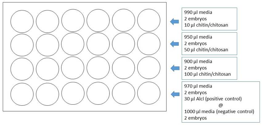
Medium has been prepared for zebrafish embryo to hatch. The recipe for media per 1 litre is 5.0 mM NaCl, 0.17 mM KCl, 0.33 mM CaCl and 0.33 mM MgSO₄. The organic solvents 0.1% dimethyl sulfoxide (DMSO) from Fisher Scientific was used. Since chitin is insoluble in water, it is needed to be diluted using this organic solvent. Healthy fertilized embryos were transferred into 24-well plate where each well contained two embryos. Treatments were carried out using two set of plates in each cycle. In total, 96 embryos were used. The total volume of each well was 1000µl, consisting of media and chitin or chitosan with respectable volume of 10µl, 50µl and 100µl. Positive control was 30µl of AlCl while and negative control was culture media itself. Figure 1 shows the volume and distribution of content in each well. All plates were incubated at 27 ± 1 °C in a temperature-maintained room.
The effects of treatments on the embryo were observed 24, 48 and 72hours post fertilization (hpf). Their mortality rates were evaluated, together with their morphological development defects. Software called ‘Danioscope’ is applied to gather all important data needed. The software is developed by a research company called Noldus Information Technology. Dinoscope can help to examine the morphology of zebrafish embryo larvae by using a few parameters including their activity, measurement on cardiovascular, blood flow and basic morphology. By using Danioscope, any developmental defects can be identified. The mortality rates were determined using the following equation: 𝑁𝑜. 𝑜𝑜𝑜 𝑑𝑒𝑎𝑑 𝑒𝑚𝑏𝑟𝑦𝑜𝑠
100 Percentage mortality(%) Total embryos x οοο =
Results
Morphological Defects of Zebrafish Embryos
Three dilutions of chitin, there dilutions of chitosan, AlCl as positive control and the media itself as negative control were applied onto the zebrafish embryos to investigate the effect of these eight different solutions on their development. Observations on morphological effects were determined using Danioscope, shown in Table 1 (chitin treatment), (Table 2) (chitosan treatment) and (Table 3) (positive/ negative control).
| Treatment | Morphological defect |
|---|---|
| 10 µl chitin | |
| Dented tail at 24 hpf | |
| Accumulation of blood in the yolk sac at 72 hpf | |
| 50 µl chitin | |
| Stunted development at 48 hpf | |
| 100 µl chitin | |
| Stunted development at 48 hpf | |
| 10 µl chitosan | |
| Stunted development at 48 hpf | |
| Yolk sac enema at 72 hpf | |
| 50 µl chitosan | |
| Accumulation of red blood cell in the head at 72 hpf | |
| Early pericardial enema at 48 hpf | |
| Stunted development at 48 hpf | |
| 100 µl chitosan | |
| No pigmentation at 72 hpf | |
| Stunted development at 72 hpf |
Table 1: Morphological observation of Danio rerio embryos and larvae that has been treated with different dilution of chitin from
| Treatment | Morphological defect |
|---|---|
| 30 µl AlCl | |
| Accumulation of red blood cell in abdomen at 24 hpf | |
| Normal embryo at 24 hpf | |
| Normal larvae at 72 hpf |
Table 2: Morphological observation of Danio rerio embryos and larvae that has been treated with 30 µl AlCl and 1000 µl media.
Mortality Rate of Zebrafish Embryos
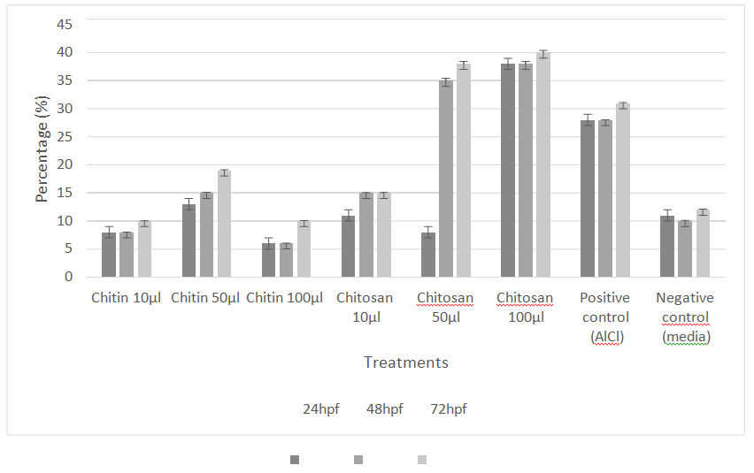
Six different dilutions of chitin and chitosan were applied onto zebrafish embryo at 0 hpf. The number of embryos that died was recorded three times, at 24 hpf, 48 hpf and 72 hpf. The percentage of mortality amongst zebrafish embryos was presented in Figure 2.
| Mean | |
| Chitin 10μl | 1.08 ± 0.289A |
| Chitin 50μl | 1.83 ± 1.267B |
| Chitin 100μl | 0.92 ± 0.669C |
| Chitosan 10μl | 1.58 ± 0.793D |
| Chitosan 50μl | 3.08 ± 3.8485E |
| Chitosan 100μl | 4.33 ± 4.097F |
| Positive control (AICI) | 3.44 ± 0.726G |
| Negative control (media) | 1.27 ± 1.223H |
Table 3: The mean of mortality developing embryos in three period (in hours post fertilization) when treated with different dilut
Table 4 showed the mean of mortality on developing embryos when treated with different dilution of chitin and chitosan. Obviously, the mortality rate is the highest on chitosan at highest concentration. There was a significant different between treatments as determined by one-way 132 ANOVA [F (7,88) =4.096, p=0.001, p<0.05].
Discussion
Morphological Defects of Zebrafish Embryos
These are overall morphological defects were observed on various dilutions of chitin and chitosan applied. Normal embryo showed when using 1000 µl media as how should it develop in optimum condition. The same treatment also showed the normal larvae that show perfect organogenesis as the organs developed well to help the larvae body system functions effectively.
Dented tail was shown by embryo that treated with 10 µl chitin, as there was kink at the tail part. This is known as kyphosis Pamanji, et al. [11]. The condition will affect its swimming ability when it hatched. Dented tail can also be observed as one of the defects. These malformations may due to decrease in collagen located in spinal column Murugesu, et al. [10] or some alteration in the composition of amino acid Celik, et al. [12] and maybe because of the inhibition of specific gene regulator named protein tyrosine kinase 7 (ptk7) [13].
Accumulation of red blood cell in the yolk sac was shown in10 µl chitin treatment, in the head when treated with 50 µl chitosan and in the abdomen when treated with 30 AlCl. This condition will of cause let to death soon due to inefficient blood circulation to the whole body. Accumulation of red blood cell also can be seen as a morphological defect that happens on zebrafish. When the accumulation occurs in the head, it may be due to serotonergic stimulation that will results in intravascular coagulation, together with cerebral haemorrhage. This was based on a study done by Andrade, et al. [14]. However, further study should be done to confirm the situation.
Stunted development was shown in embryos in many treatments including 50 µl chitin, 100 µl chitin, 10 µl chitosan and 100 µl chitosan where its development stops at certain age. These embryos were found in 72 hpf, with no eye pigmentation, less developed backbone, and delay hatching. They probably stop its development at 21-somite stage to prom-6 stage, that refers to development between 19.5 to 25 hpf. The prediction is based on comparison been done on the embryos observed with embryo sketches by Kimmel, et al. [15]. The situation occurred is known as stunted development. Yolk sac enema was shown by embryo treated with 10 µl chitosan enema occurs due to the accumulation of body fluid. In this case, the accumulation occurs in the yolk sac. Pericardial oedema can occurs due to accumulation of body fluid that originated from the failure of its osmoregulatory system [11]. If accumulation of water occurs in the head region, it will result in hydrocephalus. Murugesu, et al. [10] stated that enema can increase significantly with increase in concentration of toxicants. On the other hand, the condition of pericardial enema in the embryo was found when treated with 100 µl chitosan.
No pigmentation has also been observed as one of the morphological defects on zebrafish. Generally, pigmentation should start at retinal epithelium and the skin dorsolateral as early as 24 hpf Kimmel, et al. [15]. Pigment cell that controls colour changes in fish skin is known as melanophores.
Sugimoto, et al. [16] in their research reported that density of melanophores decreased by sporadic cell death, which also known as apoptosis. They suggested that the apoptosis process is controlled by sympathetic nervous system through sympathetic neurotransmitter, norepinephrine. The absence of pigment on zebrafish in this study probably due to the apoptosis to melanophores, due to the treatments that performed on them.
Mortality Rate of Zebrafish Embryos
Mortality can be indicated by the absence of heartbeat. Hu, et al. [9] suggested that chitosan is able to induce cellular apoptosis. The study proposed the usage of acridine orange staining, that will present a bright green fluorescent spot which indicates the occurrence of apoptosis. Chou, et al. [17] did a research on effect of low molecular weight chitosan on zebrafish. This substance showed high toxicity towards both zebrafish larvae and adults by attacking and damaging the cell membranes. Efflux of ions from the cell was induced those results in ion imbalance. Zebrafish larvae showed breakage of yolk, while the adult display damaged gill lamella that leads to hypoxia, then their death. Cheng, et al. [18] suggested that there are pores at the size of 0.17 µm² on the chorion. These pores obviously responsible for the exposure of chemicals to the embryo at their early stage.
The outcome of this research showed that both chitin and chitosan effect the zebrafish negatively although it is known that both compounds are used widely in pharmaceutical industry. Stine, et al. [19] stated that zebrafish is very sensitive at low concentration of chitin and chitosan that at last may leads to mortality. By determining the toxicity of chitin and chitosan, toxic dose that effect the development zebrafish can be recorded. This can be done using median lethal concentration (LC50). However, LC50 that can be construct using probit analysis needs a huge number of different concentrations of treatments up to 7 concentrations Wang, et al. [20] and 9 concentrations Ismail HF, et al. [21]. Since this study only use three different concentrations at 10µl, 50µl and 100µl for each compound, probit analysis cannot be constructed. Based on previous study on chitin and chitosan, LC50 has been recorded at 105 mg/L in and 548 mg/L Stine, et al. [19], 270 mg/L Wang, et al. [20], 200 mg/L [22].
This research is using chitin and chitosan with concentration of 2.5mg/mL. This concentration is very high compared to Wang, et al, (2016) [20] and Chou, et al. [17] that using 250mg/L and 0.2mg/L. This is the main factor that contributes to very high mortality rate on zebrafish in this study. Size of particles also should be taken into consideration. The lower the molecular weight of compound, the higher the rate of toxicity. Most study are using nanoparticle weight that ranging from 107 Da Wang, et al. [20] 3.6⁴Da and 1.2⁴
Da Chou, et al. [17] and size at 200 nm by Hu, et al. (2011) [9]. This study is using chitin and chitosan at 250µm in size that considered as big which gives less impact on zebrafish toxicity. Because of its toxicity at certain concentration, chitosan is widely used to eradicate tumour. A study by Jiang, et al. (2020) [23] used chitosan derivative, carboxymethyl chitosan oligosaccharide (CM-COS) as the inhibitor in growth of hepatocellular carcinoma. This study resulted in CM-COS as cytotoxic and able to inhibit tumour. At the same time, they suggested that positive charge CM-COS is attracted to negatively charged tumour cell as the main reason why this chitosan derivative only attack cancer cell but not normal cell, and it has been shown by MTT essay test conducted. Chiu, et al. (2017) [24] in their study used the combination of glycated chitosan and radiofrequency ablation on breast tumour. The combination showed a significant antitumor immune response by the rat and abled to eradicate the growth of tumour, together with suppressing its metastasis. This is supported by Zaharoff, et al. [25] whom demonstrated that chitosan enhanced humoral mediated immune response by increasing IgG production and induced cell-mediated immune response by stimulating CD4+ proliferation.
Conclusion
The effect of the different concentration of raw chitin and chitosan from horseshoe crab carapace on the developing embryo of zebrafish could be observed and recorded under a laboratory condition. Defects were shown by several characters such as reduced number of pigments, oedema and stunted development when larvae are exposed to as little as 10 µl chitin or chitosan extract. Mortality was significantly high (35-40%) after 24 hours exposure to 50 and 100 µl chitosan. This data could contribute to the knowledge gap on the concentration of chitin and chitosan used for any laboratory experiment on a model animal such as zebrafish. Further study particularly on the lethal concentration must be carried out to complete the understanding on the effect of the concentration of extract thus manipulation can be made for the target cells in study.
References
-
Tolaimate A, Desbrieres J, Rhazi M, Alagui A (2003) Contribution to the preparation of chitins and chitosans with controlled physico-chemical properties. Polymer 44(26): 7939-7952.
-
Hamed I, Ozogul F, Regenstein JM (2016) Industrial applications of crustacean by- products (chitin, chitosan, and chitooligosaccharides): A review. Trends in Food Science and Technology 48: 40-50.
-
Rinaudo M (2006) Chitin and chitosan: Properties and applications. Progress in Polymer Science 31(7): 603- 632.
-
Ismail N, Nadzari F, Ghaffar IHA, Rahman NA, Taib M, et al. (2011) Potential of Antibacterial and Antifouling From Horseshoe Crabs, Tachypleus gigas and Carcinoscorpius rotundicauda Against Gram-Positive and Negative Bacteria. Empowering Science, Technology and Innovation towards a Better Tomorrow, pp: 717-720.
-
Pati S, Chatterji A, Dash BP, Nelson BR, Sarkar T, et al. (2020). Structural characterization and antioxidant potential of chitosan by γ-irradiation from the carapace of horseshoe crab. Polymers 12(10): 2361.
-
Wardiatno Y, Riyanto B, Iskandar NA, Kleinertz S, Funch P, et al. (2021) A New Marine Biomaterial: The Shell of Mangrove Horseshoe Crabs, Carcinoscorpius rotundicauda (Latreille, 1802) Emphasizing Its Physico- Chemical Characteristics. Frontiers in Marine Science.
-
Meyer A, Ritchie PA, Erik WK (1995) Predicting developmental processes from evolutionary patterns: a molecular phylogeny of the zebrafish (Danio rerio) and its relatives. Philosophical Transactions of the Royal Society of London. Series B: Biological Sciences 349(1327): 103-111.
-
Benchoula K, Khatib A, Jaffar A, Ahmed QU, Sulaiman WMAW, et al. (2019) The promise of zebrafish as a model of metabolic syndrome. Exp Anim 68(4): 407-416.
-
Hu YL, Qi W, Han F, Shao JZ, Gao JQ (2011) Toxicity evaluation of biodegradable chitosan nanoparticles using a zebrafish embryo model. Int J Nanomedicine 6: 3351-3359.
-
Murugesu S, Khatib A, Ahmed QU, Ibrahim Z, Uzir BF, et al. (2019) Toxicity study on Clinacanthus nutans leaf hexane fraction using Danio rerio embryos. Toxicology Reports 6: 1148-1154.
-
Pamanji R, Yashwanth B, Bethu MS, Leelavathi S, Ravinder K, et al. (2015) Toxicity effects of profenofos on embryonic and larval development of Zebrafish (Danio rerio). Environmental Toxicology and Pharmacology 39(2): 887-897.
-
Celik ES, Kaya H, Yilmaz S (2012) Effects of phosalone on mineral contents and spinal deformities in common carp (cyprinus carpio, l.1758). Turkish Journal of Fisheries and Aquatic Sciences 12(2): 10.
-
Hayes M, Gao X, Yu LX, Paria N, Henkelman RM, et al. (2014) Ptk7 mutant zebrafish models of congenital and idiopathic scoliosis implicate dysregulated Wnt signalling in disease. Nature Communications 5: 1-11.
-
Andrade TS, Oliveira RD, Silva MLD, Zuben MVV, Grisolia CK, et al. (2018) Exposure to ayahuasca induces developmental and behavioral alterations on early life stages of zebrafish. Chemico-Biological Interactions 293: 133-140.
-
Kimmel CB, Ballard WW, Kimmel SR, Ullmann B, Schillingb TF (1995) Stages of embryonic development of the zebrafish. Dev Dyn 203(3): 253-310.
-
Sugimoto M, Uchida N, Hatayama M (2000) Apoptosis in skin pigment cells of the medaka, Oryzias latipes (Teleostei), during long-term chromatic adaptation: The role of sympathetic innervation. Cell and Tissue Research 301(2): 205-216.
-
Chou CM, Mi FL, Horng JL, Lin LY, Tsai ML, et al. (2020) Characterization and toxicology evaluation of low molecular weight chitosan on zebrafish. Carbohydrate Polymers 240: 116164.
-
Cheng J, Flahaut E, Shuk HC (2007) Effect of carbon nanotubes on developing zebrafish (Danio rerio) embryos. Environ Toxicol Chem 26(4): 708-716.
-
Stine JS, Harper BJ, Conner CG, Velev OD, Harper SL (2021) In vivo toxicity assessment of chitosan-coated lignin nanoparticles in embryonic zebrafish (_Danio_ _rerio_). Nanomaterials 11(1): 1-12.
-
Wang Y, Zhou J, Liu L, Huang C, Zhou D, et al. (2016) Characterization and toxicology evaluation of chitosan nanoparticles on the embryonic development of zebrafish, Danio rerio. Carbohydrate Polymers 141: 204- 210.
-
Ismail HF, Hashim Z, Soon WT, Rahman NSA, Zainudin AN, et al. (2017) Comparative study of herbal plants on the phenolic and flavonoid content, antioxidant activities and toxicity on cells and zebrafish embryo. J Tradit Complement Med 7(4): 452-465.
-
Saleh HA, Younes N, Rasool K, Younis MH, Prieto RM, et al. (2019) Impaired liver size and compromised neurobehavioral activity are elicited by chitosan nanoparticles in the zebrafish embryo model. Nanomaterials 9(1): 1-13.
-
Jiang Z, Wang S, Hou J, Chi J, Wang S, et al. (2020) Effects of carboxymethyl chitosan oligosaccharide on regulating immunologic function and inhibiting tumor growth. Carbohydrate Polymers 250: 116994.
-
Chiu HY, Leu JD, Chang CY, Lee YJ, Chen WR (2017) Combination of radiofrequency ablation and glycated chitosan as treatment on a syngeneic breast tumor model. Anticancer Res 37(6): 2965-2974.
-
Zaharoff DA, Rogers CJ, Hance KW, Schlom J, Greiner JW (2008) Immune Responses to Subcutaneous Vaccination 25(11): 2085-2094.
- Superposition of Cryo-EM and AlphaFold Predictions of Dengue Antigen-Antibody Complexes
- Jugular-Applied Coherent Low-Level Laser Therapy Enhances Systemic Mitochondrial Metabolic Function and Antioxidant Response
- Role of OMC32 Polypeptide in Acrosin-Mediated Exocytosis during the Bovine Sperm Acrosome Reaction
- Association of Galectin-3 but not Laminin in Tamoxifen-Induced Growth Suppression in Breast Cancer MCF-7 Cells
- Effect of Different Wavelengths of Light on the Rate of Photosynthesis
- Nutritional, Therapeutic, and Environmental Effect of Oyster Mushrooms: An Editorial