Haematological and Histological Examination of Oreochromis niloticus Fed Oxalic Acid Supplemented Diets Challenge with Escherichia coli
Heamatological and pathohistological evidences of Oreochromis niloticus mean 7.00g ± 0.04 fed oxalic acid supplemented diets was investigated under bio-essays laboratory culture for 90 days fed trial, the haematology indices of O. niloticus indicate that there were significant variations (p<0.05) in the haemoglobin (Hb) Pack Cell Volume PCV, with highest WBC was recorded in OAC1 and lowest RBC, the highest MCHC was significant different fron the CTR likewise MCV. This pathohistological examination indicate that the liver, kidney and intestine of fish challenged with entero-toxigenic bacteria (Escherichia coli) exhibited diffused, necrotized, hepatic variation tissues with fatty changes in the hepatic parachyma and vacuolation of hepatic parenchyma cell wall. The finding indicates that the varying inclusion level of oxalic acid in supplemented fed O. niloticus had positive effects on the health status as it increase immmonic system of the fish to fight redant the growth of Escherichia coli bacteria.
Introduction
Heamatological and pathohistological evidences have been used as important biomarkers in environmental monitoring that allows examining specific target organs in livestock production. supplementation of diets with organic acids have been reported to boost growth through improved digestion, absorption, and retention of a varieties of nutrients and minerals [1, 2]. Pathohistology analyses of internal organs stand out as a tool to indicate the health status of aquatic organism in their environment [3, 4]. Many toxicants have been shown affecting the growth parameters and reproduction with evidence of tissue damage [5, 6, 7], E. coli is bacterial infection of freshwater parasite that infecting physiological structure aquatic organism [8], thus post a tract on health status of the fish. It is only by examining appropriate tissues under high magnification that these problems can be identified. The tissues are essential before a competent histopathologist can identify genuine pathological change.
Fish sampled from different ages and species of fish will have a different appearance and these must be recognised in order to interpret sections correctly [9]. Recognition of these differences and a degree of common sense is paramount when it comes to interpretation and drawing any conclusions for any specific case examination [10].
If global aquaculture industry to continue its growth, there must be a response to the challenges that limiting the industry e.g environmental burdens, fish feed value, fish growth and health status. Research seems to be the feasible solutions to these challenges as innovations are needed in several areas to realize aquaculture potentials and to assess the haematology indices of O. niloticus fed varying levels of oxalic acid supplemented diets and investigate the immune response of O. niloticus fed oxalic acid supplemented diets and challenged with entero-toxigenic bacteria (Escherichia coli).
Materials and Methods
The research work was carried out at the Department of Fisheries and Aquaculture Technology Teaching and Research Farm, The Federal University of Technology, Akure, (FUTA), School of Agriculture and Agricultural Technology, FUTA. Haematology indices of O. niloticus fed with various supplement diets of oxalic acid was carried out on experimental fish (O. niloticus) at the Animal Production and Health Laboratory, The Federal University of Technology, Akure, Nigeria for the residue effects of oxalic diets on O. niloticus for 90 days indoor fed trial. The blood from the fish was collected from the cardiac puncture/cutting the cardiac peduncle using different 5 ml heparinized syringes, with ethylene diamine tetra acetic acid (10 ml EDTA) as anticoagulant. Fifteen blood specimens (each per tank) were taken from experimental fish for blood analysis. The heamatology parameters that were carried out is as follows.
White Blood Cells Count: The total white blood cell count was performed by taking one drop of the blood smeared on slide and air dried at room temperature. This was later fixed in 95% methanol and stained with Giemsa stain (Analar grade) for 20 minutes and mounted. White blood cells were identified using Olympus BX 50 microscope (Olympus UK).
Red Blood Cells Count: The blood cells (erythrocytes) was counted in Neubauer hemocytometer counting chamber using Olympus BX 50 microscope (Olympic UK). Number of cells counted was expressed as (106 mm-3).
Haemoglobin Concentration: This was performed with aid of Haemoglobinometer (Sigm, England). Standard Shilonometer N/10 HCL and 0.02ml pipette were used for the estimation. The graduated tube was filled to 20ml mark; 0.02ml of blood was added and mixed thoroughly until the colour matches the standard. Haemoglobin concentration was determined by the amount of solution in the graduated tube expressed in percentage.
Pack Cell Volume: Non-clotted blood was drawn by capillary action into micro-haemocrit tubes to determine the Pack Cell Volume (PCV). One end of the tube was sealed with a synthetic sealant. The sealed tube was centrifuged in a micro-haemocrit centrifuge at 10500 revolution per time (rpm). The micro-haematocrit reader was used to measure the PCV and expressed in percentage.
Mean Corpuscular Haemoglobin Concentration (MCHC): This is the concentration of haemoglobin in a unit of erythrocytes. It was calculated from the haemoglobin value (HB) in gL-1 and from the haematocrit value (PCV). MCHC=Hb/PVC x 100 Mean Corpuscular Haemoglobin (MCH): The corpuscular haemoglobin concentration expresses as the concentration of haemoglobin in unit volume of erythrocyte. It was calculated from the haemoglobin value (Hb) and from the Red Blood Cell (RBC) according to the following formula. MCH= Hb/ RBC x 100 Mean Cell Volume (MCV): This was calculated from pack cell volume (PVC) and from Red Blood Cell (RBC) according to the following formula. MCV= PVC/RBC x 100 Pathohistological Examination: Histopathological examinations were carried out to assess gills, livers, kidneys, heart and intestines of test fish for each treatment. These organs and tissues were preserved in sampling bottles containing 10% formalin before examination. These organs and tissues were cut out of the fish. The organs were dehydrated in periodic acid Schiff”s reagent (PAS) in graded levels of 50%, 70%, 90% and 100% alcohol for 3 days, to allow paraffin wax to penetrate the tissue during embedding. The organs were then cleaned and embedded in melted wax and carefully sliced into thin sections with a rotatory microtome (5μm thick). The cut sections were again cleaned by placing them in warm water (38 °C) from where they were transferred into clean slides and oven- dried at 58°C for 30 minutes to melt the wax and stained with Harris’ haematoxylin–eosin (H and E) stain. The slides containing sectioned tissues was cleaned using xylene and graded levels of 50%, 70%, 90%, 95% and 100% alcohol for two minutes each and stained in haematoxylin-eosin for ten minutes and mounted in dipterex on glass slides, to obtain their photomicrography, the stained sections were examined and photographed at different magnifications (x40, x100 and x400) by means of a binocular light microscope (Olympus Japan 312545) fitted with a digital camera (Olympus CH XSZ- 107BN), a photographic attachment (Olympus C35 AD4) and an automatic light exposure unit (Olympus PM CS5P).
Results
The haematology indices of blood of O. niloticus fed oxalic acid supplemented diets in Table 1 shown that, there were significant variations (p<0.05) in the haemoglobin (Hb) of the fish on diets, however there is different in treatment OAC2, OAC3 and OAC1. The PCV increase across from control to other treatments, but their slight relationship in the PCV treatments OAC2 and OAC4 also OAC3, OAC5 and OAC1. While highest WBC was recorded in OAC1 and lowest RBC was recorded in OAC1. The highest MCHC was recorded in OAC4 followed by OAC3, OAC2 and OAC5 which was significant different from CTR likewise highest MCV was record in the CTR. However, there were no significant variations (p>0.05) in MCH.
| OAC1 | OAC2 | OAC3 | OAC4 | OAC5 | |
|---|---|---|---|---|---|
| Hb (g/dl) | 7.20 ± 0.06a | 9.40 ± 0.21c | 8.53 ± 0.12b | 8.57±0.88b | 8.20±0.58b |
| PVC (%) | 23.00 ± 0.58a | 24.00±0.57c | 24.33±0.88 ab | 26.00±0.58bc | 25.00±0.57 ab |
| WBC(x103/mm3) | 8.10±0.15c | 5.30±0.26a | 7.13±0.23b | 7.07±0.24b | 7.00±0.36b |
| RBC (μ/l)(μ/dl) | 2.20±0.00a | 3.10 ± 0.58c | 2.7 ± 0.06b | 2.80 ± 0.03b | 2.75±0.58b |
| MCHC(g/dl) | 31.35 ± 0.95a | 33.57 ± 0.18ab | 34.14±1.17b | 35.97±0.56ab | 33.34±0.21ab |
| MCH(pg) | 328.81±14.69a | 303.19 ± 1.86a | 317.03 ±13.52a | 325.95±0.60a | 303.08±1.29a |
| MCV(fl) | 10.53 ± 0.70b | 9.03 ± 0.02a | 9.02 ± 0.15a | 9.28 ± 0.18a | 9.09±0.02a |
Table 1: Haematological profile of Oreochromis niloticus fed varying levels of oxalic acid supplemented diets (Mean±SE).
Pathohistological examination of Oreochromis niloticus fed oxalic acid supplement diets (Figures 1-6)
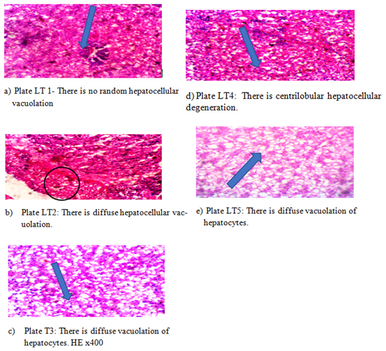
Plate LT1 – LT5: Photomicrograph of section of liver (Mag x400) Figure 1: Effects of oxalic acid on mineral availability and health of O. niloticus (7 ±0.4g).
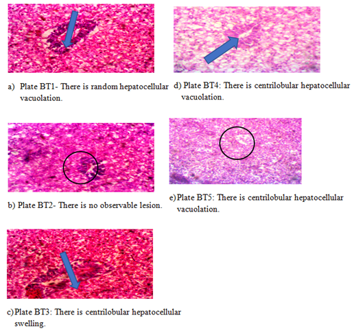
Plate BT1 – BT5: Photomicrograph of section of liver with E.coli (Mag x400) Figure 2: Effects of oxalic acid on mineral availability and health of O. niloticus before bath culture (7 ± 0.4g).
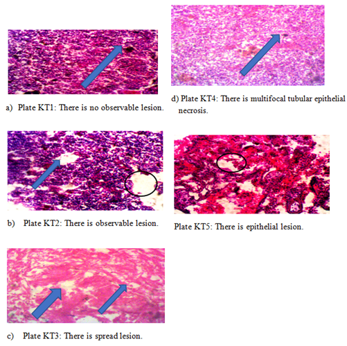
Plate KT1 – KT5: Photomicrograph of section of kidney (Mag x400) Figure 3: Effects of oxalic acid on kidney and health of O. niloticus (7 ±0.4g).
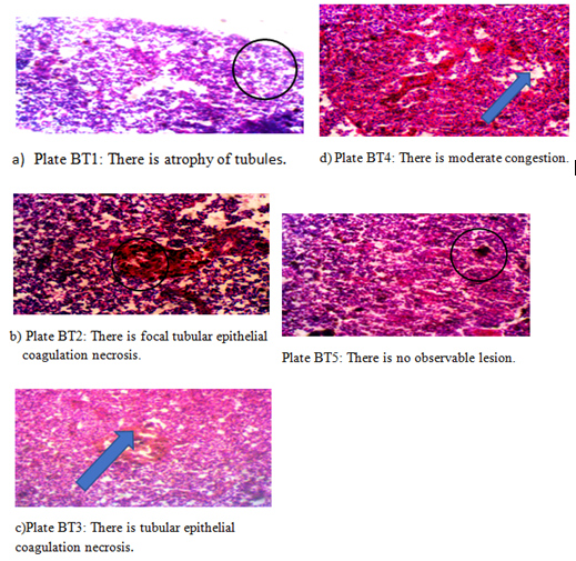
Plate KT1 – KT5: Photomicrograph of section of kidney (Mag x400) Figure 4: Effects of oxalic acid on kidney and health of O. niloticus on bath culture.
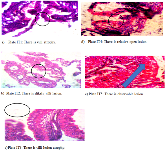
Plate IT1-IT5: Photomicrograph of section of intestine on 90 days (Mag x400) Figure 5: Effects of oxalic acid on intestine and health of O. niloticus (7 ±0.4g).
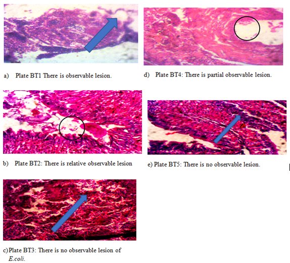
Plate BT1-BT5: Photomicrograph of section of intestine. (Mag x400) Figure 6: Effects of oxalic acid on intestine of O. niloticus of bath culture with E.coli (7 ±0.4g)
Discussion
Histological changes are generally associated with the response of hepatocytes to toxicants. The liver, kidney and intestine of fish exposed to challenge test exhibited diffused, necrotized, hepatic tissues with fatty changes in the hepatic parachyma and vacuolation of hepatic parenchyma cell wall. The liver and pancreatic tissues showed necrosis and fatty changes in the hepatic parenchyma which corroborate to the earlier observation in the used of buyric acid in O. niloticus challenged with Aeromonas sobria [11]. According to Abdel- Aziz MFA, et al. [9] and Roy, et al. [12] the hepatic tissues was damage by vacuolation and generation of inner epithical layer ranging from mild to moderate with deviation from the inner epithelial layer. Fish tissues have limited range of reactions in any particular disease process. It is imperative that as full as clinical history is provided with samples the heamatology and histology indices indicate the varying alteration in the dietary provision. This clinical information is essential in allowing the histopathologist to arrive at a diagnosis of the health status for fish.
The use of a histopathology service by fish farmers and others can allow an investigation to proceed without the requirement for an initial visit. This can be especially important for farms situated in remote areas. The only requirement is that the person taking the samples is properly trained in this critical area.
In liver, the cellular structures of hepatocytes, sinusoids, and cental vein were similar to those in control group with bath culture of E. coli, while the kidney the cellular structure of bronchial, alveoli, alveolar duct and blood vessel were normal in treatment with oxalic acid. Furthermore, signs of injury, necrosis, congestion, or haemorrhagic regions around the section or sinusoids of the intestine were not observed in bath culture. The hepatocytes arranged in cords were clearly visible. The cross-section of the liver showed no lyses in the cells, or infiltration of neutrophil, lymphocyte, or macrophage in the liver, this is in confirmed the report of [9].
As for the kidneys, histologically there was no morphological damage for the control treated group until challenge test with E. coli, the appearance of the glomerular architecture was normal similar to the control groups. The glomeruli, distal, and proximal tubules in the kidney appeared normal in treatments with dietary oxalic 1.0,1.5 and 2.0 supplementation. There was no interstitial and intraglomerular congestion or tubular atrophies. All the nephron cells were normal and showed clearly visible nucleoli with no degeneration, bleeding, necrosis, or infiltration with lymphocytes this findings is similar to Koh CB, et al. [13] whom put forward that fish fed the OAB diets had significantly lower colony forming units of adherent gut bacteria compared to the control or OTC treatments while those fed the 1.0% OAB diet had the lowest total faecal bacterial counts. Tilapia fed the 0.5% OTC or OAB diet had significantly higher resistance to S. agalactiae than those fed the control diet.
The liver and kidney are important organs, which are responsible for the metabolism, detoxification, storage, and excretion of xenobiotics and their metabolites and are susceptible to damage by external substances [12]. However, the liver as a complex organ which is comprised from several cell types performing various functions, and those cells can be damaged by different pathways. Once the hepatic cell membrane is damaged, the cytosol enzymes are released into the blood, such as aspartate aminotransferase (AST), alanine aminotransferase (ALT), and alkaline phosphatase (ALP), however, both culture and bath culture with E.coli are intracellular enzymes of which appearance in the blood is an indicative of a cellular damage, this finding is corresponding to Yacoub AM, et al. [6] and Biswadeep D, et al. [14]. Therefore, the determination in serum could be used to assess any incident organic damage in haematology examination in aquaculture particularly that there are established normal ranges of universal markers for the detection of organic damage El-Murr A, et al. [7].
Otherwise, there is no single biochemical marker that can be relied on as a universal test of liver damage (Olurin et al 2006), although AST and ALT are the serum enzymes that have been shown to be the most effective and sensitive indicators of hepatocellular injury. Unfortunately, AST also can exist in many organs including the heart and muscles; therefore, its release is not specific for acute liver diseases Limbu SM, et al. [15]. Unlike AST, ALT is primarily found in the liver Biswadeep D, et al. [14]. The serum level of histology is ubiquitous in several organs including liver, bone, kidney, intestine, and placenta and its exact role differs from one tissue to another [16, 17, 18].
Conclusion
Histopathology and heamatology evidence in liver, kidney and intestine of the sample fish were within acceptable limits and the normal functioning of respective organs base on the DTC protocol. However, the level of pollution of extogenic bacteria of this study is early warning calls for routine montoring of the pond water especially. The used of organic acid should be monitor but as an adjunct along with any number of other considerations including gross observations of fish behaviour, pattern of mortality, identification of potential factors such as haematology, histology examination of fish. In nutshell an histopathological evaluation was carried out to confirm the biochemical findings as shown to identify any structural changes. Light microscopic examination of the vital organs liver, kidney, intestine and control groups for plates did not reveal any gross pathological lesions in feed fish with supplemented oxalic acid. The photomicrographs of the liver, kidney and intestine of the control and varying dietary 4 with inclusion of 1.5g is recommended as best inclusion level-treated groups showed significant variation or morphological architecture.
References
-
Omosowone O, Dada A, Adeparusi E (2017) Comparison of dietary butyric acid supplementation effect on growth performance and body composition of Clarias gariepinus and Oreochromis niloticus fingerlings. Iranian Journal of Fisheries Sciences 17(2): 403-412.
-
Ibrahim MM (2012) Variation in parasite infracommunies of Tilapia Zilli in relation to some biotic and abiotic factors. Int Journal Tes 8(2): 59-70.
-
Abalaka SE (2017) Histopathological evaluation of Orechromis mossanbicus gills and liver as biomarkers of earthen pond water pollution. Sokoto Journal of Veterinary Sciences 15(1): 57-66.
-
Ayas Z, Ekmekci G, Ozmen M, Yerli SV (2007) Histopathological changes in the livers and kidneys of fish in Sariyar Reservoir, Turkey. Environ Toxicol Pharmacol 23(2): 242-249.
-
Olowolafe T, Olufayo MO (2018) Toxicity of aqueous extracts of bitter leaf (Vernonia amygdalina) on heamatological profile of African catfish (Clarias gariepiune) juveniles. International Journal of Fisheries and Aquatic studies 6(2): 596-600.
-
Yacoub AM, Sabra S, Al-Kourashi M (2017) Pathological changes in liver structure and function of Oreochromis niloticus experimentally exposed to Escherichia coli. Int J Biotechnol Bioeng 3: 95-106.
-
El-Murr A, Imam TS, Hakim Y, Ghonimi WAM (2015) Histopathological, immunological, hematological and biochemical effects of fipronil on Nile tilapia (Oreochromis niloticus). Journal of Veterinary Science & Technology 6(5): 2-9.
-
Ava A, Faridullah MD, Lithi UJ, Roy VC (2020) Incidence of Salmonella and Escherichia coli in fish farms and markers in Dinajpur, Bangladesh. Bangladesh Journal of scientific and industrial Research 55(1): 65-72.
-
Abdel-Aziz MFA, El Basuini MF, Teiba II, Metwally MMM, El-Dakar AY, et al. (2023) Growth performance, feed utilization, hematological parameters, and histological features of Nile tilapia (Oreochromis niloticus) fed diets with supplementary herbal extracts under prolonged water exchange. Annals of Animal Science.
-
Olurin KB, Olojo EAA, Mbaka GO, Akindele AT (2006) Histopathological responses of the gill and liver tissues of. Clarias gariepinus to the herbicide, glyphosate. African Journal of Biotechnology 5(24): 2480-2487.
-
Omosowone OO, Ayokanmi Dada A, Adeparusi E (2015) Effects of Dietary Supplementation of Fumaric Acid on Growth Performance of African Catfish Clarias gariepinus and Aeromonas Sobria Challenge. Croatian Journal of Fisheries 73(1): 13-19.
-
Roy A, Abraham TJ, Julinta Rb, Singha J, Boda S, et al. (2021) Influence of fluctuating water temperature and dietary oxytetracycline on the safty of monosex Nile tilapia Orchromics niloticus fries. Bull Environ Cintain Toxicol 107(2): 361-367.
-
Koh CB, Romano N, Zahrah AS, Ng WK (2016) Effects of a dietary organic acids blend and oxytetracycline on the growth, nutrient utilization and total cultivable gut microbiota of the red hybrid tilapia, Oreochromis sp., and resistance to Streptococcus agalactia. Aquaculture Research 47(2): 357-369.
-
Biswadeep D, Thangapalam LA, Jasmine S, Anwesda R, Sutanu K, et al. (2022) Histopathological changes and tissues resdiue concentrations of monosex Nile Tilapia (Orecgromics niloticus, L) fries exposed to Oxytetracycline. Aquaculture International 30(4): 2113- 2123.
-
Limbu SM, Chen LQ, Zhang Ml, Du ZY (2021) A global analysis on the systemic effects of antibiotics in cultured fish and their potential human health risk: A Review. Reviews in Aquaculture 13(2): 1015-1059.
-
Adebayo SF, Omosowone OO, Dada AA (2022) Effects of varying inclusion levels of oxalic acid supplemented diets on growth performance and carcass composition of Oreochromis niloticus challenged with Escherichia coli. Journal of Fisheries Sciences 16(11): 115-121.
-
Ibrahim TJ, Julinta RB, Roy A Singha J, Patil PK , Kumur Ak, et al. (2021) Dietary therapentic dose of Oxytetracycline negative influences the anitoxidieant capacity and immune-related genes expression in Nile tilapia Orechromis nilotiocus (L). Environ Toxicol Phamacol 87: 103685.
-
Martins ML, Cardoso L, Furtado W, Tancredo KR (2018) Histopathology guide for freshwater fish. Florianopolis: Editora UFSC.
- Genetic Improvement of Nile Tilapia (Oreochromis niloticus): Advances in Selective Breeding and Genomic Approaches for Sustainable Aquaculture
- Microplastics, Contaminants, and Waste Hotspots: Divergences and Faults in Prioritizing Control Efforts
- Creating a Healthier, More Vibrant Open and Closed Aquatic Environment. A Submersible, Centrifugal Magnetically Affixed Current Changing Aquarium Pump
- An Attempt to Assess Alpha Diversity and Sample Size: Using the Ostracod Assemblages off Kumamoto Port, Japan
- Assessment of the Efficiency of Common Fishing Gears and Crafts Used at Mohananda River of Chapai Nawabganj, Bangladesh
- Fish Productivity and Biodiversity Status of Sundarban Mangrove in Bangladesh