A Study on Fish Diseases in Freshwater Aquaculture at Siddipet (D) Telangana State, India
Similar to other animals, fish canal so suffer from various types of diseases. All fish carry pathogens and parasites. Usually this is at some cost to the fish, If the cost is sufficiently high, then the impact can be characterized as a disease. However, disease in fish is not understood well. What is known about fish disease after relates to aquaria fish and more recently to farmed fish. Fish can limit the impact of pathogen and parasites with behavioral or biochemical means and such fish have reproductive advantages. Interacting factors result in low grade infect ion becoming fatal diseases. In particular things that cause dress such as natural drought or pollution or predators can precipitate outbreak of disease. Fish are exposed from different environment pollutants including drugs and chemicals. The fish can also be infected or damaged by different pathogens micro-organisms or parasites The most common fish disease particularly in fresh water aquarium, include columnaris, gill disease, ick, dropsy ,tail and fin-rot fungal infections, white spot disease etc.
Sai Kumar B, Jagadeeshwara Chari T* and Ram Kumar D
Introduction
Introduction in to the environment each year, concerns remain reading our understanding of the linkages between exposure to toxic agents and potential disease. Chemical contaminants ants of aquatic environmental is of signification concern because although it is understood that aquatic system serve as major conduits for distribution and deposition of many toxic agents, relatively few methods are available which provide sufficient sensitivity, accuracy and practicality necessary for assessment of chemical toxicity [1]. As a consequence, new approaches are needed to improve the assessment of chemical toxicity. As a consequence, new approaches are needed improve the assessment of health risks associated with exposure to chemical contaminants in the aquatic environment. Similar to other animals, fish canal so suffer from various types of diseases. All fish carry pathogens and parasites. Usually this is at some cost to the fish, If the cost is sufficiently high, then the impact can be characterized as a disease [2]. However, disease in fish is not understood well. What is known about fish disease after relates to aquaria fish and more recently to farmed fish. Fish can limit the impact of pathogen and parasites with behavioral or biochemical means and such fish have reproductive advantages. Interacting factors result in low grade infect ion becoming fatal diseases. In
particular things that cause dress such as natural drought or pollution or predators can precipitate outbreak of disease. Fish are exposed from different environment pollutants including drugs and chemicals. The fish can also be infected or damaged by different pathogens micro-organisms or parasites The most common fish disease particularly in fresh water aquarium, include columnaris, gill disease, ick, dropsy ,tail and fin-rot fungal infections, white spot disease etc. Among the common fish pathogenic bacteria Streptococcus, agalactiae, Lactococcus garvieae, Aeromonas hydrophila cause infectious disease. Therefore, fish may be used as model organism in the experimental pharmacology and toxicology. Disease in fish caused bacteria are most wide spread [3].
Materials and Methods
Microscope, Spot agglutination test kit (SAT), ELISA, strips, molecular probes. Fish disease diagnosis kits and instruments play a crucial role in identification of fish diseases which help for further health management in fishes [4].
Microscope
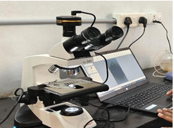
Microscope used to identify ecto parasites and endo parasites; first we had collected fluid samples and tissues from infected fish, after that prepared a slide for better visibility of specific structure, and like Giemsa stain for blood smears. The prepared slide is placed under microscope for identification of pathogens such as bacteria, fungi, and abnormal cell structures (Figure 1) [5].
Spot Agglutination Test Kit (SAT)
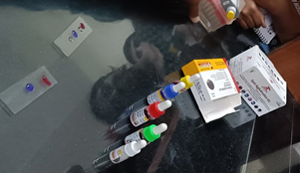
Spot Agglutination Test Kit are commonly used for rapid fish disease identification , antigen coating we have to coat the wells of a microtiter plate with specific antigens related to the suspended pathogen .the sample gets clumped in the presence of specific pathogen in sample (Figure 2) [6].
ELISA
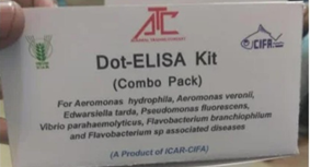
We have to collect the samples from the fish , such as blood mucus , or tissues, depending on the disease being investigated ,the collected samples tested with related antigens or antibodies Apply the prepared fish sample to a microtiter plate. The sample is typically immobilized on the plate to act as the antigen, adds a blocking agent to prevent non-specific binding on the plate. Introduce a specific antibody or antigen that will react with the target pathogen. This can be a known antibody for detecting the presence of a pathogen or an antigen to detect the presence of specific antibodies in the fish sample, Apply a substrate that the enzyme will convert, resulting in a measurable color change. The intensity of the color is proportional to the amount of bound enzyme, Measure the color change using a spectrophotometer. This provides quantitative data about the presence and concentration of the target antibodies or antigens. Determine the presence or absence of specific antibodies or antigens in the fish sample based on the colorimetric readings (Figure 3) [7].
Diseases Identified During Our Examination (Table 1)
| Disease Type | Disease Name | Causative Agent | |
|---|---|---|---|
| 1 | Bacterial | Dropsy | Pseudomonas punctata |
| 1 | Bacterial | Ulcer | Aeromonas, pseudomonas |
| 1 | Bacterial | Fin & tailrot | Aeromonas,pseudomonas,flavobacterium |
| 2 | Viral | Infectious pancreatic necrosis (IPN) | Birnavirus |
| 2 | Viral | Spring viremia of carp virus (SVCV) | Rhabdovirus, Spring viremia |
| 3 | Parasitic | Argulosis | Argulus, crustacean larvae |
| 3 | Parasitic | Lemaeosis | Anchor worm, crustacean larvae |
| 3 | Parasitic | Gill fluke (Dactylogyrus ) | Dactylogyrus |
| 4 | Protozoan | Whirling | Myxobolus cerebralis |
| 4 | Protozoan | Dermatomycosis | Saprolegnia, achyla |
| 5 | Fungal | Epizootic Ulcerative Syndrome (EUS) | Aphanomyces invades |
Table 1: Diseases Identified During our Examination.
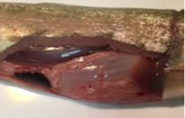
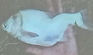
Dropsy
In fish, dropsy is a symptom rather than a specific disease. It’s characterized by the swelling or bloating of the fish’s body due to an accumulation of fluids. It’s often associated with internal issues such as organ failure or infection. If you notice dropsy in a fish, it’s crucial to address the underlying cause, which may involve isolating the affected fish, adjusting water conditions (Figure 4) [8].
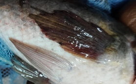
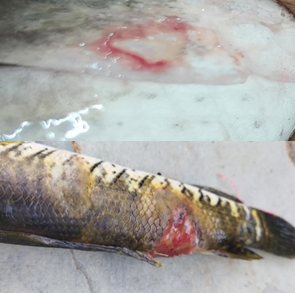
Ulcer
Ulcer disease in fish is a condition characterized by the presence of open sores or ulcers on the skin, often around the head, fins, or body. It can be caused by various factors, including bacterial infections, parasites, and environmental stressors. Common bacterial pathogens associated with fish ulcer disease include Aeromonas and Pseudomonas species (Figure 5) [9].
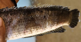
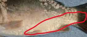
Fin & Tail Rot
Fin and tail rot in fish is a condition characterized by the deterioration or rotting of the fins and tails. It is commonly caused by bacterial infections, with species like Aeromonas and Pseudomonas being frequent culprits. Poor water quality, stress, and injuries can contribute to the development of fin and tail rot (Figure 6) [10].
Infectious Pancreatic Necrosis (IPN)
Infectious Pancreatic Necrosis is a viral disease affecting fish, causing necrosis in the pancreas and other organs. It’s caused by the IPN virus, part of the Birnaviridae family. Prevention involves strict biosecurity measures and vaccination in aquaculture (Figure 7) [11].
Spring Viremia of Carp (SVC)
Spring viremia of carp is a viral disease that affects various species of carp, including common carp and koi. The causative agent is the spring viremia of carp virus (SVCV), which is a Rhabdovirus. This disease is characterized by high mortality rates, especially in young fish. Symptoms include lethargy, erratic swimming, hemorrhages, and inflammation of internal organs. SVC is often more severe during cooler water temperatures, such as in spring and fall (Figure 8) [12].
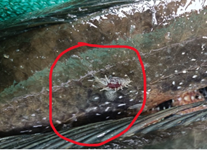
Argulosis
Argulosis also known as the common fish louse/lice, is a species of fish lice in the family Argulidae, It is “the most common and widespread native argulid in the Palaearctic “and “one of the most widespread crustacean ectoparasites of freshwater fish in the world”, considering its distribution and range of hosts. It can cause the severe disease state argulosis in a wide variety of fish species. It is responsible for epizootic outbreaks that have led to the collapse of aquaculture operations (Figure 9) [13].
Lemaeosis
Anchor Worm” disease. It’s a parasitic infection caused by a type of crustacean. Anchor worms attach themselves to fish, leading to irritation, tissue damage, and secondary infections. Treatment typically involves physical removal or chemical interventions in a separate quarantine tank (Figure 10) [14].
Dactylogyrus
Dactylogyrus commonly known as gill flukes is a parasitic flatworm that can affect fish. These microscopic parasites attach themselves to the gills of fish, causing irritation and damage. Infected fish may exhibit signs like rapid gill movement, lethargy, and reduced feeding (Figure 11) [15].

Whirling
Whirling disease is a parasitic infection in fish caused by the microscopic parasite Myxobolus cerebralis. This parasite primarily affects salmonids, such as trout and salmon. The infection can lead to skeletal deformities and damage to the nervous system, causing affected fish to swim in a whirling or corkscrew-like manner (Figure 12) [16].
Dermatomycosis
Dermatomycosis, also known as fungal infections, can affect fish and is caused by various types of fungi. These infections often occur when fish are stressed or have compromised immune systems. Symptoms include skin lesions, discoloration, and changes in behavior (Figure 13) [17].
Epizootic Ulcerative Syndrome (EUS)
Epizootic Ulcerative Syndrome (EUS) is a disease that affects fish and can result in skin lesions, ulcers, and tissue damage. It is caused by the water mold Aphanomyces invadans. EUS tends to occur in warm freshwater [18].
Result and Discussion
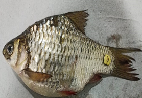
The Fish Samples are Collected From Different Ponds and Fish Farms from Siddipet District During Our Examination And This Study Period We Have Identified These Disease by performing some test like examination under microscope, dot ELISA test, spot agglutination test, and confirmed the disease by mode of infection and causative agent, Many of Aquaculture Farmers are Facing Problems with These Specific Diseases (Figure 14) [19, 20, 21, 22, 23, 24].
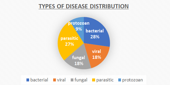
During this investigation we had find (03 diseases from bacterial infection), (02 diseases from fungal infection), (03 parasitic infection), (01 protozoan infection), (02 viral infection).
Conclusion
We here by conclude that the disease outbreaks are more in the Siddipet district area, Mostly We Have Identified There Is More Domination of Bacterial and Parasitic Disease Like, Epizootic Ulcerative Syndrome (EUS), Fin and Tail Rot, Argulosis, Lemaeosis, Branchiomycosis, Dactylogyrous. Better management practices can result in good production, maintaining water quality parameters and regular using of disinfectants during fish handling and stocking, Will run successful culture [24, 25, 26, 27, 28].
References
-
Hedrick RP, El-Matbouli M (2022) Recent Advances with Taxonomy, Life Cycle, and Development of Myxobolus cerebralis in the Fish and Oligochaete Hosts. American Fisheries Society Symposium.
-
Řehulková E, Benovics M, Šimková A (2020) Uncovering the diversity of monogeneans (Platyhelminthes) on endemic cypriniform fishes of the Balkan Peninsula: new species of Dactylogyrus and comments on their phylogeny and host-parasite associations in a biogeographic context. Parasite 27: 66.
-
Shome R, Shome BR (1999) A typical chronic form of Aeromonas hydrophila infection in Indian major carp, Catla catla, from Andaman. Current Science 76(9): 1188- 1190.
-
Ellis AE (2001) The immunology of teleosts. In: Fish Pathology. 3rd (Edn.), Viruses of lower vestebrates. Journal of Veterinary, pp: 133-150.
-
George MR, John KR, Mansoor MM, Saravanakumar R., Sundar P, et al. (2015) Isolation and characterization of a ranavirus from kos, Cyprinus carpio I experiencing mass mortalities in India. Journal of Fish Diseases 38 (4): 389- 403.
-
Gopalkrishnan V, Gupta PD (1960) An eye disease which causes mortality of theCatla catla (Ham). Current Science 29(6): 240.
-
He Y, Jiang Y, Lu L (2013) Serodiagnosis of grass camp revenus mfection in grass caip Ctenopharyngodon idella by a novel Western blot technique. Journal of Virological methods 194(1): 14-20.
-
Hemipraseuth KP, Raghavendra A, Singh R, Stadler N, Raghunath MR (2008) Efficacy of doramectin against natural and experimental infections of Lernaea exprimarea in carps. Veterinary Parasitology 156(3-4): 261-269.
-
Hemaprasanth KP, Singh R, Raghavendra A, Sridhar N, Raghunath MR, et al. (2011) Comparative ussceptibility of carp fingerlings to Lernana cyprinacea infection. Vetarmary Parasitology 178(1-2): 156-162.
-
Hoare R, Thompson KD, Herath T, Coller B, Bron JE, et al. (2016) Development, characterisation and applimtion of momeloml antibodies for the detection and quantification of Infectious salmen anaemia virus in Plasma Samples Using Luminex Bead Array Technology. PLOS ONE 11(7): e0159155.
-
Him HM, Bowser PR, Schachte JH (1991) Development and evaluation of a monoclonal antihody-based enzyme-linked immunosorbent assay for the diagnosis of Renibacterium salmoninarum infection. Journal of Aquatic Animal Health 3(3): 168-175.
-
Jeney Z, Jeney G (1995) Recent achievements in studies on dneases of common carp (Cyprinus carpio L). Aquaculture 129(1-4): 397-420.
-
Karamchandanı SJ (1952) A new species of Ergasilus from the gills of Labeo rohita (Hamilton). Records of the Indian Museum 50: 287-293.
-
Karunasagar I, Shitu MM, Girisha SK, Krohne G, Karunasagar I (2007) Brocontrol of pathogenm in slanap hatcheries using bacterioplages. Aquaculture 268(1-4): 288-292.
-
Kuan GC, Sheng LP, Rjuravanich P, Marunathu K, Ravichandran M, et al. (2013) Gold-uanoparticle based electrochemical DNA sensor for the detection of fish pathogen Aphanomyces invadons. Talanta 117: 312-317.
-
Kvamm R., Ahmed V, Parameswaran V, Sudhakatan R, Babu VS, et al. (2008) Potential use of chitosan nanoparticles for oral delivery of DNA vaccine in Asian vea bass (Lates calcarifer) to protect from Hbrio (Listonelia) anguillarum. Fick de Shellfish Inumunology 25(1-2): 47-56.
-
Laanto E, Bamford IK, Ravantti JJ, Sundberg IR (2015) The use of phage FCL-2 as an alternative te chemotherapy against columnatis disease in aquacultuse. Frontiers in Microbiology 6: 829.
-
Lakra W, Sarkar UK (2007) Freshwater fish diversity of central India In Workshop on Conservation Lasessment of Freshwater Fish Diversity for Central India (2006: CLAE, Bhopal) National Bureau of Fish Genetic Resources. Indian Council of Agrienlnural Research.
-
Leigh O, Kelly C, Praveen R, Saurabh S, Mohanty J, et al. (2012) Identification of nodoit polypepudes of the freshwater fish lace Argelus siamensis (Wahon)- prelummary fudungs. Indian Journal of Fisheries 59(1): 103-106.
-
Saurabh S, Sahoo PK, Melauty BR, Molauty J, Jena JK, et al. (2010) Modulation of the unmate umame response of roku Labeo rohita (Hamilton) by experimental freshwater lace Augulus siamensis (Wilson) infection. Aquaculture Research 41: e326-e335.
-
Seab KL, Zano X, Rappooh R (2012) Developing vacemes in the era of genomics: a decade of reverse vaccinology. Clinical Microbiology Infection 18(5): 109-116.
-
Shuralen K, Abdel-Mounu A, Rafea A, Banks H (2016) Mapping Inta Ingus representatious to feature structures of Arabic sentences. In The Challenge of Arabic for NLP/ MI International Conference, the British Computer Society: British Computer Society (BCS).
-
Shi VH, Chen I, Li CH, Lu XJ, Zhang DM, et al. (2012) Detection of bacterial pathogens in aquaculture samples by DNA microarray analysis. Aquacultura 338-341: 29- 35.
-
Axelrod HR, Untergasser D (1989) Handbook of fish diseases. Neptune NJ: TFH.
-
Andrews C (1988) The Manual of Fish Health. Stillwater. MN: Voyageur Press.
-
Cipriano RC (2001) Furunculosis and Other Diseases Caused by Aeromonas salmonicida. The Merck Veterinary Manual.
-
Kearn GC (2004) Leeches, Lice and Lampreys. In: A natural history of skin and gill parasites of fishes. Dordrecht: Springer.
-
Woo PTK (1995) Fish Diseases and Disorders: Protozoan and Metazoan Infections Cabi Series 1.
- Genetic Improvement of Nile Tilapia (Oreochromis niloticus): Advances in Selective Breeding and Genomic Approaches for Sustainable Aquaculture
- Microplastics, Contaminants, and Waste Hotspots: Divergences and Faults in Prioritizing Control Efforts
- Creating a Healthier, More Vibrant Open and Closed Aquatic Environment. A Submersible, Centrifugal Magnetically Affixed Current Changing Aquarium Pump
- An Attempt to Assess Alpha Diversity and Sample Size: Using the Ostracod Assemblages off Kumamoto Port, Japan
- Assessment of the Efficiency of Common Fishing Gears and Crafts Used at Mohananda River of Chapai Nawabganj, Bangladesh
- Fish Productivity and Biodiversity Status of Sundarban Mangrove in Bangladesh