Missing Mandibular Third Molar from Line of Fracture: A Diagnostic Dilemma
Maxillofacial fractures are clinically very significant because of functional and cosmetic importance of this region. Any misdiagnosis might result in disfigurement of the face as well as malocclusion. Conventional radiography along with clinical examination plays a vital role in diagnosis of maxillofacial fractures, however, concurring nature of facial bones and the inability to visualize the extent of fracture lines especially in multiple fractures, makes plain radiography less reliable. This report presents a trauma patient who reported in emergency department in MP Shah hospital with a misleading finding in the pre-operative Orthopantomogram when compared with the post-operative Orthopantomogram after open reduction and internal fixation of the associated fracture.
Shalender Sharma1* and Kaberi Majumder2
Introduction
Maxillofacial injuries, especially due to road traffic accidents, account for a large number of casualty cases worldwide. Restoration of facial aesthetics and function are of prime importance for a surgeon [1]. Identification of number and type of fracture depends on the degree of displacement, type of fracture, position of fracture and the imaging modality used [2, 3]. Now a days open reduction and internal fixation (ORIF) using mini-plates of facial fractures has become a mainstay of treatment for maxillofacial fractures [3].
In order to achieve good results preoperative evaluation using clinical and radiographic imaging is very important. In today’s modern world computed tomography is considered as gold standard in diagnosis and treatment planning of facial fractures [4]. Here, we present a case of bilateral mandibular fractures which was pre-operatively evaluated using Orthopantomogram (OPG) but this was found misleading when explored surgically and evaluated post-operatively using OPG.
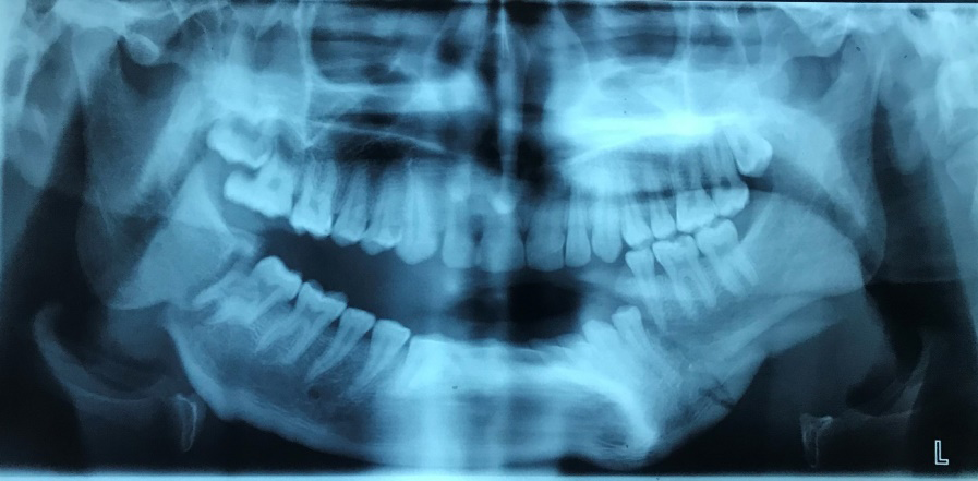
An 18 years old male patient reported to Emergency department of MP Shah hospital, with alleged history of RTA. On examination, the patient presented with multiple abrasions over left side of face, tenderness over the right angle and left body region of mandible, which was associated with restricted mouth opening. OPG was the only radiographic investigation done due to patient’s financial constraints, which revealed vertically displaced right angle with tooth in fracture line and comminuted left body fracture of mandible (Figure 1).
After pre-operative Clinical & Radiographic evaluation intermaxillary fixation (IMF) was done and Open Reduction & Internal Fixation (ORIF) of both fractures were planned under General Anaesthesia (GA).
After achieving good occlusion using IMF right angle fracture was exposed through existing laceration using Submandibular approach. Keeping in mind the impacted tooth in line of fracture and displaced fracture fragments, anatomical reduction of fracture was done followed by inferior border plating along with figure of eight wiring at inferior border to ensure sufficient fixation of the fracture segments. On intra-operative clinical examination there was no impacted teeth found in the line of fracture. Left body fracture plating was done using both Submandibular and Transoral approach. Layer by layer closure of both surgical sites were done using 3.0 Polyglactin 910 & 5.0 nylon.
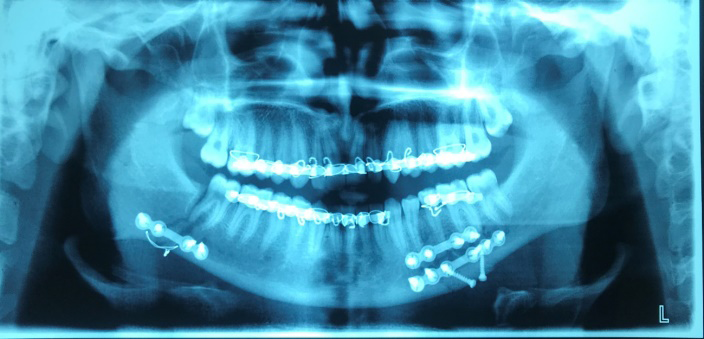
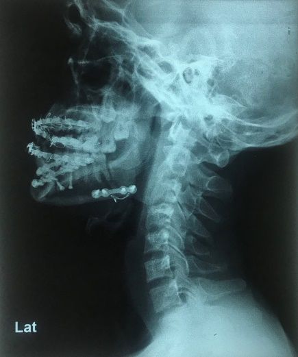
Postoperative OPG (Figure 2) showed adequate reduction and fixation of both fracture sites, but the tooth which was present in the line of fracture in the right-angle fracture was found missing. To rule out tooth displacement into the neck spaces, abdomen and lungs; lateral view of neck x ray (Figure 3), chest x-rays were advised and on radiographic interpretation there was no radiographic evidence of displacement of missing tooth.
When we compare post-operative and pre-operative OPG, pre-operative OPG revealed that there is one more image associated with crown portion of 17 and 18.
Patient is on regular follow up for 1 year with good aesthetic & functional outcomes and without any other associated fracture site complication.
Discussion
Facial injuries, especially bony fractures are very significant because of their functional and cosmetic importance. Accurate diagnosis of a maxillofacial fracture is very important to decide the treatment plan, analyse the mode of injury and anticipate the functional and cosmetic side effects [1]. The diagnostic modalities most commonly used for diagnosis are conventional radiography and Computed Tomography (CT) [4, 5]. Various studies have reported advantages of CT scan over conventional radiography due to its ability to visualize the images in three dimensions for accurate diagnosis and treatment planning of facial fractures [1, 6].
It has been observed that during a panoramic view, a patient can move in all three directional planes: horizontally, vertically, or a combination of both horizontally & vertically. Vertical motion can be detected by a vertical displacement of the maxillary structures (sinus and teeth) [7, 8]. Horizontal motion usually blurs the image of structures (teeth to a greater degree than the bone structures) and can also foreshorten or lengthen the image of mandible depending on whether the head moves towards or away from the beam, respectively [9, 10].
Small horizontal or vertical movement along with a displaced fracture creates a “step-off’ deformity that can be mistaken for an impacted tooth, especially if the movement occurs at the posterior body or angle of the mandible before maxillary structures are seen [8].
However, the most difficult motion artifact to be detected is a combination of 1 to 2 cm horizontal and 1 to 2 cm vertical movement during exposure of the posterior region of the mandible. These slight horizontal and vertical movements create a radiograph that exactly mimics an impacted tooth (Figure 1) and will be referred to as a motion pseudo impaction [9].
In the present case report, this might have happened while taking an OPG, which has led to formation of ghost image of 47 or 37 in fracture line leading to appearance of impacted third molar.
Another possible explanation for this is sagittal split of 47 at the level of cemento-enamel junction level. But this was ruled out by clinical examination of tooth, as the tooth was not mobile, and pulpal response was normal using electric pulp testing.
Since there is one more image associated with 17 and 18 along with 47, hence the most possible explanation of the tooth in line of fracture is that it is because of the pseudo image formation.
Because of tooth in line of fracture, we decide to go for extraoral approach instead of transoral approach. Extraoral approach has its own drawbacks which include injury to marginal mandibular branch of facial nerve, extraoral scar formation etc [4]. This could have been avoided by preoperative CT scan after ruling out possibility of tooth in line of fracture.
Conclusion
We can conclude that while evaluating mandibular fractures, enough time should be taken to interpret the radiographic images in detail, in correlation with the clinical examination findings. In case of doubt patient should be advised 3-Diamensional scans to rule out any missing findings and for formulation of appropriate diagnosis and proper execution of treatment plan.
Acknowledgement: None
References
-
Shah S, Uppal SK, Mittal RK, Garg R, Saggar K, Dhawan R (2016) Diagnostic tools in maxillofacial fractures: Is there really a need of three-dimensional computed tomography? Indian J Plast Surg: 49(2): 225-233.
-
Ersan N, Ilgüy M (2015) Diagnosis of unusual mandibular split fracture with cone-beam computed tomography. J Oral Maxillofac Radiol 3(2): 67-69.
-
Saheeb BD (2003) Influence of positions on the incidence and severity of maxillofacial injuries in vehicular crashes [corrected]. West Afr J Med 22(2): 146-149.
-
Tanrikulu R, Erol B (2001) Comparison of computed tomography with conventional radiography for midfacial fractures. Dentomaxillofacradiol 30(3): 141-146.
-
Mehta N, Butala P, Bernstein MP (2012) The imaging of maxillofacial trauma and its pertinence to surgical intervention. Radiolclin North AM 50(1): 43-57.
-
Smith, H, Peek ASA, Nesheim D, Nish A, Pamela Normandin, Sheryl Sahr (2012) clinical diagnosis and characteristics of facial fracture at midwestern level, trauma center. Journal of Trauma Nursing 19(1): 57-65.
-
Goel A (2015) Comparative study of clinical manifestation, plain film radiography and computed tomography for diagnosis of maxillofacial trauma. Modern Plastic Surgery 5(4): 47-49.
-
Michael J Reiter, Ryan B Schwope, Jared M (2017) The postoperative CT of the midfacial skeleton after trauma: review of normal appearances and common complications. American Journal of Roentgenology 209(4): 238-248.
-
Brad J Courter (1994) Pseudofractures of the mandible secondary to motion artifact, The American Journal of Emergency Medicine 12(1): 88-89.
-
Sandstrom CK (2013) Pseudofracture from motion artifact. In: Gunn ML, editor. Pearls and Pitfalls in Emergency Radiology: Variants and Other Difficult Diagnoses. Cambridge: Cambridge University Press 258- 266.
- Management of Chronic Insertional Achilles Tendinopathy Using Flexor Hallucis Longus Tendon Transfer in Patients Over 50 Years of Age: A Four-Case Series Following the CARE Guidelines
- Application of Induced Pluripotent Stem Cells in Bone Tissue Engineering: Current Status and Prospects
- Surgical Management of Upper Thoracic Esophageal Squamous Cell Carcinoma with Concomitant Hypersplenism: Integration of Chai's Supra-Thoracic Apex Technique with Laparoscopic Splenectomy - A Technical Innovation Case Study with Systematic Review
- Evaluation of Masticatory Functional Efficiency of Stomatognathic System in Patients Undergoing Open Reduction Internal Fixation for Treatment of Pan-Facial Trauma: A Prospective Study
- Hepatic Abscess Secondary to Appendiceal Phlegmon an Unusual Complication of Appendiceal Phlegmon
- Report of Lumboperitoneal (LP) Shunt Procedure in Over Decades Experiences, Systematic Narrative Review