Topographic and Morphometric Anatomy of Mental Foramen of Mandible of Black Bengal goat (Capra hircus) in Bangladesh with its Clinical Implication for Regional Anesthesia
The study was conducted to investigate the topographic and morphometric anatomy of mental foramen of Black Bengal goat. Total 20 mandibles from both sex of adult Black Bengal goat were studied. Mental foramen was always present at lateral aspect of the rostal part of body of each mandible with various shape, size and direction. Oval shaped and dorso-laterally directed mental foramen were predominant (85% and 80%) than the round shaped and laterally directed mental foramen (15% and 20%); respectively. The mean distance between lip commissure to mental foramen, base of body of mandible to mental foramen, 1st premolar tooth to mental foramen, lateral incisor tooth to mental foramen and caudal border of ramus of mandible to mental foramen were 2.37±0.09, 0.77±0.04, 1.46±0.09, 2.01±0.05 and 11.81±0.89 cm; respectively. Those topographic and morphometric anatomy of mental foramen of Black Bengal goat may helpful for veterinary surgeon to localize mental foramen easily for regional anesthesia during different surgical intervention of lower jaw.
Introduction
Black Bengal goat (Capra hircus) is common small sized ruminant livestock species reared all over Bangladesh. They have very high and income generating potential [1].
Mental foramen of Black Bengal goat is an opening through which the mental nerve and vessel are existed [2, 3]. It is located at the lateral aspect of the rostral part of body of each mandible. Variation in the shape, size, direction and position are noticed in mental foramen [4, 5]
of Black Bengal goat. Blockage of mental nerve under regional anesthesia during surgical interventions like impaction, fracture reduction, teeth extraction from lower jaw, epulis becomes more common practice in Black Bengal goat. The topographic and morphometric anatomy of mental foramen of Black Bengal goat is essential for veterinary surgeon to localize mental foramen easily [6, 7]. Several studies were carried out on morphometry of foramen of Iranian Native goats [4], Markhoz Goat [5]. West African Dwarf goat [8] and Gwembe Valley Dwarf goat [9]. Fewer studied were done on morphometry of mental foramen of in Black Bengal goat in Bangladesh [10]. Here, the study was planned for details investigation of topographic and morphometric anatomy of mental foramen of Black Bengal goat in Bangladesh with its clinical implication for regional anesthesia.
Materials and Methods
The study was conducted on 20 mandibles of both sex of adult Black Bengal goat from the period of 5th September to 10th November, 2017. These mandibles (along with skull) were collected from local market, Khulshi, Chittagong. Dissection was performed to expose mental foramen with mental nerve. Then the muscle and ligament from mandibles were removed and they were processed to form dried bone as per standard technique [11, 12, 13]. All mental foramen of mandibles were studied to record their topographic and morphometric features at laboratory of Department of Anatomy and Histology, Chittagong Veterinary and Animal Sciences University (CVASU), Khulshi, Chittagong, Bangladesh. The following studies were conducted on the collected and dried mandibles bones of both sex of adult Black Bengal goat.
- Mental foramen was indentified at lateral aspect of the rostral part of body of each mandible with various shape, size and direction.
- The distance between lip commissure to mental foramen were measured and recorded.
- The distance between base of body of mandible (ventral border of the mandible) to mental foramen were measured and recorded.
- The distance between lateral alveolar border of the first premolar tooth to the mental foramen were measured and recorded.
- The distance from the lateral extent of the alveolar root of lower incisor to mental foramen were measured and recorded.
- The distances from the caudal border of mandibular to the mental foramen were measured and recorded.
All measurements were expressed as mean measurements with standard deviation (Mean ± SD)
Result and Discussion
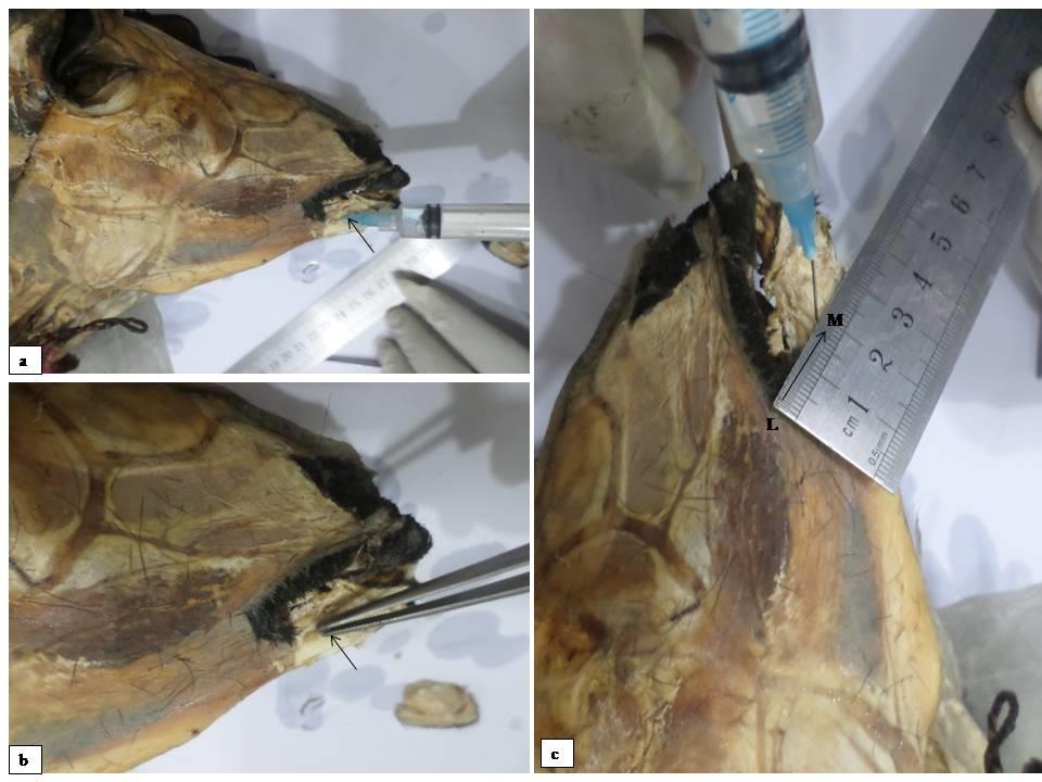
In dissected mandible of Black Bengal goat, mental foramen with mental nerve was always present at lateral aspect of the rostral part of body (Figure 1).
![Figure 2: a. Oval shaped mental foramen and b. round shaped mental foramen. In dried mandible, various shape, size, direction and position of mental foramen were noticed (Figure 2). This study was supported by the studies of Monfared, et al. (2013), Goodarzi N and Hoseini (2013), Kataba, et al. (2014) [4,5,9] where their data showed variation in shape, sized of mental foramen of goat. Oval shaped mental foramen was predominant (85%) than the round shaped mental foramen (15%). Dorso-laterally directed mental foramen was predominant (80%) than the laterally directed (20%) mental foramen (Table 1). This finding suggested, palpating the rostar portion of mandible of Black Bengal goat along the dorso-lateral direction or lateral direction; is easy to find out the mental foramen in live goat.](/fulltextimages/1813/fig_2.jpeg)
Figure 2: a. Oval shaped mental foramen and b. round shaped mental foramen. In dried mandible, various shape, size, direction and position of mental foramen were noticed (Figure 2). This study was supported by the studies of Monfared, et al. (2013), Goodarzi N and Hoseini (2013), Kataba, et al. (2014) [4, 5, 9] where their data showed variation in shape, sized of mental foramen of goat. Oval shaped mental foramen was predominant (85%) than the round shaped mental foramen (15%). Dorso-laterally directed mental foramen was predominant (80%) than the laterally directed (20%) mental foramen (Table 1). This finding suggested, palpating the rostar portion of mandible of Black Bengal goat along the dorso-lateral direction or lateral direction; is easy to find out the mental foramen in live goat.
| Parameter | Percentage | |
|---|---|---|
| Shape of mental foramen | Oval | 17 (85%) |
| Shape of mental foramen | Rounded | 3 (15%) |
| Direction of mental foramen | Dorso-lateral direction | 16 (80%) |
| Direction of mental foramen | Lateral direction | 4 (20%) |
Table 1: Variation of shape and direction of mental foramen of mandible of Black Bengal Goat.
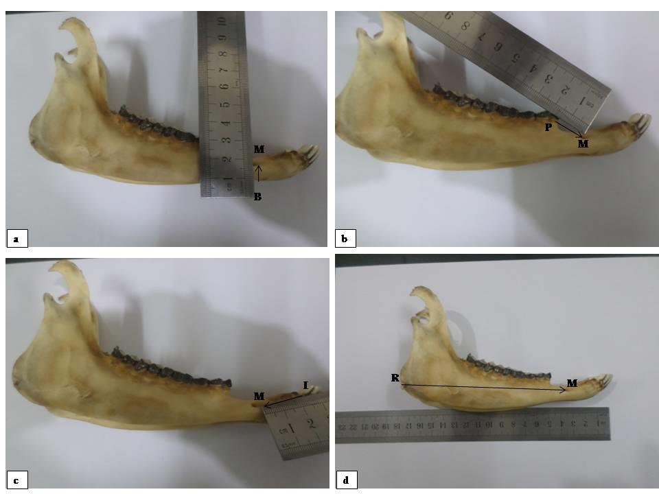
Table 1: Variation of shape and direction of mental foramen of mandible of Black Bengal Goat. The distance between lip commissure to mental foramen (LM), base of body of mandible to mental foramen (BM), 1st premolar tooth to mental foramen (PM), lateral incisor tooth to mental foramen (IM) and caudal border of ramus of mandible to mental foramen (RM) in Black Bengal Goat were indentified in (Figure 3) and data were presented in (Table 2).
| Parameter | Mean±SD(cm) | Maximum(cm) | Minimum(cm) |
| Distance between lip commissure to mental foramen | 2.37±0.09 | 2.5 | 2.2 |
| Distance between base of body of mandible to mental foramen | 0.77±0.04 | 0.8 | 0.7 |
| Distance between 1st premolar tooth to mental foramen | 1.46±0.09 | 1.7 | 1.3 |
| Distance between lateral incisor tooth to mental foramen | 2.01±0.05 | 2.1 | 1.9 |
| Distance between caudal border of ramus of mandible to mental foramen | 11.81±0.89 | 13.2 | 10.1 |
Table 2: Morphometric anatomy of mental foramen of mandible of Black Bengal goat.
Table 2: Morphometric anatomy of mental foramen of mandible of Black Bengal goat. The mean distance between lip commissure to mental foramen in Black Bengal goat was 2.37±0.09 cm, maximum was 2.5 cm and minimum was 2.2 cm. This finding suggested, palpating the rostal portion of mandible alone the cranio-ventral direction from lip commisure; is easy to find out the mental foramen in live goat. The mean distance between base of body of mandible to mental foramen in Black Bengal goat was 0.77±0.04 cm which was almost similar with previous study of Mohamed, et al. (2016) [14] where he found 0.70±0.18 cm in Barbados Black Belly Sheep. More mean distance (2.35 ± 0.26 cm) was found in the study of Kataba, et al. (2014) [9] in Gwembe Valley Dwarf goat. This difference might be due to more wideness of mandible alone dorso-ventral direction. The mean distance between 1st premolar tooth to mental foramen in Black Bengal goat 1.46±0.09 cm where 2.25±0.38 cm was found in Barbados Black Belly Sheep [14]. These findings suggested, mental foramen may be palpable along the ventro-lateral line (about 1.46 ± 0.09 cm away) from 1st premolar tooth. The mean distance between lateral incisor tooth to mental foramen in Black Bengal goat was 2.01 ± 0.05 cm; where 2.11 ± 0.17 cm was found by Uddin, et al. (2009) [10] in Black Bengal goat; 2.25 ± 0.31 cm by Mohamed, et al. (2016) [14] in Barbados Black Belly Sheep; 1.56 ± 0.22 cm by Olopade and Onwuka (2005) [8] in West African Dwarf goat and 1.58 ± 0.19 cm by Kataba, et al. (2014) [9] in Gwembe Valley Dwarf goat. The mean distance between caudal border of ramus of mandible to mental foramen in Black Bengal goat was 11.81 ± 0.89 cm which was almost similar with the previous study of Uddin, et al. (2009) [10], where he found 11.69 ± 0.4 cm and by Karimi, et al. (2012) [15], where he found 13.74 ± 0.18 cm in Mehraban Sheep. More mean distance (15.23 ± 1.46 cm) was found in the study of Mohamed, et al. (2016) [14] in Barbados Black Belly Sheep. Less mean distance (9.26±0.49 cm) was found in the study of Kataba, et al. (2014) [9] in Gwembe Valley Dwarf goat.
Conclusion
Information regarding topographic and morphometric anatomy of mental foramen of Black Bengal goat will helpful for veterinary surgeon to localize mental foramen precisely (at 2.37 ± 0.09 cm cranio-ventral direction from lip commissure; 0.77±0.04 cm from rostal base of mandible; 1.46 ± 0.09 cm ventro-lateral direction from 1st premolar tooth; 2.01 ± 0.05 cm caudal from lateral incisor tooth and 11.81 ± 0.89 cm cranial from caudal border of ramus) for regional anesthesia during different surgical intervention of lower jaw. This will also helpful for veterinary surgeon to avoid toxicity of local anesthetic agent as they can identified exact location of mental foramen during surgical procedure with short time and low cost.
Acknowledgement
Author wishes to express his deep sense of gratitude and thanks to staffs and faculty members of Department of Anatomy and Histology, Faculty of Veterinary Medicine, Chittagong Veterinary and Animal Sciences University, Khulshi, Chittagong, Bangladesh for their kindful facilitation during this work.
References
-
Chowdhury SA (2002) Goat: our natural resources and development opportunities. In: Islam, MR and Huque QME (eds.), Proceeding of the workshop on poverty alleviation through goat production: national program.
-
Getty R (1975) Session and Grossman’s The Anatomy of the Domestic Animals, 2nd (edn.), vol.1, W.B. Saunders co. Philadelphia, USA.
-
Ghosh RK (2012) Primary veterinary anatomy, 5th (edn.), Current books international, Kolkata, India.
-
Goodarzi N, Hoseini TJ (2013) Morphometric Characteristics of the Maxillofacial and Mandibular Regions of Markhoz Goat Breed and its Clinical Value for Regional Anaesthesia in Western Iran. Global Veterinaria 11(1): 107-111.
-
Monfared AL, Naji H, Sheibani MT (2013) Applied anatomy of the head region of the Iranian Native goats (_Capra hircus_). Global Veternaria 10(1): 60-64.
-
Ommer PA, Harshan KR (1995) Applied anatomy of domestic animals, 1st Edition. Jaypee brother's medical publisher, India.
-
Dyce KM, Sack WO, Wensing CJG (2002) Textbook of Veterinary Anatomy, 3rd (edn.), WB Saunders, Philadelphia, USA.
-
Olopade JO, Onwuka Sk (2005) An oestometric study of the skull of the West African Dwarf goat from South Eastern Nigeria. II- Mandibular and Maxillofacial features (Short Communication). Nigerian Veterinary Journal 27(2): 66-70.
-
Kataba A, Mwaanga ES, Simukoko H, Pares CPM (2014) Clinical anatomy of the head Region of Gwembe Valley dwarf goat in Zambia. International Journal of Veterinary Science 3(3): 142-146.
-
Uddin MM, Ahmed SSU, Islam KN, Islam MM (2009) Clinical anatomy of the head region of the Black Bengal goat in Bangladesh. International Journal of Morpholology 27(4): 1269-1273.
-
Raghavan D (1964) Anatomy of ox. Indian Council of Agricultural Research, New Delhi, India, pp: 97-117.
-
Hildebrand M (1968) Anatomical preparations. University of California Press. Berkely and Los Angeles, California, USA.
-
Merai MK (2012) Anatomical museum preparations of the skeleton and respiratory organs of some domestic animals. MVSc, Faculty of Veterinary Medicine, Beni-Suef University, Egypt.
-
Mohamed R, Drisco M, Mootoo N (2016) Clinical Anatomy of the skull of the Barbados Black Belly Sheep in Trinidad. International Journal of Current Research in Medical Science 2(8): 8-19.
-
Karimi I, Hadipour MM, Nikbakht P, Motamedi SH (2012) The Lower Jawbone of Mehraban Sheep: A descriptive morphometric approach. World's Veterinary Journal 2(4): 57-60.
- California Red-Legged Frog and Non-Listed Amphibians Response to Non-Native Fish Removal
- Industrial Standardization of the Bio-OS: Algorithmic Codification of Resilience Engineering Guidelines and Version V8 Architecture
- Climate Variability and the Sustainability of Snail Farming in Nigeria: Past Trends, Present Challenges and Potential Outlook
- The Evaluation of the Surveillance System of Anthrax in Gilgit-Baltistan, Pakistan, 2018
- Natural Decline to Extinction of A New Zealand Rabbit Population
- Mitochondrial Bio-Logistics: Steering Co-Enzyme Q10 and Lycopene Synergies within the Science 4.0 Bio-OS Framework