An In Vivo and In Vitro Trial on Layer Chicken Breed Susceptibility and Yolk Sac Infection to Escherichia Coli and Evaluation of the Immune Response in Bishoftu Poultry Farms, Central Ethiopia
Chicken colibacillosis is a bacterial disease of great concern in the layer industry causing substantial animal and economic losses worldwide. Yolk sac infection of chicks which result in high mortality in the first week of age. A challenge test was carried out to evaluate the susceptibility of three chicken breeds to E. coli pathogenic strain. Thus, a total of 48 day-old Horo, Fayoumi and Koekoek chicken breeds were allotted into the treatment and Control groups Containing 8 birds each. An experimental infection with E. coli pathogenic strain was given (104c.f.u/0.1 ml) intra yolk sac to treatment group on day-one of experiment, while the control group was kept as non-injected. The studied parameters involved examination of yolk sac weight, yolk sac, and body weight ratio and antibody titer against Newcastle disease virus (NDV). The study revealed that there was no statistically significant difference between Horo, Fayomi and Koekoek breeds in infection. However, intra yolk infection with E. coli pathogenic strain result in gross pathological change of the yolk sac, increased yolk sac body weight ratio, increased yolk sac weight and the transfer of maternal immunity in serum was not changed. In conclusion local and exotic breeds of chickens are highly susceptible to E. coli. Therefore, vaccination and therapeutic treatment should be properly used and supplementary management practices should be adopted in the farm.
Introduction
Infectious diseases are responsible for high losses of poultry industry worldwide. Most of these diseases are caused by bacterial pathogens. Chicken colibacillosis is any localized or systemic infection caused entirely or partly by avian pathogenic Escherichia coli [1]. It is a wide spread infectious disease that is a serious problem in the poultry industry. It is characterized by respiratory problems, reduced feed intake, growth retardation, uniformity reduction, and mortality [2]. Chicken yolk sac infection is an economically important disease which is characterized by mortality and poor weight gain in the first week of life. In addition, birds that adapt yolk sac infection had poor carcass quality, decreased hatchability, increased mortality and culling rate due to stunted growth. The mortality caused this infection can range from 5-10% [3, 4].
Previous reports indicated that Sub-clinical chicken yolk sac infection after oral administration of pure cultures of bacterial isolates emerged through translocation of bacteria across the gut wall of chickens some authors such as Singh, et al_. [5] conducted a research on the pathogenicity of _Escherichia coli by intra- peritoneal injection into 2-days-old chicks. Disease was induced by inoculating bacteria inside the egg shell of piped eggs and through intra yolk, intra-peritoneal, subcutaneous and oral routes. Therefore, yolk sac infection was encountered when inoculated into the yolk sac [6].
Immunoglobulin is readily transferred from chicken serum to the yolk of egg. IgA and IgM are found in albumen while IgG is found in yolk of egg. As the chick embryo develops, it absorbs some of the yolk IgG, which appears in its circulation and this would help to provide systemic protection. The maternal IgM and IgA from albumen diffused into the amniotic fluid are swallowed by embryo and present in its intestine during hatching process. These maternal antibodies effectively provide local protection and protect chicks from diseases until they disappear between 10 and 20 days after hatching. The structural change of these proteins due to microbial infection, results in immunosuppression in chickens [7].
There was dearth of investigation and study on the yolk sac infection and immune response as well as alternative treatment for chicken yolk sac infection is limited. The use of various antibiotics in treating Melese K and Urge B. An In Vivo and In Vitro Trial on Layer Chicken Breed Susceptibility and Yolk Sac Infection to Escherichia Coli and Evaluation of the Immune Response in Bishoftu Poultry Farms, Central Ethiopia. Int J Zoo Animal Biol 2019, 2(6): 000194.
colibacillosis is recommended. Conducting breed susceptibility testing based on the bacterial strain [8] and evaluating the resistance of chicken breed against any infection is essential. Therefore, the objectives of this study were,
- To evaluate the susceptibility of three chicken breeds to E. coli pathogenic strain through in vivo techniques
- To evaluate the effect of experimental yolk sac infection with E. coli on maternal immunity through in vivo techniques
- To appreciate and characterize lesions on yolk sac of chicken
Materials and Methods
Study Area
The study was conducted in Bishoftu Agricultural Research Center poultry farm during the period from 2015 to 2017. Bishoftu is located 47 km Southeast of Addis Ababa at an altitude of about 1900 m.a.s.l with (38o 58′′ E 08o 44′′ N). It receives an annual rainfall of 1115.6 mm with two rainy seasons. The short rainy season extends from March to May, while the main rainy season extends from June to September. The average maximum and minimum temperatures are 30.5oC and 8.5oC, respectively [9].
Study Chickens and Experimental Design
A 48 day-old Koekoek, Horoo and Fayomi breeds of chickens were reared under good management conditions in Experimental house. Feed and water were provided with adlibitum. A single factorial randomized experimental design was used to determine the relative resistance of three chicken breeds to E .coli strain.
Sampling Size
The sample size for this experiment was determined based upon the formula developed by Dell, et al. [10], Where 1- ß represents is the power and p represents the proportion of chickens in the experimental colony that are not infected. The power of an experiment is the probability that the effect of study will be detected. It is arbitrarily set to 0.8 or 0.9 (80 or 90% chance of finding significance effects). Besides this, 1-power, symbolized as ß, is the chance of obtaining a false-negative result (if the experiment failed to reject an untrue null hypothesis or to detect the specified treatment effect). The proportion of not infected is used in the formula. Accordingly, 50% was Copyright© Melese K and Urge B.
taken as the probability of infection and a 95% chance of detecting that infection, and then the number of chickens that need to be sampled (N) = log ß/log p, N = log 0.05/ log 0.5 N = 4.32 so with an approximate number, 5 chickens were taken from each treatment group. However, to increase the accuracy of the experiment, 8 chickens were taken from each breed for the treatment and 8 chickens for the control with a total of 48 chickens (24 for treatment and 24 for control) for in vivo experiment.
Chicken House Preparation and Challenging Experiment
First two houses (one for the experimental and the other for control) 3x3 m with 3 m high was prepared in National veterinary institute. It was cleaned and fumigated using potassium permanganate (20 g) and formalin (30 ml) for one cubic meter and closed for three days. The houses were designed for poultry research purpose. It was ventilated with meshes at the top of their walls. All the materials used were fumigated together. Day old chickens of three breeds (Local, Fayomi and Koekoek) were taken from Bishoftu Agricultural Research Center to National veterinary institute. All chickens were tagged on their one wing and the number in the tag was registered. They were feed with chicken starters feed formulated in Bishoftu Agricultural Research Center. Both water and feed were given as an adlibitum. At the first day of age, half of all the three breeds (8-Local, 8-Fayomi and 8- Koekoek) were randomly selected and the treatment groups were allocated at one house and the control groups of chickens were at another house.
Inoculum Preparation
Pathogenic strain of E. coli was isolated from the birds suspected for Colibacillosis, was taken from National Animal Health Research and Investigation center and identification of the organism was done by morphological, cultural and staining characteristics, sugar fermentation and biochemical and the total viable count was done by plate count methods [11, 12, 13]. After making serial dilution, the isolate of E. coli (104c.f.u/0.1 ml) was inoculated into the yolk sac of each chick using sterilized insulin syringe [14]. Chicks of control group were injected nutrient broth (0.1ml/chick) on day-one of age.
Sample Collection
Two chicken were slaughtered from each group at different interval on the 3rd , 5th ,7th and 9th days of post Melese K and Urge B. An In Vivo and In Vitro Trial on Layer Chicken Breed Susceptibility and Yolk Sac Infection to Escherichia Coli and Evaluation of the Immune Response in Bishoftu Poultry Farms, Central Ethiopia. Int J Zoo Animal Biol 2019, 2(6): 000194.
inoculation from each breed as well as control and treatment groups were taken randomly and slaughtered at 48 hours intervals until all the chickens were removed. Sterilized slaughtering materials were used for individual chicks and its yolk sac to prevent cross contamination. Aseptically method was used to take yolk sac and blood sample from brachial wing vain of each chicken.
Statistical Data Analysis
All data collected were coded and entered into Microsoft Excel spreadsheet 2007 computer program and analyzed using Statistical Package for Social Science (SPSS)-Version 19 or 20. In all cases, p-value less than 0.05 held at 95% confidence intervals was considered for significance level.
Results and Discussions
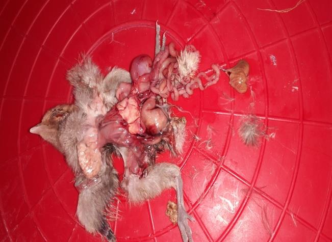
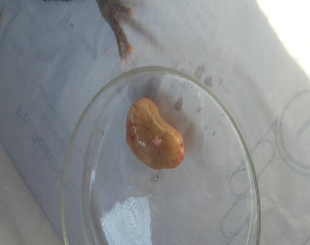
The findings of the present study indicated that body weight of infected chicken was lower than that of control chicken. This result was similar with the study of Khan, et al. [12] who reported reduced weight gain due to yolk sac infection. This might be due to refusal of feed intake by chicken during infection. The study also revealed that yolk sac body weight ratio in infected chicken was higher than in the control group (Tables 1-3). This is comparable with the findings of Deeming, et al. [15] who reported that yolk sacs of infected chicks were bigger than the uninfected yolk sacs from chickens of same age. Other authors such as Sander, et al. [7] and Khan, et al. [12] also reported similar findings. This higher yolk sac body weight ratio is justified by the fact that decreased yolk absorption in infected chicks due to protein alteration, binding or decreased permeability of the yolk sac membrane. Furthermore, reduced weight gain and high yolk sac weight resulted in higher yolk sac body weight ratio in E. coli infected group as compared with control groups. Similar studies in E. coli infection were also reported by Khan, et al. [12]. Examination of yolk sac revealed that the yolks of infected chicks were discolored, having abnormal consistency (watery in initial stage and hard in latter stage) and congested yolk sac blood vessels. This result is similar with the findings reported by Khan, et al., Jordan, et al. and Anjum, et al. [12, 15, 16]. The haemagglutination inhibition (HI) titers of serum against Newcastle disease virus were highest throughout the experimental period and this showed that there was protective antibody level with some exceptions. These results were contradicted with the findings of Sander, et al. [7]. The geometric mean titers against Newcastle Copyright© Melese K and Urge B.
disease virus in serum and yolk were higher in the control groups than in the infected chicken [17] (Figures 1-4).
Breed of chickens Groups Susceptibility Pearson χ2 D.F p-value Susceptible Resistant Fayomi Treatment 2 (25.0) 6 (75.0) 2.286 1 0.131 Control 0(0.0) 8(100.0) Horro Treatment 1 (12.5) 7(87.5) 2.618 1 0.106 Control 4 (50.0) 4(50.0) Kokok Treatment 2(28.6) 5 (71.4) 0.603 1 0.438 Control 1(12.5) 7(87.5) D. F. =Degree of Freedom, χ2= Chi-Square. Table 1: The association between Breed and Susceptibility of Yolk sac body weight ratio of the three chicken breeds.
| Sum of Squares | D.F. | Mean Square | F | Sig. | |
| Between Breeds of chicken | 11.845 | 2 | 5.923 | 1.364 | 0.266 |
| Within Breeds of chickens | 191.031 | 44 | 4.342 | ||
| Total | 202.876 | 46 |
Table 1: The association between Breed and Susceptibility of Yolk sac body weight ratio of the three chicken breeds.
| for header rows | for data cells | Mean Difference (I-J) | Std. Error | Sig. |
| Fayome | Horo | -0.3711503 | 0.7366824 | 0.617 |
| Fayome | Kokok | 0.8400664 | 0.7488598 | 0.268 |
| Horo | Kokok | 1.2112167 | 0.7488598 | 0.113 |
Table 2: ANOVA Statistics of chicken breed to yolk sac body weight ration.
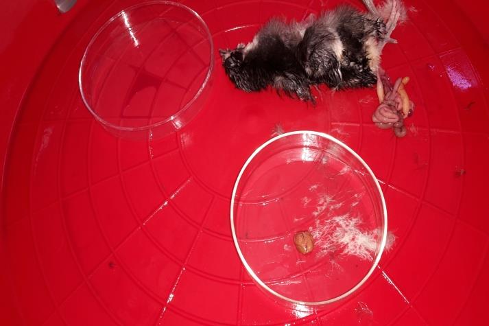
Figure1: Yolk sac control day 3.
Melese K and Urge B. An In Vivo and In Vitro Trial on Layer Chicken Breed Susceptibility and Yolk Sac Infection to Escherichia Coli and Evaluation of the Immune Response in Bishoftu Poultry Farms, Central Ethiopia. Int J Zoo Animal Biol 2019, 2(6): 000194.
Copyright© Melese K and Urge B.
Conclusion and Recommendations
The present study showed that there is no relative resistance or susceptibility difference among the three poultry breeds in terms of yolk sac weight, yolk sac body weight ratio, Gross pathological lesion and maternal immunity transfer. The study also stated that Escherichia coli (0157 H7) have the potential to invade and results in invasive infection or it results in local and systemic infection in chickens. Therefore further study should be conducted to know the resistance ability of E coli and breed susceptibility and resistance potential of chickens.
Melese K and Urge B. An In Vivo and In Vitro Trial on Layer Chicken Breed Susceptibility and Yolk Sac Infection to Escherichia Coli and Evaluation of the Immune Response in Bishoftu Poultry Farms, Central Ethiopia. Int J Zoo Animal Biol 2019, 2(6): 000194.
References
-
Barnes HJ, Vaillancourt JP, Gross WB (2003) Colibacillosis. Diseases of Poultry. 12th (Edn.), In: Barnes HJ, Fadly AM, Glisson JR, McDougald LR, Swayne DE, Saif YM, et al. (Eds.), Iowa State University Press, Ames pp: 631-656.
-
Ask B, Van Der Waaij EH, Stegeman JA, Van Arendonk JAM (2006) Genetic variation among broiler genotypes in susceptibility to colibacillosis. Poult Sci 85: 415-421.
-
Corts C, Isaies G, Cuello C, Floes J, Campos C, et al. (2004) Bacterial isolation rate from fertile eggs, hatching eggs, neonatal broilers with yolk sac infection. Rev Latenoamericana de Microbiologia 46: 12-16.
-
Ulmer FAM (2011) Yolk Sac Infections in Broiler Chicks: Studies on _Escherichia coli_, Chick Acquired Immunity and Barn Microbiology. University of Alberta Edmonton, Alberta pp: 1-197.
-
Singh MP, Gupta RS, Singh SP (1997) Pathogenicity of _Escherichia coli_ isolated from _Colibacillosis_ in chicks and their antibiotic spectra. Indian J Comparative Microbiol Immunol Infec Dis 18(2): 142-146.
-
Seigo S, Iwamori H, Hirai K (1970) Studies on the organisms isolated from chicks with omphalitis. Japanese Poult Sci 4: 57-61.
-
Sander JE, Willinghan EM, Wilson JL, Thayer SG (1998) The effect of inoculating _Enterococcus faecalis_ into the yolk sac on chick quality and maternal antibody absorption. Avian Dis 42(2): 359-363.
-
(2014) Merck Veterinary Manual (2011) Omphalitis. Accessedonline.
-
(2005) National Meteorological Service Agency: Rainfall and temperature data, Addis Ababa, Ethiopia.
-
Dell R, Holleran S, Ramakrishnan R (2000) Sample Size Determination. ILARJ 43(4): 207-213.
-
Jalil H, Das P (2001) Biochemical and serological characterization of _Escherichia coli._ Copyright© Melese K and Urge B.
-
Khan KA, Khan SA, Hamid S, Aslam A, Rabbani M (2002) A study on the pathogenesis of yolk retention in broiler chicks. Pakistan Vet J 22(4): 175-180.
-
Collins CH, Lyne PM, Grange JM (1995) Collin’s and Lyne’s Microbiological Methods, 8th (Edn.), Butterworth Heinemann, Oxford, UK pp: 129-131.
-
Kloryga MA (1986) Pathogenicity of five rough mutants of _Escherichia coli_ isolated from broilers_._ Avian Pathol 15(4): 749-759. Melese K and Urge B. An In Vivo and In Vitro Trial on Layer Chicken Breed Susceptibility and Yolk Sac Infection to Escherichia Coli and Evaluation of the Immune Response in Bishoftu Poultry Farms, Central Ethiopia. Int J Zoo Animal Biol 2019, 2(6): 000194.
-
Jordan FTW (1990) Poultry Diseases. 3rd (Edn.), ELBS and Bailliere Tindall, London, UK pp: 40.
-
Anjum A (1997) Poultry Diseases, Vet Ag Publications, Faisalabad-Pakistan pp: 178-180.
-
Fuller R, Jayne Williams DJ (1968) The origin of bacteria recovered from the peritoneum and yolk sac of healthy chickens. British Poult Sci 9: 159-163. Copyright© Melese K and Urge B.
- California Red-Legged Frog and Non-Listed Amphibians Response to Non-Native Fish Removal
- Industrial Standardization of the Bio-OS: Algorithmic Codification of Resilience Engineering Guidelines and Version V8 Architecture
- Climate Variability and the Sustainability of Snail Farming in Nigeria: Past Trends, Present Challenges and Potential Outlook
- The Evaluation of the Surveillance System of Anthrax in Gilgit-Baltistan, Pakistan, 2018
- Natural Decline to Extinction of A New Zealand Rabbit Population
- Mitochondrial Bio-Logistics: Steering Co-Enzyme Q10 and Lycopene Synergies within the Science 4.0 Bio-OS Framework