The Pathophysiology and Neurobehavioral Effects of Chlorfenapyr Insecticide in Lactating Female Sprague Dawley Rats and in HepG2 Cell Line
Chlorfenapyr (CFP) is good candidate insecticide for control of vectors blood borne diseases like malaria. As little information about CFP toxicity and possibility of its residue’s presence in food stuffs, milk and environment, we explore the postnatal toxic effects of CFP in female Sprague dawley rats and its pups. CFP was given orally at doses of 0,54 and 108 mg/kg to female albino rats immediately at first day after delivery till 21 days of lactation. All dams and its pups were weighted, euthanized and blood was separated for serum separation and tissues were preserved either at 4c as tissue homogenate for measurements of oxidant/antioxidants levels or in buffered formalin for histopathological examination. The highest dose of CFP induced hepatorenal toxicity in dams and its pups with evidence of increase liver enzymes and creatinine level when compared to control groups. Also, CFP displayed histopathological changes in liver, kidney, brain and spleen tissues of dams as well as rats’ pups after 21 days of treatment. The toxic effects resulted from secretion of CFP in milk and increased the free radicals’ production and oxidants like MDA in tissues of rats’ pups. Also, CFP had a cytotoxic effect on HepG2 cells indicated by induction of oxidative stress and lethality to cell line. Taken collectively, chlorfenapyr is a good candidate insecticide in vector control but had a cytotoxic effect in female albino rats, its pups, and in HepG2 cells.
Introduction
Chlorfenapyr is a pro-insecticide used since 1995 for control of agriculture pest. Human toxicity from CFP was little as only few cases were reported with nausea, vomiting, fever, rhabdomyolysis and nervous system toxic manifestationafter ingestion of current insecticide [1, 2, 3, 4, 5].
CFP is a member of a new class of insecticide, of pyrroles group (chemical name: 4-bromo-2-(4-chlorophenyl)- 1-(ethoxymethyl)-5-(trifluoromethyl)-1H-pyrrole-3- carbonitrile; trade name: Pylon miticide-insecticide) [6]. The uses of the CFP were removal of mites, caterpillar pests, thrips, and fungus gnats by foliar spray on ornamental crops in greenhouses and its mode of action was through inhibition of oxidative phosphorylation in the mitochondria, resulting in reduction of ATP production, cellular death, and ultimately, death of the organism [7].
The CFP is a light tan or light-yellow solid powder. While CFP toxicity has not yet been studied in humans and animals resulted that classification of CFP toxicity as category III chemical. Recently, In few studies recorded that CFP induced developmental and maternal toxicity in female albino rats [7, 8].
CFP sources or residues were recorded in environment, in food products, water, animal derived foods and in tissue of treated rats, analyzed by different methods of chromatography like GLC and HPLC. The rationale of this study to explore the Pathophysiology effects of chlorfenapyr in female albino rats and its pups [9, 10, 11, 12, 13, 14, 15].
Materials and Methods
Animals
Sprague Dawley rats obtained from Experimental Unit in the Faculty of Pharmacy, Mansoura University; Animals weighed about 250 ±10 gm and were obviously healthy then grouped and housed in plastic cages with soft wood shavings as a bedding material then adapted for about 2 weeks and maintained on a balanced ration before the experiment.
Tested Chemicals
CFP is a light green wettable powder (WP) with slight chlorine odor and kindly obtained from Central Agricultural Pesticide Laboratory, Ad Doki, Giza, Giza Governorate after HPLC analysis to confirm the percentage. HepG2 (85011430, sigma, USA) was gifted to us was provided to us from faculty of medicine, Mansoura university, Egypt
Experimental Design
Eighteen (18) pregnant female Sprague Dawley rats were separated into three groups with six females for each. CFP was given orally and daily at doses of 0,54 and 108 mg/ kg (equivalent to 1/20 and 1/10 of LD50) to female albino rats immediately at first day after delivery till 21 days of lactation. The neonates litter size recorded and both dams and neonates weighed daily and kept under observation until weaning.
Clinical Signs and Behavioral Test
The treated dams and neonates observed daily throughout the experimental period for any abnormal behavior, findings or alteration. All rats were trained for behavioral assessment of gait movement to its cage with marking of its toe with ink and then all rats were tested after end of treatment and before sacrifice.
Maternal And Neonatal Body Weight Gain
The initial body weight determined from the day of parturition for both dams and neonates and then throughout the experiment the body weight calculated before each administration. The body weight gain % determined according to the following formula [16].
Sample Collection
At day 21 postpartum, dams and weaned rats euthanized with thiopental Na (40 mg/Kg i.p). For hematological examinations fresh blood sample collected from the heart with a sterile syringe and then collected in centrifuge tubes contain K3EDTA as anticoagulant.
The fresh blood collected in gel tube (not contain anticoagulant) for serum separation in centrifuge at 3000 rpm for 15 minutes then 20. Also, the liver tissue sample removed from dams and weaned rats and washed with saline solution then one gram of tissues homogenized in falcon tube with 9 ml ice cold phosphate puffer (PBS) PH7.4 through homogenizer then centrifuged at 3000 rpm for about 15 minutes at 4, the supernatant separated, collected and 20 in Eppendorf tubes [17].
The liver, kidney, spleen and brain specimen from both dams and weaned rats collected and kept in 10% neutral buffered formalin for histopathlogical processing, and analysis.
Hematological Examination
Blood sample analysis carried out by Mindray BC-1800 hematological analyzer whereas hemoglobin (Hb), red blood cell count (RBC), hematocrit (HCT), mean corpuscular volume (MCV), mean corpuscular hemoglobin concentration (MCHC) and mean corpuscular hemoglobin (MCH) evaluated besides total and differential white blood cells were also measured [18].
Biochemical Analysis
Gamma glutamyl transferase activity, Alanine aminotransferase activity, Glucose, serum total protein, albumin, creatinine, urea and cholesterol level were measured in serum of treated and control group.
Antioxidant and Oxidative Stress Biochemical Analysis
Liver homogenate of all treated groups and pups were analyzed for GSH, GST, SOD, CAT and MDA levels.
Histopathologic Examination
Specimens from liver, kidney, spleen and brain were fixed in 10% formalin and 5μ thickness sections of specimens prepared then stained with hematoxylin and eosin (H&E) and examined microscopically.
Cytotoxicity of CFP On HepG2 Cells
The stock solution of CFP (100 mmol/L) was prepared in ethanol and stored at 4°C. Working solutions were prepared by dissolving the stock solution in the culture medium. We exposed HepG2 cells to different concentrations (0, 10,20, 40 ng/mL) of CFP for 24 hours to determine its toxic effects. The HepG2 cells were cultured according methods described earlier [19].
Cell Viability and Oxidative Stress Tests
The viability of cells was detected by quantification of formed formazan salt. In this regard, HepG2 cells (2 × 10⁵ cells) were seeded in 96-well plates. Later 24 h, the medium solution was changed with other medium containing different concentrations (0, 10, 20,40 ng/mL) of chlrfenapyr and solvent (ethanol) were added for 24. MTT (50 µg/mL) was added to each well. After 4 h incubation at 37°C, the later solution was discarded and formed formazan crystals was dissolved in DMSO (100 µL). The color developed was measured at 570 nm using a multiplate reader (Synergy HT, Bio-Tek, Winooski, Vermont).
Additionally, the cell extract was centrifuged (10000 g, 10 min, 4ºC) and supernatant was used for oxidative stress assays such as glutathione (GSH), superoxide dismutase (SOD), and MDA.
Statistical Analysis
Statistical analyses were carried out using SPSS software program (13, USA). Homogeneity of the groups was tested by Kruskal Walis test. One-way ANOVA was used to define significance between groups at p< 0.05 (20).
Results
Postnatal Maternal and Pups’ Body Weight Gain% upon Exposure to CFP
A relative significant decrease in the body weight gain in all treated group, lactating dams throughout the study in respect to the control group especially groups of 1/10 LD50 of CFP results illustrated in Table 1. Additionally, there was a significant decrease in the body weight gain of pups in all treated groups in respect to the control group (Tables 1 & 2). Additionally, it was noticed that CFP had toxic effect on neurons indicated by abnormal movement especially at highest dose when compared to control group (data not shown).
| Mean Initial body weight | Mean Final body weight | Body weight gain % | |
|---|---|---|---|
| Group 1 (Control) | 158.67±2.33 | 206.67±0.88a | 30.25 |
| Group 2 | 156.67±2.03 | 180.23±1.53e | 15.04 |
| Group 3 | 158.21±0.58 | 189.67±1.76c | 19.88 |
Table 1: The initial and final body weight mean and body weight gain % in lactating female rats administered orally different dos
| Mean Initial body weight | Mean Final body weight | Body weight gain % | |
|---|---|---|---|
| Group 1 (Control) | 5.47±0.12 | 34.83±0.73a | 536.75 |
| Group 2 | 5.37±0.29 | 24.13±0.58c | 349.35 |
| Group 3 | 5.87±0.12 | 30.67±0.88b | 422.49 |
Table 2: The initial and final body weight mean and body weight gain % in pups of maternally treated dams orally with different d
Postnatal Maternal and Pups’ Biochemical Analysis
Metabolic, Liver and Kidney Functions’ Biomarkers in Lactating Dams and Pups a) Metabolic, Liver and Kidney Functions’ Biomarkers in Lactating Dams: In lactating dams, the results showed that a significant decrease in glucose, cholesterol and total protein in all treated groups in respect to the control group especially group of 1/10 LD50 of CFP equivalent to 108 mg/kg Bw. Also, the group of 1/10 LD50, 1/20 LD50 of CFP equivalent to 108 mg/kg Bw. and 54 mg/kg Bw respectively showed a significant decrease of albumin and globulin levels in comparison to control group (Table 3).
In table 3, there was a significant increase of all biomarkers (ALT, AST and GGT) in most of treated groups in comparison to the control group especially higher doses groups (1/10 LD50 of CFP equivalent to 108 mg/kg Bw.). In addition, there was a significant increase in blood urea nitrogen in most of treated groups in comparison to the control group especially groups 1/10 LD50 of CFP equivalent to 108 mg/kg Bw. On the other hand, only in groups 1/10 LD50 of CFP equivalent to 108 mg/kg Bw. showed a significant increase creatinine level.
| ALT (U/I) | AST (U/I) | GGT (U/I) | Glucose (mg/dl) | Cholesterol (mg/dl) | Protein (g/dl) | Albumin (g/dl) | Globulin (g/dl) | Urea (mg/dl) | Creatinine (mg/dl) | |
|---|---|---|---|---|---|---|---|---|---|---|
| Control | 24.06±1.97c | 34.67±1.7d | 16.63±2.49c | 152.47±2.66a | 93.9±1.84a | 8.53±0.52a | 5.77±0.45a | 2.77±0.28a | 39.33±0.85c | 0.56±0.02b |
| Group 1 | 44.14±2.04a | 64.4±2.46b | 37.01±2.86a | 92.77±2.83c | 72.8±3.59c | 4.93±0.09c | 3.32±0.63cd | 1.61±0.31b | 67.97±4.36a | 1.33±0.23a |
| Group 2 | 34.5±2.47b | 40.1±1.14cd | 25.23±3.19b | 143.03±0.9b | 74.13±2.58c | 7.07±0.21b | 4.32±0.78bc | 2.73±0.23a | 54.29±3.32b | 0.72±0.04b |
Table 3: The postnatal maternal biochemical metabolic, liver and kidney biomarkers changes after administration of different dose
b) Liver and Kidney Functions’ Biomarkers in Pups: Liver and kidney functions estimated in pups of maternally treated dams for the same groups as illustrated in table 4.
In table four, there was a significant increase in ALT and GGT in all maternally treated groups in comparison to the control group especially maternally treated groups of 1/10 LD50 of CFP equivalent to 108 mg/kg Bw. Also, there was a significant increase in blood urea nitrogen and creatinine in all treated groups in comparison to the control group especially group of 1/10 LD50 of CFP equivalent to 108 mg/ kg Bw.
| ALT (U/I) | GGT (U/I) | Urea (mg/dl) | Creatinine (mg/dl) | |
|---|---|---|---|---|
| Control | 35.27±1.22c | 24.30±0.96c | 47.33±1.65c | 0.69±0.02c |
| Group 1 | 60.74±3.73a | 53.97±2.92a | 77.62±2.12a | 1.82±0.08a |
| Group 2 | 44.43±2.47b | 33.34±1.44b | 59.02±1.56b | 0.86±0.04b |
Table 4: The biochemical liver and kidney biomarkers changes in maternally treated pups with different doses of CFP orally from 0
Oxidative Stress Biomarkers in Lactating Dams and Pups a) Oxidative Stress Biomarkers in Lactating Dams: The lactating dams shown a significant decrease in GSH, GST and CAT in most of treated groups when compared with the control group especially higher doses groups (1/10 LD50 of CFP equivalent to 108 mg/kg Bw. The treated groups (1/10 LD50 CFP equivalent to 108 mg/kg Bw. showed significant decrease in level of SOD. All treated groups showed a significant increase MDA oxidant in respect to the control group especially groups of 1/10 LD50 of CFP equivalent to 108 mg/kg Bw (Tables 5 & 6).
| Groups | GSH mg/g. tissue | GST U/g. tissue | SOD U/g. tissue | CAT U/g. tissue | MDA nmol/g. tissue |
|---|---|---|---|---|---|
| Group 1 (Control) | 28.69±0.95a | 10.78±0.81a | 27.42±1.04a | 17.96±0.94a | 30.97±0.93c |
| Group 2 | 15.91±1.42b | 6.05±0.47c | 16.08±1.53b | 10.94±0.71c | 73.96±1.90a |
| Group 3 | 19.67±1.29b | 7.69±0.44b | 24.82±1.39a | 13.54±0.76b | 61.14±2.72b |
Table 5: The biochemical oxidative stress biomarkers changes in lactating dams after administration of different doses of CFP gro
b) Oxidative Stress Biomarkers in Pups: The pups of maternally treated dams displayed a significant decrease in GST, GSH, SOD and CAT in most of treated groups when compared with the control group especially higher doses groups (1/10 LD50 of CFP equivalent to 108 mg/kg Bw. On the other hand, all maternally treated groups in respect to the control group especially groups of 1/10 LD50 of CFP equivalent to 108 mg/kg Bw. showed a significant increase in MDA level when compared to control group.
| GSH mg/g. tissue | GST U/g. tissue | SOD U/g. tissue | CAT U/g. tissue | MDA nmol/g. tissue | |
|---|---|---|---|---|---|
| Group 1 (Control) | 18.94±1.10a | 8.04±0.44a | 14.63±1.17a | 9.37±0.51a | 49.45±0.96c |
| Group 2 | 10.11±0.39c | 4.37±0.45b | 8.74±1.13b | 5.49±0.29b | 87.38±1.03a |
| Group 3 | 14.33±1.06b | 5.53±0.29b | 13.73±0.64a | 8.84±0.71a | 69.29±0.65b |
Table 6: The biochemical oxidative stress biomarkers changes in pups of maternally treated dams with different doses of CFP orall
Hematological Finding in Lactating Dams
There was a significant decrease in the Hb content in the groups received 1/10 LD50 of CFP equivalent to 108 mg/kg Bw in comparison to the control group. Also, total leukocytic count showed a significant increase in the groups treated with 1/10 LD50 of CFP equivalent to 108 mg/kg Bw. in comparison to the control group. On the other hand, there was no significance change in all treated groups in PCV, MCV, MCH and MCHC levels in respect to the control values (Table 7).
| RBCs (million cells/uL) | Hb (g/dL) | PCV (%) | MCV (fL) | MCH (pg) | MCHC (g/ dL) | TOTAL WBCs (1000 cells/uL) | |
|---|---|---|---|---|---|---|---|
| Group 1 (Control) | 8.42±0.02a | 14.76±0.05a | 45.75±0.05a | 54.34±0.20 | 17.53±0.10 | 32.26±0.10 | 7.67±0.10b |
| Group 2 | 8.22±0.15a | 14.22±0.12b | 44.94±0.03a | 54.68±0.97 | 17.32±0.22 | 31.65±0.25 | 8.82±0.07a |
| Group 3 | 8.42±0.14a | 14.56±0.10 ab | 45.76±0.10a | 54.36±0.98 | 17.30±0.16 | 31.83±0.28 | 7.63±0.17b |
Table 7: The hematological finding in lactating dams after administration of different doses of CFP orally from 0th - 21th days p
Histopathological Findings
The histopathological changes were observed in lactating dams and pups of maternally treated dams with different doses of CFP (108 mg/kg Bw. and 54 mg/kg Bw.) in comparison to the control group and the results showed that there were severe pathological changes especially at the higher doses groups.
Liver: The lactating dams treated with different doses of CFP displayed degenerative changes and intralobular histiocytic infiltration with intralobular fibroblastic proliferation in the hepatic tissue. While the pups of treated dams with different doses of CFP shown a focal histiocytic and lymphocytic infiltration besides congestion of portal vein and margination of leukocytes in a dose dependent manner in respect to the control group, results illustrated (Figure 1).
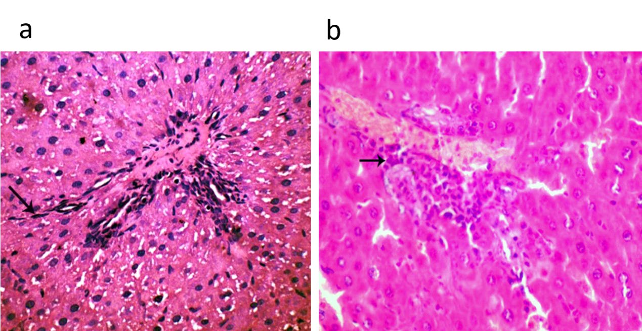
Figure 1: The depicted figure shown (a) Liver of lactating dams treated with 1/10 LD50 of CFP orally from 0th - 21th days postpartum showing intralobular fibroblastic and histiocytic infiltration in the hepatic tissue (arrow) in (HE, 400x). (HE, 400x) (b) Liver of suckling pups of treated dams with 1/10 LD50 of CFP orally from 0th - 21th days postpartum showing mild lymphocytic infiltration in hepatic tissue (arrow) (HE, 400x).
Brain: The lactating dams treated with different doses of CFP there was neuronal necrosis, neuronophagia and astrocytosis in the brain tissue with degenerative changes of purkinje cells in cerebellum and the pathological changes were more sever in respect to the control group, results illustrated in Figures 2a & 2b.
The pups of treated dams with different doses of CFP there was focal edema in the neutrophils with focal hemorrhagic areas in the brain tissue in a dose dependent manner in respect to the control group, results illustrated in Figure 2c.
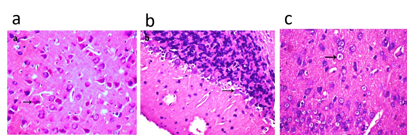
Figure 2: The depicted figure shown (a) Brain of lactating dams treated with 1/10 LD50 of CFP orally from 0th - 21th days postpartum showing central chromatolysis and neuronal necrosis in the brain tissue (arrow) (HE, 400x) (b) Brain of lactating dams treated with 1/20 LD50 of CFP orally from 0th - 21th days postpartum showing degenerative changes of purkinje in cerebellum (arrow) (HE, 400x) (c) Brain of suckling pups of treated dams with 1/10 LD50 of CFP orally from 0th - 21th days postpartum showing cytotoxic edema of neurons (arrow) in (HE, 400x).
Kidney: The lactating dams treated with different doses of CFP displayed fibrous tissue proliferation of renal glomeruli with degenerative changes in renal tubular epithelium results illustrated in Figure 3a. while the pups of treated dams with different doses of CFP shown a degeneration in the renal tubular epithelium and interstitial lymphocytic infiltration with congestion of the renal glomeruli in a dose dependent manner in respect to the control group, results illustrated in Figure 3b.
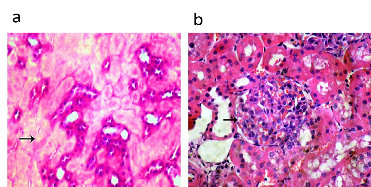
Figure 3: The depicted figure shown a) kidney of lactating dams treated with 1/10 LD50 of CFP orally from 0th - 21th days postpartum showing degenerative changes and necrosis of renal tubular epithelium (HE, 400x). (b) Kidney of suckling pups of treated dams with 1/10 LD50 of CFP orally from 0th - 21st days postpartum showing congestion of renal glomeruli (arrow) (HE, 400x).
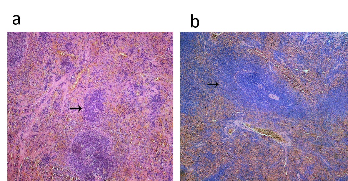
Spleen: For lactating dams treated with different doses of CFP there was marked lymphoid depletion in the splenic tissue and the pathological changes were more sever in the dose of highest dose of CFP in respect to the control group, results illustrated in Figure 4a.
The pups of treated dams with different doses CFP displayed a severe lymphoid depletion with congestion of the splenic sinusoids in a dose dependent manner in respect to the control group, results illustrated in Figure 4b.
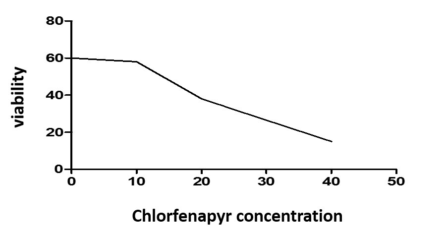
Notably, the chlorfeapyr treatment to HepG2 cells at highest concentration of clorfenapyr at 20 and 40 is cytotoxic as increased numbers of dead cells versus viable HepG2 cells figure 6. Moreover, the cell lysis shown a significant reduction of glutathione superoxide dismutase, and increased MDA level especially at high concentration of chlorfenpayr at doses of 20 and 40 ng/ml Table 8 & Figure 5.
| GSH mg/g. tissue | SOD U/g. tissue | MDA nmol/g. tissue | |
|---|---|---|---|
| 0 | 20.69±1.095a | 8.42±0.054a | 14.97±0.93c |
| 10 ng/ml | 18.69±1.09a | 7.42±0.034a | 13.97±0.93c |
| 20 | 10.69±1.03b | 3.42±0.054b | 33.97±0.93b |
| 40 | 8.91±1.042b | 2.08±0.53b | 75.96±1.90a |
Table 8: Effect of CFP on oxidative stress in (HepG2) cells.
Discussion
CFP has a household and agriculture uses but also have a hazardous effects on animals and human health (1- 4) especially when contaminated the environment [9, 10], or remained as residues in food products (11-13) , water, animal derived foods [14, 15].
CFP, a pyrrole insecticide has displayed a potential role to control of parathyroid resistant insects [21] and it was considering as a candidate insecticide for targeting malaria vectors that were resistant to pyrethroids [22]. Even chlorfenaapyr considered a good candidate’s insecticides, it had developmental and maternal toxicity reported earlier [7, 8].
Here, we are the first report of postnatal effects of chlofenapyr in female albino rats and its pups through hepatorenal toxicity, increased the oxidant, reduction of antioxidant levels in tissue of dams and pups and pathological changes in both tissue of dams and its pups. In this regard, CFP could pass through placental barrier as it evidence its presence in milk as residues [15]. In consistent to current study, maternal toxicity of oral exposure CFP at doses of 1/10, 1/20 of LD50 in female albino rats recorded earlier with evidence of induction of pathological features in liver, kidney, placenta, increased activity of liver enzymes, urea and creatinine levels, MDA as oxidant and reduced the levels of antioxidants [8]. Notably, CFP induced a significant inhibition in the activity of GST in the antioxidant in CHOK1 cells which retain GST levels after treatment with vitamin C or vitamin E [23]. Additionally, CFP had a cytotoxic effect in the different antioxidant assays. CFP is a pyrrole pro- insecticide; it is bio transformed by phase 2 oxidative elimination of N-ethoxymethyl group, which induced ablation of the mitochondrial ATP production through uncoupling of the mitochondrial oxidative phosphorylation that might enhanced the reactive oxygen radicals’ production [23, 24].
Notably, the cytotoxic effect of CFP recorded on CHO-K1 cell line as well as indicated cases of human intoxicated with CFP [25]. Finally, the pathological effect of CFP on brain was translated on neurobehavioral changes. In conclusions, chlorfeapyr is a good candidate insecticide in vector control but had a cytotoxic effect in female albino rats, its pups, and in HepG2 cells
Acknowledgment
All authors thank dr Mohamed Fawzy for help us in pathological examination and comments of all lesion in this research work. In addition, we thank for all staff member of our department for support us during experimental research.
Conflict of Interest: All authors have no conflict of interest
References
-
Kang C, Kim DH, Kim SC, Kim DS (2014) A patient fatality following the ingestion of a small amount of CFP. Journal of emergencies trauma and shock 7(3): 239-241.
-
Endo Y, Tachibana S, Hirano J, Kuroki Y, Ohashi N, et al. (2004) Acute CFP poisoning.
-
Hoshiko M, Naito S, Koga M, Mori M, Hara K, et al. (2007) Case report of acute death on the 7th day due to exposure to the vapor of the insecticide CFP. The Japanese journal of toxicology 20(2): 131-136.
-
Choi JT, Kang GH, Jang YS, Ahn HC, Seo JY, et al. (2010) Fatality from acute CFP poisoning. Clinical Toxicology 48(5): 458-459.
-
Kwon J, Kim H, Han H, Kim J, Park J (2012) A case of CFP intoxication with central nervous system involvement. J Clin Toxicol 2(8): 147.
-
Lee J, Lee JH, Baek JM, Lee DS, Park IY, et al. (2013) Toxicity from intra-abdominal injection of CFP. Case reports in emergency medicine.
-
Sleem F, Elalfy MM, Allah AAA, Hamed MF, Abomosallam M (2019) Developmental and Ultrastructure Toxicity of Greenhouse Insecticide CFP in Rat Fetuses. American- Eurasian Journal of Toxicological Sciences 11(1): 1-10.
-
Elalfy M, Abomosallam M, Allah A, Mahmoud H, Eleuony O (2019) Maternal and Cytotoxic Effects of CFP as a Pro- Insecticide in Pregnant Female Albino Rats American- Eurasian Journal of Toxicological Sciences 11(2): 29-38.
-
Cao Y, Chen J, Wang Y, Liang J, Chen L, et al. (2005) HPLC/ UV analysis of CFP residues in cabbage and soil to study the dynamics of different formulations. Science of the Total Environment 350(1-3): 38-46.
-
Mai YL, Zhong GH, Hu MY, Liu XQ (2004) Determination of CFP residues in cabbage and soil by gas chromatography. Pesticides-Shenyan 43(5): 233-235.
-
Mai Yl, Liu HM, Liu CL, Chen WT, Hu MY (2007) Study on Residue Dynamics of CFP in Cucumber and Amaranth [J]. Journal of South China Agricultural University.
-
Wu D, Luo XT, Pan HJ, Nie XY, Fan LJ (2010) GC determination of CFP residue in cucumber s and apples. Food Science 31(10): 268-271.
-
Huang Fu, Chen G, Yang T (2007) Residue detection of CFP in vegetables. Acta Agriculturae Zhejiangensis 19(2): 119.
-
Wu Y, Zhou Z (2008) Application of liquid-phase microextraction and gas chromatography to the determination of CFP in water samples. Microchimica Acta 162(1-2): 161-165.
-
Hamilton DJ (2018) A data-fitted mathematical sequence to calculate depuration rates for pesticide residues in milk from dairy cow feeding studies. Computational Toxicology 6: 1-8.
-
Bhardwaj S, Srivastava M, Kapoor U, Srivastava L (2010) A 90 days oral toxicity of imidacloprid in female rats: morphological, biochemical and histopathological evaluations. Food and chemical toxicology 48(5):1185- 1190.
-
Fernandez Botran R, Gorantla V, Sun X, Ren X, Perez- Abadia G, et al. (2002) Targeting of glycosaminoglycan- cytokine interactions as a novel therapeutic approach in allotransplantation1. Transplantation 74(5): 623-629.
-
Agbasi PUAN, Onye JJ, Ibeawuchi C, Uzoechi SC, Alagwu EA, et al. (2015) The effect of subchronic low dose of DDVP and sodium azid on the hematological indices of albino rats. toxicology physiology and biochemistry 4: 103-110.
-
AlKahtane AA, Alarifi S, Al Qahtani AA, Ali D, Alomar SY, et al. (2018) Cytotoxicity and genotoxicity of cypermethrin in hepatocarcinoma cells: a dose-and time-dependent study. Dose-Response 16(2): 1559325818760880.
-
Snedecor GW, Cochran WG (1989) Statistical Methods, eight edition. Iowa state University press, Ames, Iowa.
-
Ngufor C, N Guessan R, Boko P, Odjo A, Vigninou E, et al. (2011) Combining indoor residual spraying with CFP and long-lasting insecticidal bed nets for improved control of pyrethroid-resistant Anopheles gambiae: an experimental hut trial in Benin. Malaria journal 10(1): 343.
-
N’guessan R, Boko P, Odjo A, Akogbeto M, Yates A, et la. (2007) CFP: a pyrrole insecticide for the control of pyrethroid or DDT resistant Anopheles gambiae (Diptera: Culicidae) mosquitoes. Acta tropica 102(1): 69-78.
-
Al Sarar AS, Abobakr Y, Bayoumi AE, Hussein HI (2015) Cytotoxic and genotoxic effects of abamectin, CFP, and imidacloprid on CHO K1 cells. Environmental Science and Pollution Research. 22(21): 17041-17052.
-
Black BC, Hollingworth RM, Ahammadsahib KI, Kukel CD, Donovan S (1994) Insecticidal action and mitochondrial uncoupling activity of AC-303,630 and related halogenated pyrroles. Pesticide Biochemistry and Physiology 50(2): 115-128.
-
Chomin J, Heuser W, Nogar J, Ramnarine M, Stripp R, et al. (2018) Delayed hyperthermia from CFP overdose. The American journal of emergency medicine 36(11): 2129.e1-2129.e2.
- California Red-Legged Frog and Non-Listed Amphibians Response to Non-Native Fish Removal
- Industrial Standardization of the Bio-OS: Algorithmic Codification of Resilience Engineering Guidelines and Version V8 Architecture
- Climate Variability and the Sustainability of Snail Farming in Nigeria: Past Trends, Present Challenges and Potential Outlook
- The Evaluation of the Surveillance System of Anthrax in Gilgit-Baltistan, Pakistan, 2018
- Natural Decline to Extinction of A New Zealand Rabbit Population
- Mitochondrial Bio-Logistics: Steering Co-Enzyme Q10 and Lycopene Synergies within the Science 4.0 Bio-OS Framework