Pain Assessment Using Various Local Anesthetics in Black Bengal Goats Undergoing Rumenotomy
The present study was aimed to evaluate the comparative efficacies of incisional local anesthetics following rumenotomy in Black Bengal goats. Goats (N=40) were randomly allocated equally into LID, LID+A, BUP and SAL groups. After overnight fasting, goats were injected with 0.5 mg kg-1 meloxicam subcutaneously and 1 mg kg-1 diazepam intramuscularly. Left paravertebral analgesia were achieved in goats of aforementioned groups using 5 ml of 2% lidocaine, 2% lidocaine plus adrenaline, 0.5% bupivacaine and 0.9% NaCl. The rumenotomy was performed and before closing the skin, 2% lidocaine (7 mg kg-1), 2% lidocaine plus adrenaline (7 mg kg-1) and 0.5% bupivacaine (2.5 mg kg-1) were infiltrated incisionally in goats of LID, LID+A, BUP groups, respectively. However, the goats of SAL group were infiltrated incisionally with 5 ml of 0.9% saline. Pain and wound tenderness were evaluated by pricking and manual pressing around the wound whereas sedation was assessed by observing behavioral manifestations using dynamic interactive visual analogue scale before premedication, 0.5, 1, 2, 4, 8 and 22 hours after surgery. Data were analyzed using one-way ANOVA followed by Bonferroni test. Goats of LID and LID+A groups showed moderately less pain up to 4 hours. BUP groups exhibited the least pain at 4 and 8 hours. LID group measured lower (P
Introduction
All surgical procedures inflict tissue injury and thereby elicit postoperative pain in animals [1]. Pain is generally manifested through behavior and vocalization in animals. Perception of pain in animals may not be the same as in man [2]. The pain perception depends on neuronal input and its interpretation based on prior experience and present state of mind. Although often considered bad, it most frequently serves as a warning device so as to avoid the potentially harmful stimulus.
Postoperative pain can result in many undesirable effects such as decreased food intake, exacerbated protein catabolism, lactic acidosis, gastrointestinal ileus, respiratory depression, cardiac dysrhythmias, central hypersensitivity and chronic pain [3, 4]. All these changes delay the wound healing; prolong the period of hospitalization and mortality [5, 6, 7, 8]. Alleviating of pain not only prevents the above problems but also results in faster recovery [9].
Pain management has been widely recognized as an essential component of veterinary care [10]. Postoperative pain is very difficult to assess thereby animals are often undertreated for pain. Veterinarians must rely on interpretation of physiological and behavioral manifestations for the assessment of pain because the animals cannot verbalize their pain. The pain can be managed effectively when its signs can be assessed reliably and accurately [7]. The different methods of pain a measurement in animal models has been described very recently [11]. There is no any ideal method that validates and measures the pain objectively. Ideal pain assessment scale should be ease for use, efficacious, repeatable and non-biased. The wound infiltration with local anesthetics is inconsistently and randomly implicated in human beings and small animals [12]. At present, the wound infiltration with local anesthetics for postoperative pain relief are gaining popularity in veterinary medicine because of its simplicity, safety and low cost. Local anesthetic blocks the sensory nerve impulse and prevents the development of central sensitization. Moreover, its recommended dose is virtually free from side effects [13]. The present work was therefore initiated in the Black Bengal goats after rumenotomy to evaluate the efficacies and comparative analgesic effects of different incisional local analgesics.
Materials and Methods
Experimental Animals
Forty Black Bengal goats (16 males and 24 females), aged 8.30±1.97 months and weighing 8.50±1.70 kg were used in this study. The goats were maintained without medication, considered apparently healthy and were free from any painful conditions. This study followed the guidelines of the Ethics Committee of the Faculty of Veterinary Medicine, Bangladesh Agricultural University (BAU), and Mymensingh-2202. All goats were cared for in accordance with guidelines provided by the Animal Care and Use Facility at the Veterinary Clinic, BAU.
Study Design
The order of treatments was determined by a random number generator prior to the start of the study. Each goat was assigned to one of the treatment groups on the day of surgery: lidocaine (LID), lidocaine plus adrenaline (LID+A), bupivacaine (BUP) or saline (SAL). Pre-and post-procedural pain, wound tenderness and scoring were performed by using the Dynamic Interactive Visual Analogue Scale (DIVAS) by one observer (MKS) who was blinded to the treatment status of the goats. Incision length was measured upon completion of surgery.
Premedication, Anesthesia and Surgery
Surgeries were performed on early hours of the day. Food, but not water, was withheld for 12 hours prior to premedication. All goats were clinically examined prior to premedication. Baseline values were recorded for heart rate (HR), respiratory rate (RR), rectal temperature (RT) and temperament of the animal. The body weight (kg) was recorded for each goat before premedication. Goats were injected with 0.5 mg kg-1 SC meloxicam (Melonex, Intas Pharmaceuticals Ltd., India) as a preemptive analgesic and premedicated with 1 mg kg-1 IM diazepam (Sedil, Square, Bangladesh). After 30 minutes of premedication, paravertebral analgesia was achieved by injecting 5 ml of 2% lidocaine, 2% lidocaine plus adrenaline, 0.5% Bupivacaine (Ultracaine, Jayson Pharmaceuticals Ltd.) and 0.9% normal saline in divided doses for last thoracic (2 ml for T13) and the first two lumbar spinal nerves (1.5 ml for each L1 and L2) distal to their emergence from intervertebral foramina into the goats of LID, LID+A, BUP and SAL groups, respectively [14, 15]. Rumenotomy was performed in all goats following standard surgical procedure. The incised ruminal edges were cleaned thoroughly with normal saline and sutured in double layers of cushing pattern using catgut ‘1’ (Catgut, Johnson & Johnson Ltd., Mumbai, India). The peritoneum was sutured with catgut ‘0’ (Catgut, Johnson & Johnson Ltd., Mumbai, India) in simple continuous pattern and all muscle layers were sutured together with catgut ‘1’ employing the lock stitch.
Just prior to the closure of the skin, 2% Lidocaine hydrochloride (7 mg kg-1) or 2% Lidocaine plus adrenaline (7 mg kg-1) or 0.5% Bupivacaine (2.5 mg kg-1) was infiltrated incisionally into the wound. In the control group, the area was infiltrated incisionally with normal saline. Finally, the skin incision was sutured with nylon thread in a horizontal mattress pattern. Upon completion of the surgery, the goats were placed in sternal recumbency for 10 minutes. Once recovered from anesthesia, the goats were fed on dried grass supplemented with concentrated ration and availed ad libitum drinking water. The surgical wounds were dressed daily with povidone iodine and Ampicillin (Acipillin, ACI Pharmaceuticals Ltd., Bangladesh) at a dose of 10 mg kg-1 was administered IM four times a day up to 5 consecutive days.
Scoring of Pain, Wound Tenderness and Sedation
DIVASs were used for scoring the pain, wound tenderness and sedation before premedication and at 0.5, 1, 2, 4, 8 and 22 hours after the surgery. It was recorded by placing a cross mark on a 100-mm scale between ‘no pain’ and ‘worst imaginable pain’ for pain scores (DIVAS-pain).
Similar methods were applied for wound tenderness (DIVAS- wound tenderness) and sedation (DIVAS-sedation). Goats were also observed for the signs of local anesthetic toxicities such as sedation, nausea, and seizures. Data were obtained by measuring the distance on the DIVAS from ‘0’ to the cross marked point in mm.
Statistical Analysis
Data are presented as mean ± SD. Statistical analyses were performed using the SPSS information system for windows (SPSS V 16, SPSS Institute Inc. USA). Data were analyzed using ANOVA followed by Bonferroni test. P values less than 0.05 were considered significant.
Results
No adverse events or reactions to the treatments given were observed through the course of the study.
Divas-Pain Scores
Overall, goats showed the severe pain at 1 hour, thereafter decreased gradually through other observational periods and measured the least at 22 hours after surgery (Figure 1). The goats of LID, LID+A groups exhibited lower pain scores (P<0.01) than SAL groups at 0.5, 1, 2 and 4 hours postoperatively. The goats of the BUP group were observed with lower pain (P<0.01) than SAL or LID group at 0.5 hour, but with higher pain (P<0.05) than that of LID+A group at 1 hour. The LID group showed lower pain scores than the LID+A group, whereas BUP group resulted in higher pain scores than LID and LID+A groups at 2 hour. Inadvertently, the goats of BUP groups exhibited the least pain at 4 and 8 hours among the groups. The goats of LID+A group also exhibited lower pain scores than SAL group at 8 hours after surgery. There were no remarkable differences in measured pain scores among the groups at 22 hours of surgery.
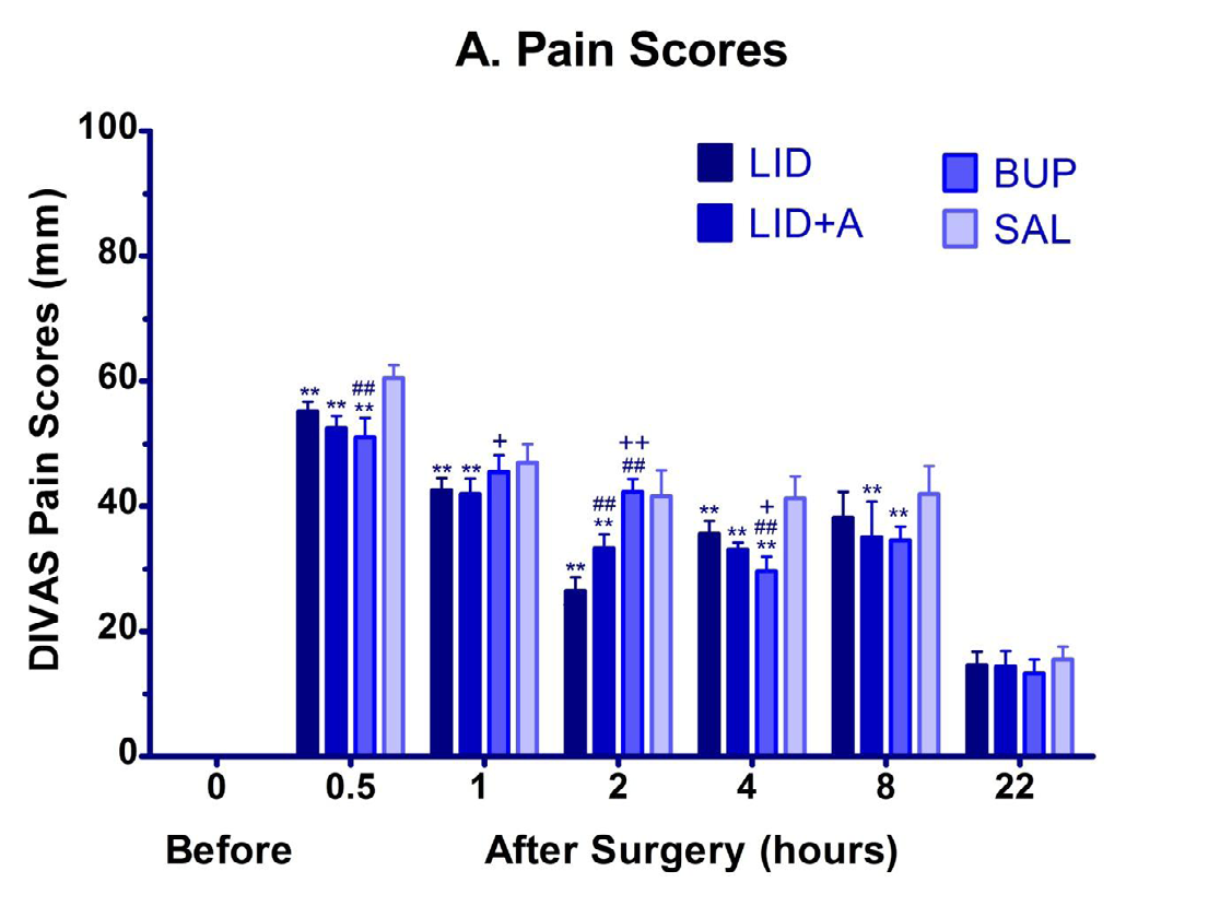
Figure 1: Postoperative pain was scored using a DIVAS in 40 goats for 22 hours following rumenotomy. Goats were preemptively injected with meloxicam and premedicated with diazepam. Paravertebral anesthesia was achieved with lidocaine, lidocaine+adrenaline, bupivacaine and saline. Rumenotomy was performed and incisional infiltration of lidocaine, lidocaine+adrenaline, bupivacaine and saline before suturing the incised skin. Basal pain scores are not shown because the goats were not in pain prior to surgery and their scores were zero. **P < 0.01, *P < 0.05; LID/ LID+A/BUP group vs. SAL group. ##P < 0.01, #P < 0.05; LID+A /BUP vs. LID group. ++P < 0.01, +P < 0.05; BUP vs. LID+A group; One-way ANOVA followed by Bonferroni’s post-test.
Divas-Wound Tenderness Scores
Figure 2 shows the WT scores that increased gradually irrespective of groups. The WT scores decreased (P<0.01) in the LID group as compared to SAL and BUP groups at 0.5 hour. All local anesthetics treated goats exhibited less WT scores than SAL group at 1, 2, 4, 8 and 22 hours after surgery.
However, the WT scores decreased (P<0.01; 0.05) in goats of LID+A group than that of LID group at 2 and 4 hours. But the LID+A group showed higher WT scores than the BUP group at 2 hour. The goats of LID+A and BUP groups exhibited lower (P<0.01) WT scores than the SAL group at 4, 8 and 22 hours. The BUP group showed the higher WT scores than the LID+A group at 4, 8 and 22 hours of surgery.
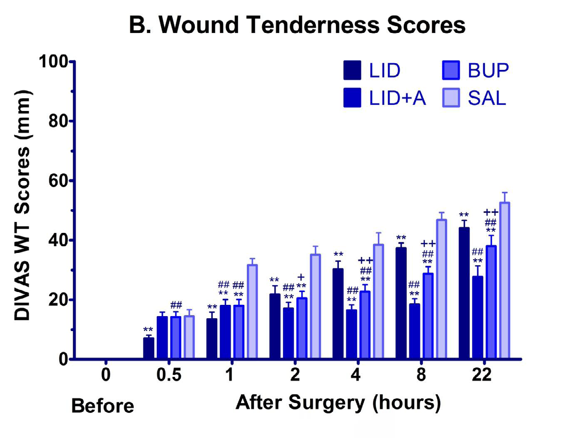
Figure 2: Wound tenderness scores in goats of different treatment groups at each assessment time. Basal DIVAS-wound tenderness scores are not shown because the goats were not having the wound prior to surgery and their scores were zero. **P < 0.01, *P < 0.05; LID/ LID+A/BUP group vs. SAL group. ##P < 0.01, #P < 0.05; LID+A /BUP vs. LID group. ++P < 0.01, +P < 0.05; BUP vs. LID+A group; One-way ANOVA followed by Bonferroni’s post-test.
Divas-Sedation Scores
Goats showed no signs of sedation before premedication. The sedation scores were highest at 0.5 hour and decreased thereafter in all treatment groups (Figure 3). Specifically, the goats of LID were sedated more (P<0.01) at 1, 2 and 8 hours of surgery than SAL goats. The goats of BUP group resulted in lower sedation scores (P<0.05) than the LID group at 1 hour but higher (P<0.01) at 4 and 8 hours of surgery. Also, the goats of LID+A group sedated higher (P<0.01) than goats of LID group at 4 hours of surgery. No significant changes in sedation scores were reported among the groups at 22 hours of surgery.
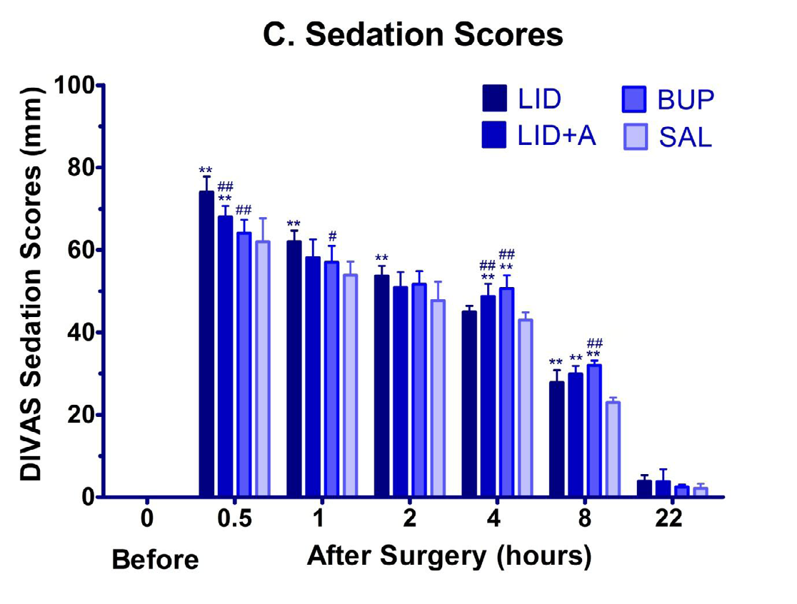
Figure 3: Sedation scores in different treatment groups at different assessment times. Basal DIVAS-sedation scores are not shown because the goats were not sedated before premedication and their scores were zero. **P < 0.01, *P < 0.05; LID/ LID+A/ BUP group vs. SAL group. ##P < 0.01, #P < 0.05; LID+A /BUP vs. LID group. ++P < 0.01, +P < 0.05; BUP vs. LID+A group; One-way ANOVA followed by Bonferroni’s post-test.
Discussion
The ability to quantify the degree of pain experienced by small ruminants is now considered as an important component in the assessment of animal welfare. Pain is the result of a cascade of physiological, immunological, cognitive, and behavioral effects that may make it uncontrollable. The responsive or interactive behaviors after surgical wound palpation are recently considered as a vital determinant for assessing the degree of postoperative pain. Awareness regarding the pain assessment and its proper treatment has been increased recently in animals [16]. Among several pain scales, the VAS has been proved sensitive, reliable and easy to use by an experienced observer in both human and veterinary practices to quantify the degree of pain and sedation [8, 12, 17, 18, 19]. In the present study, DIVAS [20] after slight modification of consequent IVAS [21] or VAS was used to quantify the pain, wound tenderness and sedation. In earlier study, Firth AM, et al. [22] reported the widespread observer variation in measuring the pain scores with VAS. Therefore, a single observer scored the pain, wound tenderness and sedation at each time points throughout the study. The responsive or interactive behaviors including biting of the wound, painful bleating, falling on the ground towards the opposite side of the wound, re-standing and statue posture showed after manipulation of surgical wounds. Interestingly, these behavioral manifestations varied accordingly with the severity of pain. Janyaro H, et al. [23] developed a dedicated 0-4 scale including the above interactive behavioral signs to grade the pain in goats. Accumulating set of experimental studies [24, 25, 26] used electromyography for measuring the visceral pain objectively in the rats and goats. However, it is not feasible to employ the electromyography for pain measurement in routine veterinary practice.
Overall, the postoperative pain scores were increased in Black Bengal goats of all groups as compared to their baseline scores indicating that pain is provoked after surgery if it is not managed properly. Multimodal analgesic modules constituting local anesthetics are gaining popularity for treating the postoperative pain. In the present study, goats were injected with meloxicam as a preemptive analgesic and diazepam as a sedative in combination with incisional infiltrations of local analgesics. The local anesthetics block the sodium influx into the nerve axon and thereby inhibit the action potential that ultimately disrupts the generation and transmission of nerve impulses to the central nervous system. The goats were observed with peak pain at 0.5 hour and gradually decreasing at the subsequent time points in this study. The dogs premedicated with acepromazine and butorphanol along with intraperitoneal and incisional splash of local anesthetics exhibited also the severe pain at 0.5 hour after ovariohysterectomy [12]. A study of Flecknell P, et al. [2] reported that the combined use of preemptive analgesics provides optimum analgesia than the single analgesic agent. Because the constituents of balanced regimens act differently in order to potentiate their analgesic effect and lessen the untoward effects [27].
The goats of LID, LID+A groups were observed with lower pain at 0.5, 1, 2 and 4 hours which indicates lidocaine provided excellent analgesia up to 2 hour of surgery. Earlier study also reported that lidocaine has relatively rapid onset (5 to 10 minutes) and intermediate duration (1 to 2 hours) of action [12]. However, bupivacaine followed lidocaine plus adrenaline attenuated pain efficiently at 4 and 8 hours. The effect of lidocaine may be prolonged due to added adrenaline which not only slowed onset but also prolonged the analgesic effects at the infiltrated sites caused by vasoconstriction [14]. The BUP group showed comparatively higher pain scores at initial time points because of its slower onset of action however it persisted longer up to 8 hours of surgery. Carpenter, et al. [12] reported that the intraperitoneal and incisional administration of bupivacaine resulted in efficacious analgesia than lidocaine following ovariohysterectomy in dogs.
In this study, the goat evinced the moderate pain because the saline treated goats measured an average of 60.5 out of 100 on the DIVAS-pain. So, no goats were further supplemented with analgesic drugs. This moderate level of pain corresponds with the findings of Zimmermann M, et al. [28] who reported the mild to moderate level of pain after abdominal surgery in farm animals. The incisional infiltration of local anesthetics also relieved the pain provoked due to abdominal surgeries in human beings as these patients exhibited the lower pain scores (25 to 50 mm) with VAS [29]. Several studies also reported similar findings in ovariohysterectomized dogs [12, 22, 23, 24, 25, 26, 27, 28, 29, 30]. However, the variation in pain responses should be accepted from the species to species as well as between individuals and within species. Presently, electro acupuncture has been proved effective for attenuating both acute and chronic pain in goats because it down-regulates the protease activated receptor-2 mediated calcitonin gene related peptide release in the spinal cord [31].
Irrespective of groups, the WT scores increased gradually across the assessment time. The lidocaine treated goats measured the least WT scores at 0.5 hour. Thereafter, all local anesthetics treated goats exhibited lower WT scores than SAL group. The WT scores of LID+A group were higher than BUP group at 2 hours but lower than LID group at 2 and 4 hours. Subsequently, the goats of BUP group showed the higher WT scores than the LID+A group at 4, 8 and 22 hours. So far no rigid explanation has been reported regarding the assessment of wound tenderness in animals. The reason may be the variation in onset as well as duration of action of individual local anesthetics. Moreover, the dogs that received post-incisional infiltration of bupivacaine revealed significantly less erythema and complications as excessive inflammation, splenic laceration and herniation at 24 hours after surgery [32].
Whether or not the sedation is employed as an adjunct with local analgesia will depend on the doses, species, temperament, and magnitude of the procedure and health status of the animal. The role of diazepam in the pain modulation is still controversial. In the present study, goats were injected with diazepam which facilitated for the reduction of fear and liability to sudden movement. The sedation was characterized by interactive bleating, decreased response to noise, mild relaxation of jaw, drowsiness, salivation, reduced response to pin pricks, slight incoordination and muscle relaxation, drooping of the upper eyelids and sluggish pedal reflexes which correspond with earlier findings in goats and yaks [33, 34].
All goats were severely sedated at 0.5 hour and decreased thereafter in all groups. Specifically, the goats of LID were sedated most at 1 and 2 hours. However, the goats BUP and LID+A groups resulted in higher sedation at 4 and 8 hours of surgery. On the contrary, Carpenter RE, et al. [12] reported that the dogs in the SAL group were sedated the most followed by LID and BUP at each assessment time. It has been reported that local analgesic drugs may also exert a sedative action when they are absorbed from the site of administration [35]. In the current study, we measured the sedation scores to ascertain whether the sedative effects were due to systemic absorption of local anesthetics. The goats treated with lidocaine were sedated most up to 2 hours whereas the bupivacaine treatment resulted in peak sedation at 4 and 8 hours. The reason may be the systemic absorption of the different local anesthetics. In consistent with present findings, Carpenter RE, et al. [12] confirmed that the dogs of SAL group also received supplemental sedatives and analgesia. It appears that if local analgesics were not used for postoperative pain control, there would not be any differences in sedation scores among the groups throughout the study periods. Earlier studies of Waterman Pearson, et al. [27, 36] also reported that there were no significant differences in degree of sedation between the groups at any time and the level of sedation did not influence the differences in pain scores in cats and dogs.
Conclusions
It is essential to consider the postoperative incisional infiltration of local anesthetics for the control of pain along with good postoperative care in Black Bengal goats after rumenotomy. The incisionally infiltrated lidocaine provided excellent analgesia for a short period whereas lidocaine plus adrenaline and bupivacaine provided analgesia for a longer period of time.
Clinical Relevance
A single dose of postoperative incisional local analgesic is unlikely to provide postoperative analgesia and therefore the multimodal analgesic approach with incisional local analgesic is suggested for postoperative pain control in small ruminants.
Acknowledgements
This study was supported by Advanced Studies and Research, Bangladesh Agricultural University, Mymensingh-2202. The authors thank laboratory attendants for technical help and the nursing staff for assistance with pre-anesthetic medication.
References
-
Tsai TY, Chang SK, Chou PY, Yeh LS (2013) Comparison of postoperative effects between lidocaine infusion, meloxicam, and their combination in dogs undergoing ovariohysterectomy. Vet Anaesth Analg 40(6): 615-622.
-
Flecknell P (1999) Pain-assessment, alleviation and avoidance in laboratory animals. Anzccart News 12: 1-8.
-
Grisneaux E, Pibarot P, Dupuis J, Biais D (1999) Comparison of ketoprofen and carprofen administered prior to orthopedic surgery for control of postoperative pain in dogs. J Am Vet Med Assoc 215(8): 1105-1110.
-
Morton DB, Griffiths PH (1985) Guidelines on the recognition of pain, distress and discomfort in experimental animals and an hypothesis for assessment. Vet Rec 116(16): 431-436.
-
Carroll GL, Howe LB, Slater MR, Haughn L, Martinez EA, et al. (1998) Evaluation of analgesia provided by postoperative administration of butorphanol to cats undergoing onychectomy. J Am Vet Med Assoc 213(2): 246- 250.
-
AVTRW (1986) Guidelines for the recognition and assessment of pain in animals. Prepared by a working party of the Association of Veterinary Teachers and Research Workers. Vet Rec 118(12): 334-338.
-
Holton LL, Scott EM, Nolan AM, Reid J, Welsh E, et al. (1998) Comparison of three methods used for assessment of pain in dogs. J Am Vet Med Assoc 212(1): 61-66.
-
Lascelles BDX, Cripps PJ, Jones A, Waterman Pearson AE (1998) Efficacy and kinetics of carprofen, administered preoperatively or postoperatively, for the prevention of pain in dogs undergoing ovariohysterectomy. Vet Surg 27(6): 568-582.
-
Johnson JM (1991) The veterinarian’s responsibility: assessing and managing acute pain in dogs and cats. II. Compend Contin Educ Pract Vet 13(6): 911-921.
-
Taylor JS, Vierck CJ (2003) Effects of ketamine on electroencephalographic and autonomic arousal and segmental reflex responses in the cat. Vet Anaesth Analg 30(4): 237-249.
-
Regmi B, Shah MK (2020) Possible implications of animal models for the assessment of visceral pain. Anim Model Exp Med 3(3): 215-228.
-
Carpenter RE, Wilson D V, Evans AT (2004) Evaluation of intraperitoneal and incisional lidocaine or bupivacaine for analgesia following ovariohysterectomy in the dog. Vet Anaesth Analg 31(1): 46-52.
-
Lemke KA, Dawson SD (2000) Local and regional anesthesia. Vet Clin North Am Small Anim Pract 30(4): 839-857.
-
Hall LW, Clarke KW, Trim CM (2001) General principles of local analgesia. In: 10th (Edn.), Veterinary Anaesthesia pp: 225-245.
-
Hussain SS (1995) Local and regional anaesthesia in veterinary practice-A review. Indian J Vet Surg 16(1): 1-9.
-
Shrestha S, Shah MK (2020) Current practices of Nepalese veterinarians for the clinical management of pain in animals. J Agric For Univ 4: 225-230.
-
Bianconi M, Ferraro L, Ricci R, Zanoli G, Antonelli T, et al. (2004) The Pharmacokinetics and Efficacy of Ropivacaine Continuous Wound Instillation after Spine Fusion Surgery. Anesth Analg 98(1): 166-172.
-
Dix P, Sandhar B, Murdoch J, MacIntyre PA (2004) Pain on medical wards in a district general hospital. Br J Anaesth 92(2): 235-237.
-
Mathews KA (2000) Pain assessment and general approach to management. Vet Clin North Am Small Anim Pract 30(4): 729-755.
-
Shah MK, Gurung YB (2013) The Effect of Local Analgesics for Postoperative Pain Control after Ovariohysterectomy (OVH) in Dogs. J Univ Grants Comm 2: 14-26.
-
Cambridge AJ, Tobias KM, Newberry RC, Sarkar DK (2000) Subjective and objective measurements of postoperative pain in cats. J Am Vet Med Assoc 217(5): 685-690.
-
Firth AM, Haldane SL (1999) Development of a scale to evaluate postoperative pain in dogs. J Am Vet Med Assoc 214(5): 651-659.
-
Janyaro H, Wan J, Tahir AH, Shah MK, Li XJ, et al. (2016) Visceral pain triggered by traction on the ileocecal ligament with ileitis. J Pain Res 9: 745-755.
-
Shah MK, Wan J, Janyaro H, Tahir AH, Cui L, et al. (2016) Visceral Hypersensitivity Is Provoked by 2,4,6-Trinitrobenzene Sulfonic Acid-Induced Ileitis in Rats. Front Pharmacol 7: 214.
-
Tahir AH, Wan J, Shah M, Janyaro H, Li XJ, et al. (2016) A novel model for studying ileitis-induced visceral hypersensitivity in goats. Acta Vet Scand 58(1): 72.
-
Wan J, Ding Y, Tahir AH, Shah MK, Janyaro H, et al. (2017) Electroacupuncture attenuates visceral hypersensitivity by inhibiting JAK2/STAT3 signaling pathway in the descending pain modulation system. Front Neurosci 11: 644.
-
Slingsby LS, Waterman Pearson AE (2001) Analgesic effects in dogs of carprofen and pethidine together compared with the effects of either drug alone. Vet Rec 148(14): 441-444.
-
Zimmermann M (1986) Behavioural investigation of pain in animals. Assess Pain Farm Anim Proc pp: 16-29.
-
Moiniche S, Mikkelsen S, Wetterslev J, Dahl JB (1998) A qualitative systematic review of incisional local anaesthesia for postoperative pain relief after abdominal operations. Br J Anaesth 81(3): 377-383.
-
Hardie EM, Hansen BD, Carroll GS (1997) Behavior after ovariohysterectomy in the dog: What’s normal?. Appl Anim Behav Sci 51(1-2): 111-128.
-
Shah MK, Ding Y, Wan J, Janyaro H, Tahir AH, et al. (2020) Electroacupuncture intervention of visceral hypersensitivity is involved in PAR-2-activation and CGRP-release in the spinal cord. Sci Rep 10(1): 11188.
-
Fitzpatrick CL, Weir HL, Monnet E (2010) Effects of infiltration of the incision site with bupivacaine on postoperative pain and incisional healing in dogs undergoing ovariohysterectomy. J Am Vet Med Assoc 237(4): 395-401.
-
Pratap K (1997) Clinical evaluahdn of different preanaesthetics in goats : An experimental study. Indian Vet J 74: 897-898.
-
Kumar A, Nigam JM, Sharma SK (1999) Diazepam sedation in yaks. Indian Vet J 76: 211-213.
-
Hall LW, Clarke KW, Trim CM (2001) Principles of sedation, analgesia and premedication. In: 10th (Edn.), Veterinary Anaesthesia 75-112.
-
Slingsby LS, Waterman-Pearson AE (1998) Comparison of pethidine, buprenorphine and ketoprofen for postoperative analgesia after ovariohysterectomy in the cat. Vet Rec 143(7): 185-189.
- Natural Decline to Extinction of A New Zealand Rabbit Population
- Mitochondrial Bio-Logistics: Steering Co-Enzyme Q10 and Lycopene Synergies within the Science 4.0 Bio-OS Framework
- Hymenoptera Specimens from the Caño Negro Wetland, of the National Museum Collection, Costa Rica
- Science 4.0: Comprehensive Architecture of the Biological Operating System (Bio-OS) A Framework for Systemic Resilience and Industrialized Bio-Governance
- Rabbit on, or Hare Back? Understanding Climate Change
- Clinical Validation of Science 4.0: Flow Steering and Epigenetic Drift Inversion on a 76-Year-Old Hybrid System