Clinical Management of Burn in Stray Bull: A Case Report
The present case report shares successful management of burn injuries in a stray bull. Beside the proper wound care procedures, the bull was also treated for hypovolemia and prevention of secondary bacterial infection. The animal recovered uneventfully followings post-operative care with application of ointment and good nutritional supplement.
Introduction
Burn is an injury of integuments and underlying tissues, occurring due to high temperature or chemical substances. Burn may be defined as tissue change that occur on excessive absorption of heat by skin. Based upon the depth of tissue changes, burn of the body are classified into four degrees such as, first degree burn, second degree burn, third degree burn, fourth degree burn. In severe cases burn will produce a local and a systemic response. When compared to other diseases and disorders, the number of cases of burn in animals is generally very less. In first degree burn, only epidermis is affected whereas in second degree burn, the whole thickness of the skin is involved more or less completely but not the subcutaneous fat and muscle as in third degree burn.
In third degree burn, heat penetrates into the dermis causing destruction of not only epidermis but also dermis and its components. In fourth degree burn, changes are similar to those described in the third degree burn but extend to the sub-cutaneous fascia and deeper tissue. This present case study report a burn injury in stray bull and its successful therapeutic management [1].
Case History and Observation
One case of burn injury was reported in stray bull age of 8-9 years old in area of Jirania, West Tripura district, (Tripura, India) on 1st June, 2023. Cause of this burn injury was unknown. A team from Mobile Veterinary Unit (MVU), Jirania went to the site immediately on getting call from the local people.
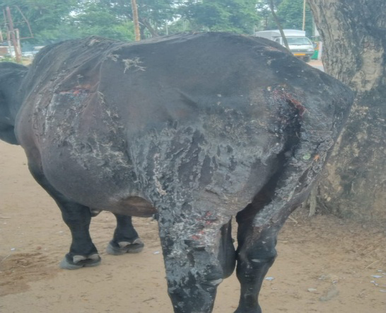
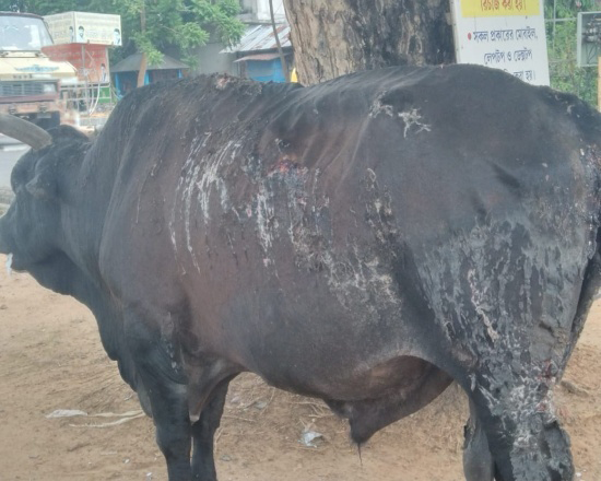
On observation, the rectal temperature, respiration rate and heart rate were 102.4º F, 40 breaths per minute and 72 beats per minute respectively, which were within normal physiological limit. An extensive burnt area was observed on the outer right and left side of abdomen, the outer side of both the hind limb, rectal muscle and the tail. As the injury was 4-5 days old there were presence of maggotic infestation in affected part of rectum and tail. The affected portion was red with peeling of skin covering the full thickness of skin layers (Figure 1).
Treatment and Discussion
After proper examination of the animal, the bull was injected with Ringer’s lactate @ 30 Ml/kg body weight to prevent hypovolaemic shock. The animal was also administered with inj. Melonex @ 0.2 mg/kg b.wt IM for 3 days and inj. Strepto-penicillin @ 20,000 U/kg b.wt IM for 7 days respectively to reduce pain and to control secondary bacterial infection respectively. Inj. Ivermectin @ 0.3 mg/kg b.wt SC was also administered to control maggotic infestation in affected part and advised to repeat after 7 days. Then maggotic infestated lesion were cleaned with turpentine
oil, all the burnt lesion were dressed with povidone iodine 5% liquid and applied Silver Sulfadiazine ointment topically over the affected area. The bull was administered with inj. Vitamine A,D3, E 10 ml IM twice a week for 30 days and inj. Pheneramine maleate 10 ml IM for 7days as a supportive therapy. Topicure spray was also used to prevent fly infestation.
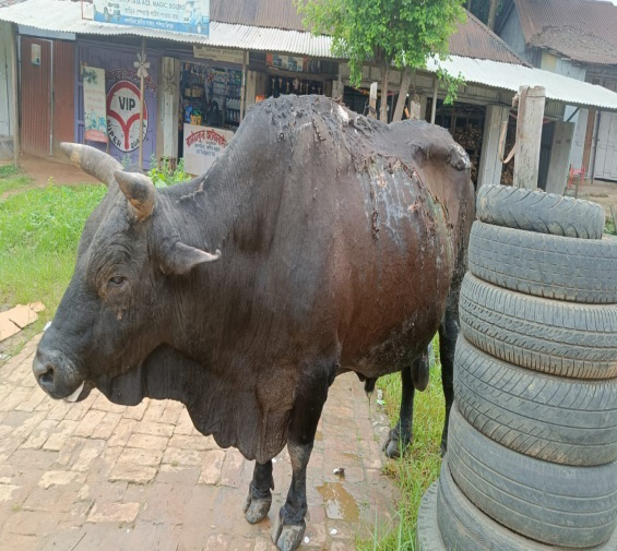
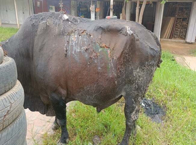
The burnt skin started peeling off from the 7th day onwards exposing underlying tissue Development of melanin pigment was also noticed on the 34th days (Figure 2).
Burn injury are very rare in Veterinary patients. Most severe burns will produce a local and systemic response, both of which must be appropriately treated to increase patient’s chances of survival [2]. A multi-dimensional treatment protocol should be followed to treat the bull. A better result can be achieved with correction of body fluid balance by restoration of ionic balance, reduction of hypovolaemia, and prevention of secondary bacterial infections [3] as applied in the case. The main aim of burn therapy are to save animal’s life, relieve pain, early wound closure and minimize deformities [4].
Conclusion
Burn in bull could successfully be treated by following a protocol of Ringer’s lactate, antibiotic therapy, povidone- iodine dressing along with topical application of silver sulfadiazine ointment which is readily available.
Conflict of Interest
The author declared no conflict of interest.
References
-
Venugopalan A (2004) Essentials of Veterinary Surgery. Burns and Scalds. Chapter 8. Oxford and IBH Publishing Co.Pvt.Ltd, New Delhi, India, pp: 70-74.
-
Geiser DR, Walker RD (1984) Management of thermal injuries in large animals. Vet Clin North Am Large Animal Pract 6(1): 91-105.
-
Sudheerbabu G, Divya T and Raja K (2018) Therapeutic management of second degree burns in buffalo. Int J Sci Environ Technol 7(2): 482-485.
-
Sagar PV, Rajesh K, Kavitha KL and Suresh K (2010) Clinical management of second degree burns in a she buffalo:A Case Report. Buffalo Bull 29(1): 65-68.
- Mitochondrial Bio-Logistics: Steering Co-Enzyme Q10 and Lycopene Synergies within the Science 4.0 Bio-OS Framework
- Hymenoptera Specimens from the Caño Negro Wetland, of the National Museum Collection, Costa Rica
- Science 4.0: Comprehensive Architecture of the Biological Operating System (Bio-OS) A Framework for Systemic Resilience and Industrialized Bio-Governance
- Rabbit on, or Hare Back? Understanding Climate Change
- Clinical Validation of Science 4.0: Flow Steering and Epigenetic Drift Inversion on a 76-Year-Old Hybrid System
- Seeds Planted by another Mind