Revisiting Acanthocephalus dirus (Van Cleave, 1931) (Acanthocephala: Echinorhynchidae) Using SEM and Energy Dispersive X-Ray Analysis (EDXA) for the First Time
Acanthocephalus dirus (Van Cleave, 1931) Van Cleave and Townsend, 1936 is the most widely distributed and studied species of acanthocephalans in North America. Acanthocephalus jacksoni Bullock, 1962 from New England and Acanthocephalus parksidei Amin, 1975 from Wisconsin are junior synonyms. Line drawings were used in these and all subsequent descriptive accounts since. We are reporting SEM images of A. dirus for the first time from 2 Wisconsin populations from Catostomus commersoni Lacépède showing new details that could not have been seen only with optical microscopy. The new SEM observations include new features that were not previously reported such as apical sensory pores, micropores variably distributed on the trunk and on the proboscis and hooks, hooks of variable size, and bursa with 2 circles of sensory papillae and 2 pedunculated suckers. We are also providing a detailed elemental analysis of middle hooks using Energy Dispersive x-ray analysis (EDXA) showing high levels of sulfur in all samples especially hook tips.
Omar M Amin1*, Matheus Augusto Gabriel de Souza Menezes2, Solinus Farrer2, Nataliya Y Rubtsova1
¹Institute of Parasitic Diseases, USA
high levels of sulfur in all samples especially hook tips.
Introduction
Van Cleave [1] first described Acanthocephalus dirus as Echinorhynchus dirus from the freshwater drum Aplodinotus grunniens Rafinesque in the Yazoo River, Mississippi before Van Cleave and Townsend [2] reassigned it to the genus Acanthocephalus Koelreuter, 1771 after validating the posterior position of the cerebral ganglion and re-evaluating the length of the lemnisci. Van Cleave and Townsend [2] reported the wider distribution of A. dirus in the Ohio and Illinois rivers and tributaries from other species of fish, and noted that this is the first occurrence of a species of the genus Acanthocephalus in North America. Amin [3] accounted for the distribution of A. dirus in the Mississippi River drainage system, or waters previously connected to it in the Mobile Bay drainage, Great Lakes, and in New
England. Acanthocephalus dirus has the “widest host (65 fish species in 16 families) and geographical distribution (in 10 states) and exhibits the greatest morphological variability compared to the 2 other species of Acanthocephalus in North America, A. tahlequahensis Oetinger and Buckner, 1976, and A. alabamensis Amin and Williams, 1983 (see Amin [3], p. 210). About the same time, Amin [4] provided an expanded diagnosis of A. dirus within which he assigned Acanthocephalus jacksoni Bullock, 1962 from New England and Acanthocephalus parksidei Amin, 1975 from Wisconsin and Lake Michigan as junior synonyms based on the use of (1) comparative morphometrics of populations from Mississippi (2 collections), Kentucky (2 collections), Illinois (3 collections), West Virginia, Wisconsin, and Ohio, (2) stratified histograms, (3) canonical discriminant function analysis, and (4) cluster analysis. Within the Mississippi River populations, Amin [4] identified clinal variations showing that the more northern populations had the largest hooks and fewer hooks per row compared to the more southern Mississippi populations.
Our studies of A. dirus began with investigating its Wisconsin population from various fishes in waters of southeastern Wisconsin tributaries of the Mississippi River and southwestern Lake Michigan. The Wisconsin population is most intensively studied morphologically, ecologically, biologically, developmentally, and from an evolutionary and cladistic standpoint, compared to other populations and species of the genus Acanthocephalus in North America. The following is a chronological review of research on the Wisconsin population of A. dirus since its initial description as A. parksidei from 11 species of fish in the Pike River where Amin [5] (Table 1, p. 304) distinguished between it and A. jacksoni Bullock [6] from New England and Van Cleave’s [1] Mississippi population using distinct variations in trunk dimensions and proboscis armature; line drawings of 17 figures were included. In the same year, Amin [7] described the variability in size and shape of trunk, proboscis, receptacle, lemnisci, eggs, number, arrangement and size of hooks, and reproductive structures in males and females, as well as abnormalities in hooks and body wall, and monorchidism. “Most of these variations were related to worm sex and age as well as to host species” (Amin [7], p. 307). Ecological associations in 11 species of fish were found to be related to host species, age and sex, concurrent infections, collection site, and season (Amin [8] p. 318) who also found that parasite distribution in host intestine to be related to intensity of infection, posterior migration and concurrent infection. An additional collection of helminths from 26 species of fish in 10 families from 2 streams in SE Wisconsin included moderate to heavy infections of A. dirus in 11 fish species (Amin [9], p. 226). A study of 8 species of helminth parasites of 18 species of SW Lake Michigan fishes revealed the presence of a viable population of A. dirus there, mostly in the white sucker Catostomus commersoni (Lacépède) (Catostomidae) (the type host of A. parksidei) and slimy sculpin Cottus cognatus Richardson (Cottidae) (Amin [10]); mixed infections with Echinorhynchus salmonis Müller, 1784 were common. Cystacanth infections and developmental cycle appeared to be associated with those of the isopod intermediate host Caecidotea militaris (Hay) (Crustacea) in a SE Wisconsin stream with 1 breeding season observed suggesting that A. dirus has a well-defined life cycle of 1 year in both intermediate and definitive hosts (Amin et al. [9]). Amin et al. [11] noted that cystacanths were over-dispersed fitting a negative binomial or truncated negative binomial distribution. Amin [4] provided an expanded description of A. dirus including the full spectrum of variabilities in all populations in North America encompassing 775 worms in 10 collections using various computer-based analyses justifying the designation of A. parksidei and A. jacksoni as junior synonyms, with the Mississippi River population being the most ancient and diversified. Amin, et al. [12] demonstrated interspecific variability in the genus Acanthocephalus using stratified histograms contrasting means of 20 variables and included a key to the species. In his discussion of the hosts and geographical distribution of Acanthocephalus in North America, Amin [3] Table 2 further differentiated between the 3 known species of Acanthocephalus, elaborated on the origin, dispersal, and diversification of A. dirus, and declared that “the discovery of species of Acanthocephalus in new hosts from the Mississippi River drainage system which might have evolved through host isolation is not unlikely” (p. 218). “Cladistic analysis provided greater understanding and further support of the evolutionary relationships among the species and populations of the genus Acanthocephalus from North American freshwater fishes” Amin [13]. A survey with line drawings of abnormalities in some helminth parasites of fish including body form, body wall, and proboscis anomalies in A. dirus from Wisconsin was reported by Amin [14]. The seasonal distribution of A. dirus from 9 species of fish in 5 families in Tichigan Lake exhibited considerably higher parameters in 1984 than in 1977-1978 leaving the question open as to whether A. dirus “undergoes annual cycles in fecundity and abundance” [15].
| Element* | Whole hook | Site of sample | Longitudinal sections | ||||
|---|---|---|---|---|---|---|---|
| Hook tip | Hook middle | Hook base | Hook tip | Hook middle | Hook base | ||
| Magnesium (Mg) | 0.48 | 0.00-0.23 | 0.14-0.19 | 0.13-0.15 | 0.08 | 0.09 | 0.06 |
| Sodium (Na) | 0.76 | 0.00-0.01 | 0.00-0.13 | 0.09-0.13 | 0.03 | 0.00 | 0.00 |
| Phosphorous (P) | 0.46 | 0.45-1.15 | 0.86-3.94 | 0.60-1.31 | 0.38 | 0.43 | 0.42 |
| Sulfur (S) | 8.1 | 9.72-23.15 | 9.09-11.35 | 6.28-12.25 | 19.18 | 11.59 | 9.00 |
| Calcium (Ca) | 0.7 | 0.25-0.26 | 0.14-0.19 | 0.15-0.38 | 0.27 | 0.28 | 0.25 |
Table 1: ** Chemical localization of elements in various parts of middle hooks of females of _Acanthocephalus dirus_ from _Catost
*Palladium (Pd) and Gold (Au) were used to count the specimens and the Gallium for the cross cut of the hooks. These and other elements (C, O, N) common in organic matter are omitted. Data is reported in weight (WT%). Table 1: Chemical localization of elements in various parts of middle hooks of females of Acanthocephalus dirus from Catostomus commersoni collected in the Pike River, SE Wisconsin.
The above review addresses only the Wisconsin population of A. dirus which is being investigated herein for SEM-based description and Energy Dispersive x-ray analysis (EDXA). We have studied many species of acanthocephalans using X-ray scans (EDXA) of FIB-sectioned hooks and spines for metal composition. The biological significance of EDXA as a diagnostic tool is exemplified by the observation that populations of an acanthocephalan species will consistently have similar EDXA spectra irrespective of host species or geography [16]. Results of the X-ray analysis of the FIB- sectioned hooks (dual beam SEM) of A. dirus show differential composition and distribution of metals in different hook parts characteristic of the species being examined.
Ten surveys and reports from 7 states along the Mississippi River drainage system providing host and locality data and some descriptive information are included in Amin [3, 4]. Kennedy [17] reviewed the evolutionary ecology of A. dirus based on some relevant publications by Amin and Amin et al., quoted above. Our most recent work on A. dirus from the Wisconsin material by Amin, et al. [18] is a cross sectional study of males and females showing many internal anatomical details not previously reported.
Materials and Methods
Collections of the Wisconsin Population of A. dirus and Locations
A total of 4159 ethanol-fixed and preserved specimens of A. dirus were originally collected from 737 examined fishes of 11 species and 6 families seined from 7 sites on the Pike River in Racine and Kenosha counties, Wisconsin (42°38′40″N,87°51′09″W) in 1972, 1973 [5]. A total of 1583 additional specimens were collected from 117 infected of 845 examined fish of 16 species and 7 families in Tichigan Lake, a relatively large eutrophic impoundment on the Fox River which is a tributary of the Mississippi River in Racine County (42°48′20″N,88°13′00″W) in 1977, 1978, and 1984 [15]). A total of 224 acanthocephalans were collected from 12 species of fish gill-netted or trawled in the coastal waters of Lake Michigan off Kenosha County at the Wisconsin- Illinois state line (42°34’33.59”N, 87°50’26.39”W) in 1980- 1981 (Amin [10]). In a separate Mississippi River collection, White, et al. [19] provided specimens of A. dirus from their 1005 specimens reported as A. jacksoni collected in 1973 from Catostomus commersoni from tributaries of the Kentucky and Ohio rivers (Madison and Fayette counties) (37°43’12.00”N,84°16’48.00”W) in Kentucky Amin [4]. Our specimens from the Pike River and Tichigan Lake have direct continuity with those from the Mississippi River which connects with Lake Michigan via the Illinois River.
A total of 60 specimens of A. dirus from 2 collections from white suckers, C. commersoni: 30 specimens (15 males, 15 females) in each collection from the Tichigan Lake (coll. M-98a in 1984), and from the Pike River (coll. M-11, 1985) were used for SEM and EDXA studies. All males had everted bursae.
Scanning Electron Microscopy (SEM)
Specimens that had been fixed and stored in 70% ethanol were processed for SEM following standard methods Lee [20]. These included critical point drying (CPD) (Tousimis Automandri 931.GL; Rockville, Maryland) and mounting on aluminum SEM sample mounts (stubs) using conductive double-sided carbon tape. Samples were sputter coated with an 80%-20% gold-palladium target for 3 minutes using a sputter coater (Quorum (Q150T ES) www.quorumtech.com) equipped with a planetary stage, depositing an approximate thickness of 20 nm. Samples were placed and observed in an FEI Helios Dual Beam Nanolab 600 (FEI, Hillsboro, Oregon) Scanning Electron Microscope (FEI, Hillsboro, Oregon). Samples were imaged using an accelerating voltage of 5 kV, and a probe current of 86 pA, at high vacuum using a SE detector.
Focused Ion Beam (FIB) Sectioning of Hooks
A dual-beam SEM with gallium (Ga) ion source (GIS) is used for the LIMS (Liquid Ion Metal Source) part of the process. The gallium beam (LIMS) is a gas injection magnetron sputtering technique whereby the rate of cutting can be regulated. Hooks were sectioned at two positions (Tip and Middle) using the FEI Helios Dual Beam Nanolab mentioned above. The dual-beam FIB/SEM is equipped with a gallium (Ga) Liquid Ion Metal Source (LIMS). The hooks of the acanthocephalans were centered on the SEM stage and cross-sectioned using an ion accelerating voltage of 30 kV and a probe current of 2.7 nA. Following the initial cut. The time of cutting is based on the nature and sensitivity of the tissue. The sample also goes through a cleaning cross-section milling process to obtain a smoother surface. Cuts were analyzed with an X-ray normally at the tip, middle, and base of hooks for chemical ions with an electron beam (Tungsten) to obtain an X-ray spectrum. The intensity of the GIS was variable according to the nature of the material being cut. Results were stored with the attached imaging software then transferred to a USB for future use.
Energy Dispersive X-Ray Analysis (EDXA)
The Helios Nanolab 600 is equipped with an EDXA (Mahwah, NJ) TEAM Pegasus system with an Octane Plus detector. The sectioned cuts were analyzed by EDXA. Spectra of selected areas were collected from whole specimens and from the center and the edge of each cross-section. EDXA spectra were collected using an accelerating voltage of 15 kV, and a probe current of 1.4 nA. Data collected included images of the displayed spectra as well as the raw collected data. Relative elemental percentages were generated by the TEAM software.
Results and Discussion
Amin [5] described and measured 100 males (from C. commersoni and Semotilus atromaculatus Mitchill) and 130 females (from 5 host species) of the Wisconsin population of A. dirus, originally named A. parksidei, during November and May from the Pike River, Kenosha County. A detailed description was provided with 7-line drawings of 2 males, male and female reproductive systems, an egg, and 2 proboscides. Six other line drawings comparing the body form of A. parksidei with A. jacksoni were also included. The following is an SEM description of specimens of A. dirus from this collection in Wisconsin revisiting external features first seen with SEM that were not possible to observe with light microscopy that was used for the original line drawings. Detailed anatomical description of internal structures and measurements reported by Amin [5] are not repeated herein.
New Morphological SEM Description of A. dirus from Wisconsin
Specimens cylindrical, small to medium in length, wider near middle, and tapering towards both ends. Males (Fig. 1) smaller than females. Proboscis nearly cylindrical, usually directed ventrad, with 10-16 (10-14 in males) rows of 8-11 hooks each (Figs. 2, 3). Proboscis with well-defined apical sensory pore (Fig. 4) and micropores (Fig. 10). Middle hooks most robust (Fig. 6) gradually becoming more slender and smaller anteriorly (Fig. 5) and posteriorly (Fig. 7). Apical and basal hooks smallest. All hooks with thin cortical layer and thick core (Fig. 8) and micropores (Fig. 9) comparable to those of the proboscis. Anterior trunk integument with micropores (Fig. 17) that vary from those in the middle and posterior trunk (Fig. 18) as well as from those on the proboscis and hooks in diameter and distribution. Bursa normally tilting ventrad (Fig. 11), with large spheroid base and the multi- layered corrugated distal end housing the partially retracted elevated inner wall with sensory papillae (Figs. 12-13, arrows). Extended inner bursal wall shows 2 clusters of sensory papillae (Fig. 14, arrows) and source attachment of suckers to ventral aspect of bursa. Two adjacent inflatable suckers (Fig. 15) pedunculated at ventral aspect of bursa characterize A. dirus males. High magnification of 1 sucker (Fig. 16) demonstrating 1 sensory papilla (Fig. 16, arrow). Female gonopore terminal with plump vulva (Fig. 19) with prominent orifice. Eggs elongate with dense network of fibrils (Fig. 20).
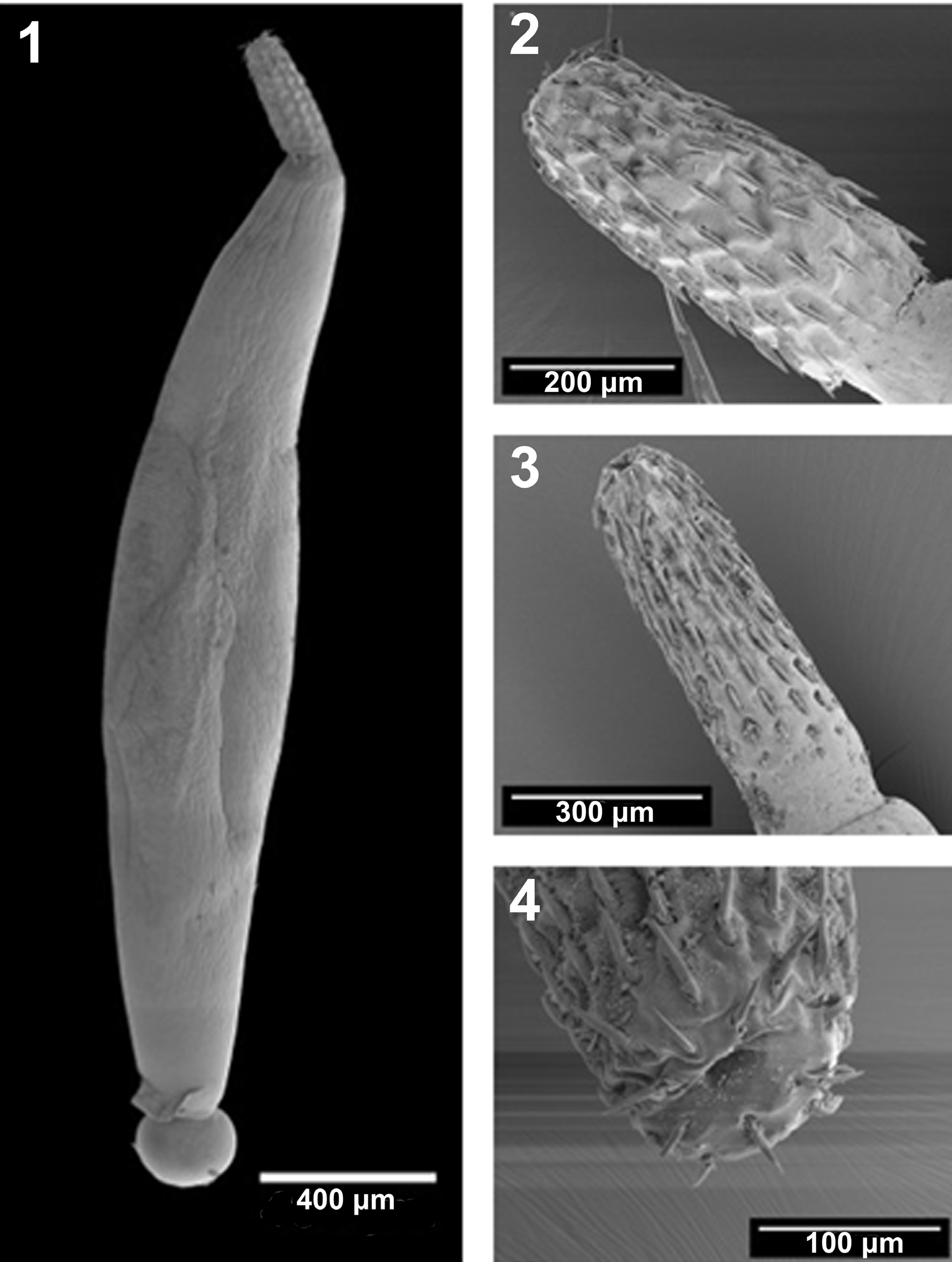
Figures 1-4: SEM of specimens of Acanthocephalus dirus from the Wisconsin population originally obtained from Catostomus commersoni in the Pike River.
- A whole male. Note the overall shape and the size of proboscis and bursa compared to the trunk.
- A proboscis with 8 hooks per row.
- Another proboscis with 10 hooks per row. The number of hooks per row in this population varied between 8 and 11 (usually 9 or 10).
- The anterior end of a proboscis showing the recessed apical sensory pore.
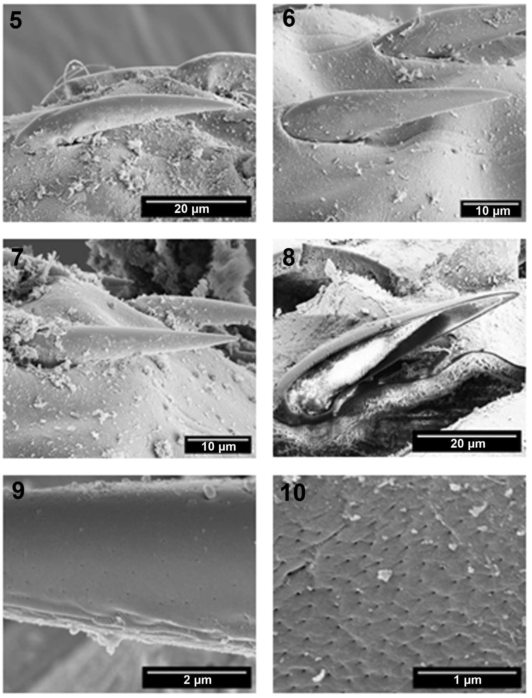
Figures 5-10: SEM of specimens of Acanthocephalus dirus from the Wisconsin population originally obtained from Catostomus commersoni in the Pike River. The proboscis. 5-7. Detail of sub-apical, middle, and pre-basal hooks, respectively, showing differences in size and thickness. Middle hooks appeared more robust. 8. A Gallium-cut section of a middle hook with a thin cortical layer and thick separated core. The simple elongate root is masked by the thick tegumental layer. 9. A highly magnified hook showing micropores continuous with micropores on the proboscis proper. 10. Micropores on the surface of the proboscis.
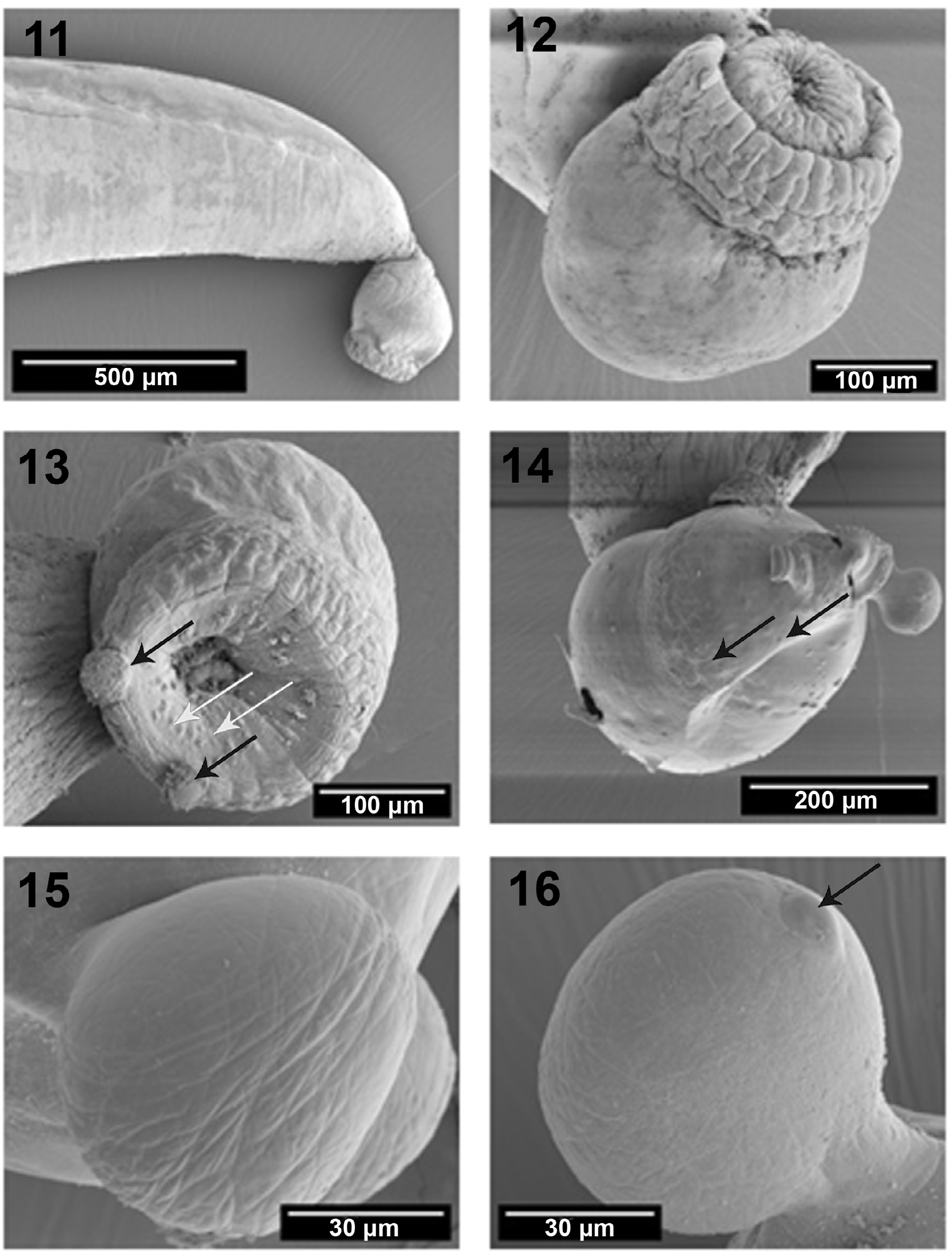
Figures 11-16: SEM of specimens of Acanthocephalus dirus from the Wisconsin population originally obtained from Catostomus commersoni in the Pike River. The bursa. 11. The terminal bursa in its usual ventrally oriented position. 12. A closer perspective of a bursa with the large spherical base and the multi-layered corrugated distal end housing the partially retracted elevated inner wall with sensory papillae. 13. A terminal perspective of another bursa showing the inner sensory papillae (white arrows) and the two suckers (black arrows). 14. A lateral perspective of a bursa with totally evaginated inner wall showing the distribution of the two zones of sensory papillae (arrows) and the two pedunculated suckers; one is broken. 15. Lateral view of the two suckers. 16. An inflated pedunculated sucker and its sensory papilla from the bursa proper (arrow).
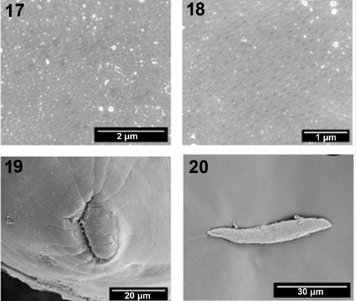
Figures 17-20: SEM of specimens of Acanthocephalus dirus from the Wisconsin population originally obtained from Catostomus commersoni in the Pike River. 17, 18. The differential distribution of micropores in the Anterior vs. the posterior integument, respectively. 19. The shape of the female genital opening with the vulva extended. 20. A ripe egg. Note the subcutaneous filaments not fully emerged.
Micropores
Micropores normally found in the various trunk regions in variable distributions and diameter, were also unusually detectable on the hooks and proboscis (Figs. 9, 10).
Energy Dispersive X-Ray Analysis (EDXA)
The spectra of EDXA summarized in Table 1 indicate that sulfur was the only dominant element in all sample sites of all hook parts and in longitudinal sections from the same parts reaching 23.15 WT% in hook tips. Calcium was negligible from all sites and the WT% of phosphorous was very low reaching a maximum of 3.94 from the middle of 1 hook of 1 specimen (Table 1).
Morphology
This is the first morphological account of A. dirus from Wisconsin, or from anywhere, using SEM. The first description by Amin [5] was quite detailed but the light microscopy account and the resulting line drawings were limited and missed many features observable only by SEM. We have demonstrated the presence of an apical sensory pore on the proboscis of A. dirus for the first time. Our Wisconsin specimens had proboscides with 8-11 hooks per row (Figs. 2, 3) which appears to be at the middle of a clinal variation between the northern New England populations of A. jacksoni with 7-10 hooks per row and the southern Mississippi population of A dirus with 11-13 hooks per row. Bullock (1962, line drawings Figs. 12-18) reported intraspecific variability in proboscis armature of A. jacksoni as did Amin (1975b, line drawings Figs. 1-8) [7] in A. parksidei. Intraspecific variability by age, sex, host, and geographical location has been documented in Amin [7]. The layers of hooks and the presence of micropores not only on the hooks and the proboscis but also on the different regions of the trunk at variable densities and diameters indicate that practically all the integument surface participate in the process of nutrient absorption; see below. The morphology of the bursa is delineated for the first time showing the presence of 2 clusters of sensory papillae and the true appearance of the characteristic 2 suckers with their own sensory papillae. Only Amin, et al. [21] and Doyle and Gleason [22] previously showed microscopic images of A. dirus suckers, compressed under cover slips, appearing as concentric double walls first reported by Doyle and Gleason [22] from the northern hogsucker Hypentelium nigricans Lesueur in Gasper River, Kentucky. The morphology of the female genital orifice is seen for the first time when extended. The fibrillar matrix of the ellipsoid eggs is evident through the thin outer membrane (Fig. 20). Bullock [6] and Oetinger and Nickol [23] recognized the fibrillar coat on the inner surface of the outer egg membrane of A. jacksoni in egg suspensions. The latter authors [23] (p. 1056) related the function of the filaments to “entangling and clumping… of eggs (for) enhancing infection of the intermediate host or they might retard passage through the isopod alimentary canal giving acanthors additional time in which to hatch.” Whitfield [24] described the ultrastructure of filaments from other acanthocephalan species.
Micropores
We have reported micropores in a large number of acanthocephalan species [25] and in a few more since, and demonstrated the tunneling from the cuticular surface into the internal crypts by TEM. The micropores of A. dirus, like those reported from other species of the Acanthocephala, are associated with internal crypts and vary in diameter and distribution in different trunk regions, proboscis, and hooks, corresponding with differential absorption of nutrients. Amin, et al. [26] gave a summary of the structural-functional relationship of the micropores in various acanthocephalan species. Wright and Lumsden [27] and Byram and Fisher [28] reported that the peripheral canals of the micropores are continuous with canalicular crypts. These crypts appear to “constitute a huge increase in external surface area . . . implicated in nutrient up take.” Whitfield [29] estimated a 44-fold increase at a surface density of 15 invaginations per 1 µm² of M. moniliformis tegumental surface. The micropores and the peripheral canal connections to the canaliculi of the inner layer of the tegument were demonstrated by transmission electron micrographs in Corynosoma strumosum (Rudolphi, 1802) Lühe, 1904 from the Caspian seal Pusa caspica (Gmelin) in the Caspian Sea (Figs. 19, 20 of Amin, et al. [30]) and in Neoechinorhynchus personatus Tkach, Sarabeev, Shvetsova, 2014 from Mugil cephalus Linn. in Tunisia (Figs. 26, 29, 30 in Amin, et al. [21]).
Energy Dispersive X-Ray Analysis (EDXA)
Our studies of acanthocephalan worms have usually involved X-ray scans (EDXA) of FIB-sectioned hooks and spines [31, 32, 33, 34]. Sulfur is usually seen at the outer edge of large hooks and Calcium and Phosphorus are major ions in the base and middle of hooks where tension and strength are paramount for hook function. The following results of the X-ray analysis of the FIB-sectioned hooks (dual beam SEM) are limited to those from our Wisconsin specimens of A. dirus. The tip of middle hooks of A. dirus showed the highest level of Sulfur reaching 23.25 WT%. Comparable or higher levels of sulfur were observed in other species of acanthocephalans. For instance, Cavisoma magnum (Southwell, 1927) Van Cleave, 1931 from Mugil cephalus in the Arabian Sea, has a similar pattern but considerably higher levels of sulfur in hook tips (43.51 wt. %) and edges (27.46 wt. %) (Amin, et al. [35]). Our results are comparable to those of mammalian teeth enamel. The chemical elements present in the hooks are typical for other acanthocephalans [32, 33]. X-ray scan analysis provides insight into the hardened components, e.g., calcium, sulfur, and phosphorus, of acanthocephalan hooks. The EDXA appears to be species-specific, as in fingerprints. For example, EDXA is shown to have significant diagnostic value in acanthocephalan systematics. For example, Moniliformis cryptosaudi Amin, Heckmann, Sharifdini, Albayati, 2019 from Iraq is morphologically identical to Moniliformis saudi Amin, Heckmann, Mohammed, Evans, 2016 from Saudi Arabia, and it was erected based primarily on its distinctly different EDXA pattern (Amin, et al. [36]) as a cryptic species. Our methodology for the detection of the chemical profile of hooks in the Acanthocephala has also been used in other parasitic groups including the Monogenea [37, 38] and Cestoda [39].
Biological Significance of EDXA
The taxonomic identity of species is deep-seated at the genetic level which is expressed by the organism’s morphology and biochemistry as revealed, in part, by its elemental spectra. Amin, et al. [16, 40, 41, 42] discussed in detail the biological significance of EDXA as a diagnostic tool exemplified by the observation that populations of an acanthocephalan species will consistently have similar EDXA spectra irrespective of host species or geography. Metal analysis of hooks has become a diagnostic standard since hooks have the highest level of elements compared to the mid- and posterior trunk regions of the acanthocephalan body (Heckmann et al., [33]). Specifically, the Sulfur content in the proboscis is paramount in the composition of disulfide bonds in the thiol groups for cysteine and cystine of the polymerized protein molecules [43]. The formed disulfide bonds are direct by-products of the DNA-based process of protein synthesis which makes up the identity of a biological species. Accordingly, the level of sulfur in our EDXA profiles will indicate the number of sulfur bonds that along with the levels of calcium phosphates, will characterize the elemental identity of a species based on its nuclear DNA personality. Variations in chemical compositions probably indicate differences in allele expression. The DNA-generated sulphide bonds evident in our EDXA profiles have an important role in the stability and rigid nature of the protein accounting for the high sulfur content of the proboscis. The above processes explain the observed species-specific nature of EDXA profiles noted in our many findings.
Compliance with Ethical Standards
- Conflict of Interest: The authors declare conflicts of interest none.
- Ethical Approval: The authors declare that they have observed all applicable ethical standards.
Acknowledgments
This project was supported by the Department of Biology, Brigham Young University (BYU), Provo, Utah, and by an Institutional Grant from the Parasitology Center, Inc. (PCI), Scottsdale, Arizona. The EDXA and SEM methodologies used in this project have been pioneered by Dr. Richard A. Heckmann (BYU) (deceased) to whom we shall always dedicate our highest gratitude. We thank Michael Standing, Electron Microscopy Facility (EMF), BYU for his technical help and expertise and for the use of the instrumentation of the EMF.
References
-
Van Cleave HJ (1931) New Acanthocephala from fishes of Mississippi and a taxonomic reconsideration of forms with unusual numbers of cement glands. Trans Amer Microscop Soc 50: 348-363.
-
Van Cleave HJ, Townsend LH (1936) The assignment of _Echinorhynchus dirus_ to the genus _Acanthocephalus_. Helminthol Soc Wash 3: 63.
-
Amin OM (1985) Hosts and geographic distribution of _Acanthocephalus_ (Acanthocephala: Echinorhynchidae) from North American freshwater fishes, with a discussion of species relationships. Proc Helminthol Soc Wash 52(2): 210-220.
-
Amin OM (1984) Variability and redescription of _Acanthocephalus dirus_ (Van Cleave, 1931) Van Cleave and Townsend, 1936 (Acanthocephala: Echinorhynchidae) from freshwater fishes in North America. Proc Helminthol Soc Wash 51: 225-237.
-
Amin OM (1975a) _Acanthocephalus parksidei_ sp. n. (Acanthocephala: Echinorhynchidae) from Wisconsin fishes. J Parasitol 61(2): 301-306.
-
Bullock WL (1962) A new species of _Acanthocephalus_ from New England fishes, with observations on variability. J Parasitol 48: 442-451.
-
Amin OM (1975b) Variability in _Acanthocephalus_ _parksidei_ Amin, 1974 (Acanthocephala: Echinorhynchidae). J Parasitol 61: 307-317.
-
Amin OM (1975c) Host and seasonal associations of _Acanthocephalus parksidei_ Amin, 1974 (Acanthocephala: Echinorhynchidae) in Wisconsin fishes. J Parasitol 61: 318-329.
-
Amin OM (1977a) Distribution of fish parasites from two southeast Wisconsin streams. Trans Wis Acad Sci, Arts, Lett 65: 225-230.
-
Amin OM (1977b) Helminth parasites of some southwestern Lake Michigan fishes. Proc Helminthol Soc Wash 44: 210-217.
-
Amin OM, Burns LA, Redlin MJ (1980) The ecology of _Acanthocephalus parksidei_ Amin, 1975 (Acanthocephala: Echinorhynchidae) in its isopod intermediate host. Proc Helminthol Soc Wash 74: 37-46.
-
Amin OM, Huffman DG (1984) Interspecific variability in the genus _Acanthocephalus_ (Acanthocephala: Echinorhynchidae) from North American freshwater fishes, with a key to species. Proc Helminthol Soc Wash 51(2): 238-240.
-
Amin OM (1986) On the species and populations of the genus _Acanthocephalus_ from North American freshwater fishes: cladistic analysis. Proc Biol Soc Wash 94: 574-579.
-
Amin OM (1989) Abnormalities in some helminth parasites of fish. Trans Am Microsc Soc 108: 27-39.
-
Amin OM (1991) Helminth parasites from some Tichigan Lake fishes in southeast Wisconsin. J Helminthol Soc Wash 58: 255-260.
-
Amin OM, Lisitsyna O, Heckmann RA (2022c) Evaluating energy dispersive X-ray analysis (EDXA) as a diagnostic tool in acanthocephalan taxonomy as evidenced in Palaeacanthocephala and Archiacanthocephala. Syst Parasitol 100(1): 43-57.
-
Kennedy CR (2006) Ecology of the Acanthocephala. Cambridge Univ Press, Cambridge, pp: 249.
-
Amin OM, Rubtsova NYu (2023) The anatomy of _Acanthocephalus_ _dirus_ (Van Cleave, 1931) (Acanthocephala: Echinorhynchidae) in histological sections. Sci Parasitol 24: 31-44.
-
White G, Harley JP (2020) Helminth parasites of the white sucker (Pisces: Catostomidae) in the Kentucky River drainage. Transactions of the Kentucky Academy of Science 35: 24-26.
-
Lee RE (1992) Scanning electron microscopy and X-ray microanalysis. Prentice Hall, Englewood Cliffs, New Jersey, USA, pp: 464.
-
Amin OM, Heckmann RA, Sharifdini M, Rubtsova N, Chine HJ (2020b) On the _Neoechinorhynchus agilis_ (Acanthocephala: Neoechinorhynchidae) complex, with the description of _Neoechinorhynchus ponticus_ n. sp. from _Chelon auratus_ Risso in the Black Sea. Parasite 27: 48.
-
Doyle LR, Gleason LN (1991) Suckers and other bursal structures of _Pomphorhynchus bulbocolli_ and _Acanthocephalus dirus_ (Acanthocephala). J Parasitol 73: 437-440.
-
Oetinger DF, Nickol BB (1974) A possible function of the fibrillar coat in _Acanthocephalus_ _jacksoni_ eggs. J Parasitol 60(6): 1055-1056.
-
Whitfield PJ (1973) The egg envelopes of _Polymorphus_ _minutus_ (Acanthocephala). Parasitol 66: 387-403.
-
Heckmann RA, Amin OM, El Naggar AM (2013) Micropores of Acanthocephala, a scanning electron microscopy study. Sci Parasitol 14: 105-113.
-
Amin OM, Heckmann RA, Radwan NA, Mantuano JS, Alcivar MAZ (2009) Redescription of _Rhadinorhynchus_ _ornatus_ (Acanthocephala: Rhadinorhynchidae) from skipjack tuna, _Katsuwonus pelamis_, collected in the Pacific Ocean off South American, with special reference to new morphological features. J Parasitol 95: 656-664.
-
Wright RD, Lumsden RD (1969) Ultrastructure of the tegumentary pore canal system of the acanthocephalan _Moniliformis dubius_. J Parasitol 55: 993-1003.
-
Byram JE, Fisher Jr FM (1973) The absorptive surface of _Moniliformis dubius_ (Acanthocephala). I. Fine structure. Tissue and Cell 5(4): 553-579.
-
Whitfield PJ (1979) The biology of parasitism: an introduction to the study of associating organisms. Univ Park Press, Baltimore, Maryland, USA, pp: 277.
-
Amin OM, Heckmann RA, Halajian A, El-Naggar AM (2011) The morphology of an unique population of _Corynosoma_ _strumosum_ (Acanthocephala, Polymorphidae) from the _Caspian seal_, _Pusa caspica_, in the land-locked Caspian Sea using SEM, with special notes on histopathology. Acta Parasitol 56: 438-445.
-
Heckmann RA (2006) Energy dispersive x-ray analysis (EDXA) in conjunction with electron optics, a tool for analyzing aquatic animal parasite diseases and deaths, an update. Proc Parasitol 41: 1-18.
-
Heckmann RA, Amin OM, Standing MD (2007) Chemical analysis of metals in Acanthocephalans using energy dispersive X-ray analysis (EDXA, XEDS) in conjunction with a scanning electron microscope (SEM). Comp Parasitol 74(2): 388-391.
-
Heckmann RA, Amin OM, Radwan NAE, Standing MD, Eggett DL, et al. (2012) Fine structure and energy dispersive X-ray analysis (EDXA) of the proboscis hooks of _Radinorynchus ornatus_, Van Cleave 1918 (Rhadinorynchidae: Acanthocephala). Sci Parasitol 13: 37-43.
-
Standing MD, Heckmann RA (2014) Features of Acanthocephalan hooks using dual beam preparation and XEDS phase maps. Microscopy and Microanalysis 20(S3): 1328-1329.
-
Amin OM, Heckmann RA, Bannai MA (2018) _Cavisoma magnum_ (Cavisomidae), a unique Pacific acanthocephalan redescribed from an unusual host, _Mugil cephalus_ (Mugilidae), in the Arabian Gulf, with notes on histopathology and metal analysis. Parasite 25: 5.
-
Amin OM, Heckmann RA, Sharifdini M, Albayati NY (2019) _Moniliformis cryptosaudi_ n. sp. (Acanthocephala: Moniliformidae) from the Long-eared Hedgehog _Hemiechinus auritus_ (Gmelin) (Erinaceidae) in Iraq; A Case of Incipient Cryptic Speciation Related to _M. saudi_ in Saudi Arabia. Acta Parasitol 64: 195-204.
-
Rubtsova NY, Heckmann RA, Smit WS, Luus-Powell WJ, Halajian A, et al. (2018) Morphological studies of developmental stages of _Oculotrema hippopotami_ (Monogenea: Polystomatidae) infecting the eye of _Hippopotamus amphibius_ (Mammalia: Hippopotamidae) using SEM and EDXA with notes on histopathology. Korean J Parasitol 56(5): 463-475.
-
Rubtsova NY, Heckmann RA (2019) Structure and morphometrics of _Ancyrocephalus paradoxus_ (Monogenea: Ancyrocephalidae) from _Sander lucioperca_ (Percidae) in Czechia. Helminthol 56(1): 11-21.
-
Rubtsova NY, Heckmann RA (2020) Morphological features and structural analysis of plerocercoids of _Spirometra_ _erinaceieuropaei_ (Cestoda: Diphyllobothriidae) from European pine marten, _Martes_ _martes_ (Mammalia: Mustelidae) in Ukraine. Comp Parasitol 87(1): 109-117.
-
Amin OM, Chaudhary A, Singh HS (2022a) Description of immature _Southwellina hispida_ (Van Cleave, 1925) Witenberg, 1932 (Acanthocephala: Polymorphidae) from the body cavity of the paratenic host _Gillichthys_ _mirabilis_ Cooper (Gobiidae) in California. Acta Parasitol_._ https.://doi.org/10.1007/s11686-022-00552-2
-
Amin OM, Ahmed M, Chaudhary A, Heckmann RA, Singh HS (2022) The morphological and molecular description of _Neoechinorhynchus_ (_Neoechinorhynchus_) _poonchensis_ n. sp. from _Schizothorax richardsonii_ (Gray) in Poonch, Jammu and Kashmir, India. Folia Parasitologica 69: 001.
-
Amin OM, Rodriguez SM, Rubtsova N, Heckmann RA, Peña C, et al. (2022d) A comparative assessment of the morphology of _Profilicollis altmani_ (Acanthocephala, Polymorphidae) from crustaceans and shore birds in Peru, with special notes on hook elemental analysis (EDXA), SEM imaging, histopathology, and molecular profile. Parasite 29: 9.
-
Stegman JK (2005) Stedman’s Medical Dictionary for the Health Professions and Nursing. In: 5th (Edn.), World Cat, Lippincott, Williams and Wilkins philadelphia, Baltimore, Maryland, USA, pp: 1597.
- California Red-Legged Frog and Non-Listed Amphibians Response to Non-Native Fish Removal
- Industrial Standardization of the Bio-OS: Algorithmic Codification of Resilience Engineering Guidelines and Version V8 Architecture
- Climate Variability and the Sustainability of Snail Farming in Nigeria: Past Trends, Present Challenges and Potential Outlook
- The Evaluation of the Surveillance System of Anthrax in Gilgit-Baltistan, Pakistan, 2018
- Natural Decline to Extinction of A New Zealand Rabbit Population
- Mitochondrial Bio-Logistics: Steering Co-Enzyme Q10 and Lycopene Synergies within the Science 4.0 Bio-OS Framework