Case Report of Hemangiopericytoma Development After Prolonged Use of Oclacitinib Maleate in a Dog with Atopic Dermatitis and Iatrogenic Hyperadrenocorticism
The canine atopic dermatitis is a chronic allergic disease that affects dogs genetically predisposed. Clinically, dogs present pruritus symptoms that lead to self-trauma. The most commonly used drugs are glucocorticoids to control pruritus, however, due to the genetic nature of the disease, there is a need for continuous and lifelong use of medication, which can lead to the development of iatrogenic hyperadrenocorticism. Therefore, others drugs such as cyclosporine, oclacitinib maleate and lokivetmab are recommended. The oclacitinib maleate is an inhibitor of the intracellular signaling pathway mediated by enzymes of the Janus kinase family, responsible by the activation of T lymphocytes and synthesis of cytokines with proinflammatory and pruritic activity and cytokines that participate in survival, development, growth and differentiation of T lymphocytes and cytokines that participate in survival, development, growth and Its use is restricted to dogs with atopic dermatitis that do not present an infectious or tumoral condition. This case report describes a 15-year-old female Lhasa Apso dog initially treated with glucocorticoids who developed iatrogenic hyperadrenocorticism. After replacement with oclacitinib maleate and its prolonged use, the dog presented the growth of three large nodules defined by histological analysis as hemangiopericytoma.
Abbreviations
JAK: Janus Kinase; ALT: Alanine Aminotransferase; AP: Alkaline Phosphatase.
Introduction
The canine atopic dermatitis is a chronic inflammatory allergic skin disorder that affects animals with a genetic predisposition, which may present clinical signs between 6 months and 7 years of age, which initially include erythema and papules. With progression of disease and chronicity of inflammatory process, pustules, macules, crusts, hyperpigmentation, lichenification and alopecia may appear [1].
The classic treatment of canine atopic dermatitis involves the use of glucocorticoids, but their prolonged use can lead to the emergence of iatrogenic hyperadrenocorticism [2]. Therefore, others drugs such as cyclosporine, oclacitinib maleate and lokivetmab are recommended.
Oclacitinib maleate, an inhibitor of enzyme Janus kinase 1 and Janus kinase 2 (JAK1 and JAK2), has recently been introduced to the treatment of canine atopic dermatitis. The Janus kinases belong to a family of non-receptor intracellular tyrosine kinases (JAK1, JAK2, JAK 3 and TYK2) that transduce signals through the JAK-STAT intracellular signaling pathway, which stimulates gene expression of molecules and cytokines that promote leukocyte mobilization, cytokine synthesis and inflammatory response [3].
The oclacitinib maleate inhibits the secretion of cytokines interleukin-2 (IL-2), interleukin-15 (IL-15), interleukin-18 (IL-18), interleukin-31 (IL-31) and interferon gamma (IFN-γ) by helper T lymphocytes, and their associated inflammatory response [4, 5]. The most common drug reactions associated with oclacitinib maleate include vomiting and uncomplicated diarrhea; however, long-term treatment may further the development of hyperlipidemia, urinary tract infections and worsening of neoplastic conditions [6, 7].
Hemangiopericytoma is a malignant soft tissue tumor of mesenchymal origin of pericytes, which affects the cutaneous and subcutaneous tissue; in dogs, is located mainly in locomotor limbs [8, 9]. Its diagnosis is established through histopathological analysis, and its subtypification through immunohistochemical analysis [10]. This case report describes an association between long-term use of oclacitinib maleate and the development of hemangiopericytoma.
Case Report
In July 2024, a fifteen-year-old female Lhasa Apso, castrated and vaccinated, was attended. According to the owner, the dog had an atopic allergy since the first two years of life, being initially treated with corticosteroids (prednisolone), and over the years developed iatrogenic hyperadrenocorticism, being treated since then with trilostane (1.5 mg, BID, orally). Due to the pruritic condition associated with atopic dermatitis, the dog began to be treated with oclacitinib maleate (0.6 mg/kg, SID, orally). After four years of continuous treatment with oclacitinib maleate, the dog began to present multiple skin nodules.
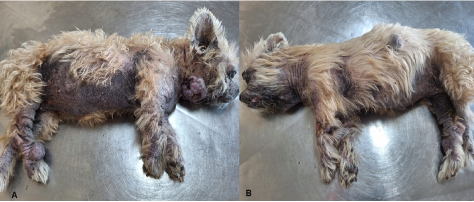
During clinical examination, the dog presented an alopecic area in abdomen, hyperpigmentation, hyperkeratosis, lichenification and three large nodules in the skin, located in lower right cervical region, distal end of right pelvic limb and left flank (Figure 1). In addition, the dog had bilateral ocular keratoconjunctivitis sicca.
A complete blood count, measurement of alanine aminotransferase (ALT), alkaline phosphatase (AP), glucose, triglycerides, cholesterol, type I urine, dexamethasone suppression test, abdominal ultrasound and doppler echocardiography were performed. A biopsy of the skin nodules was also performed for histopathological analysis.
In the blood count, a smaller number of erythrocytes (5.08 million/mm3; normal values=5.7-7.4 million/ mm3), and small decrease in hematocrit (37.0%; normal values=38.0-47.0%) was observed.
The dosages of ALT (252 U/L; normal values=9-92 U/L), AP (350; normal values=10-155 U/L), glucose (155 mg/dL; normal values=60-118 mg/ dL), cholesterol (367 mg/dL; normal values=135-270 mg/dL) and triglycerides (292.0; normal values=32.0 a 125.0 mg/dL) were elevated.
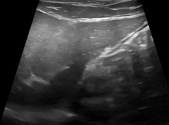
Type I urinalysis was performed by collecting urine through ultrasound-guided cystocentesis, and biochemical examination showed absence of bilirubin, glucose, ketone bodies and occult blood. In sedimentoscopy, erythrocytes, leukocytes and absence of bacteria were observed. In dexamethasone suppression test, the basal cortisol value was 4.45 (normal values= 1.0 a 4.6 μg/dL), and the cortisol value 8 hours after dexamethasone was 0.86 (normal value below 0.90 μg/dL). The abdominal ultrasound revealed a increase in the size of adrenal glands and liver (Figures 2 & 3). The doppler echocardiography and color flow mapping not shown any alteration.
The result of histopathological analysis of samples obtained from the three skin nodules was hemangiopericytoma (Figure 4).
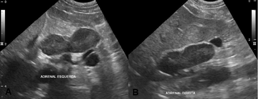
Figure 2: Abdominal ultrasound. A. Left adrenal gland with rounded contours and increased dimensions, measuring 2.8 x 1.0 x 0.98 cm (length x thickness of caudal pole x thickness of cranial pole) with reduced echogenicity and preserved echotexture. B. Right adrenal gland with rounded contours and increased dimensions, measuring 2.9 x 1.0 x 0.98 cm (length x thickness of caudal pole x thickness of cranial pole) with reduced echogenicity and preserved echotexture.
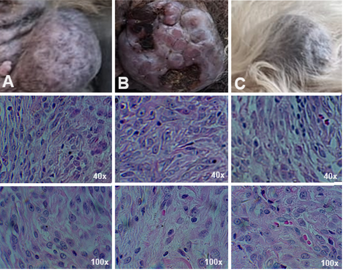
The histopathological analysis of the three nodules using a 40 and 100 objective demonstrated the presence of spindle cells in a sarcomatous pattern, with imprecise limits, nuclear pleomorphism and evident nucleoli (hematoxylin/eosin staining). A. Subcutaneous mass located in the distal end of right pelvic limb. B. Nodule located in lower right cervical region formed by multiple small nodules and foci of necrosis. C. Nodule located on left flank.
Discussion
The canine atopic dermatitis is a genetic disease in which animal is sensitive to allergens present in environment through inhalation, ingestion or contact, being considered a highly pruritic chronic inflammatory disease [1]. The main clinical sign in dogs with canine atopic dermatitis is pruritus, which leads to the appearance of skin lesions caused by trauma, recurrent opportunistic infections and a chronic inflammatory process with thickening of corneal layer (hyperkeratosis), hyperpigmentation, lichenification and alopecia [2]. Animals can develop recurrent and chronic otitis externa, conjunctivitis with secondary blepharitis and keratoconjunctivitis sicca [11, 12].
The most commonly used therapy for controlling pruritus involves the use of oral or cutaneous corticosteroids, such as prednisolone and methylprednisolone, that have rapid action and low mineralocorticoid effect. However, due to need for their use for long periods, these medications lead to development of adverse effects that affect liver, adrenal glands and gastrointestinal tract leading to decrease of quality and longevity life these animals [2]. In the dog of this case study, the problems resulting from prolonged use of corticosteroids were the development of iatrogenic hyperadrenocorticism, with the appearance of bilateral adrenal hyperplasia, anemia, hepatic steatosis and increased serum levels of cholesterol, triglycerides and alkaline phosphatase.
The oclacitinib maleate is a drug used to control pruritus in dogs with canine atopic dermatitis [13, 14]. Its mechanism of action involves synthesis blocking of cytokines with pruritic and pro-inflammatory activity, through the inhibition of enzymes from Janus Kinase (JAK) family, such as JAK1 and JAK2 [15].
The oclacitinib maleate has the ability to clinically reduce pruritus, but it does not have the ability to reverse its pathogenesis, requiring continuous use. The protocol indicated for the treatment of atopic dermatitis in dogs uses two cycles, the first through daily administrations with a
12-hour interval for 14 days and the second with a 24-hour interval. Its prolonged use is common in veterinary clinical practice, including the use of daily cycle every 12 hours [16]. In some animals, pruritus returns after changing between cycles by switching to once-daily administration or stopping it [17].
One of the major problems with its continued use is the increased risk of serious infections in animals with an ongoing infection or that have some hidden or present neoplasia. However, the literature does not provide direct evidence of the association between the use of oclacitinib maleate and the development of cancer in dogs [18]. The literature describes the use of oclacitinib maleate associated with depletion of TCD4 and TCD8 lymphocytes in dogs in vitro, of T and B lymphocytes in lymphoid tissues in a murine experimental model, and decreased in lymphoproliferative activity and cytokine synthesis (IFN-γ, IL-2 and IL-15), factors that contribute to reduce the ability of immune system to control the growth of tumor cells [19, 20, 21]. The drug’s leaflet warns about its action on the immune system and its promotion of tumor progression. In the dog in this case study, a diagnosis of hemangiopericytoma was made, a tumor originating from pericytes (cells that surround the capillaries) with slow growth [8, 9]. It was suggested by tutor that prolonged use of oclacitinib maleate was the emergence cause of hemangiopericytoma, but he was unable to inform whether the animal had any lesions prior to the use of this drug. Since the dog presented continuous use of glucocorticoids and hyperadrenocorticism, also capable of causing immunosuppression, it was not possible to establish a plausible association regarding the emergence of hemangiopericytoma.
Conclusion
The canine atopic dermatitis is a skin disease that affects dogs with a genetic predisposition mainly to aeroallergens. Due to the appearance of pruritus and self-trauma, treatment involves drugs that modulate the immune response, such as glucocorticoids. Since its long-term use can lead to the emergence of iatrogenic hyperadrocorticism, other drugs have been developed, such as oclacitinib maleate. Although this drug is capable of controlling the pruritic condition, it interferes with the immunosurveillance system, favoring the progression of already established tumors. Therefore, it is essential that the veterinarian is alert about its use in animals with canine atopic dermatitis.
References
-
Drechsler Y, Dong C, Clark DE, Kaur G (2024) Canine atopic dermatitis: prevalence, impact, and management strategies. Vet Med (Auckl) 15: 15-29.
-
Olivry T, DeBoer DJ, Favrot C, Jackson HÁ, Muelller RS, et al. (2015) Treatment of canine atopic dermatitis: 2015 update guidelines from the International Committee on Allergic Diseases of Animals (ICADA). Vet Res 11: 210.
-
De Caro Martins G, da Costa-Val AP, Coura FM, Diamantino GML, Nogueira MM, et al. (2022) Immunomodulatory effect of long-term oclacitinib maleate therapy in dogs with atopic dermatitis. Vet Dermatol 33(2): 142-e40.
-
Banovic F, Tarigo J, Gordon H, Barber JP, Gogal RM, et al. (2019) Immunomodulatory in vitro effects of oclacitinib on canine T-cell proliferation and cytokine production. Vet Dermatol 30(1): 17-e6.
-
de Mello Souza CH, Shiomitsu K, Hwang B (2022) Cytokine production and the effects of oclacitinib in three canine mast cell tumor lines. Vet Dermatol 33(2): 159-e46.
-
Simpson AC, Schissler JR, Rosychuk RAW, Moore AR (2017) The frequency of urinary tract infection and subclinical bacteriuria in dogs with allergic dermatitis treated with oclacitinib: a prospective study. Vet Dermatol 28(5): 485-e113.
-
Ashton LV, Weishaar KM, Seguin B, MacNeill AL (2023) Oclacitinib and myxoma virus therapy in dogs with high- grade soft tissue sarcoma. Biomedicines 11(9): 2346.
-
Stefanello D, Avallone G, Ferrari R, Roccabianca P, Boracchi P (2011) Canine cutaneous perivascular wall tumors at first presentation: clinical behavior and prognostic factors in 55 cases. Vet Intern Med 25(6): 1398-1405.
-
Avallone G, Stefanello D, Ferrari R, Roccabianca P (2020) The controversal histologic classification of canine subcutaneous whorlig tumors: The path to perivascular wall tumors. Vet Comp Oncol 18(1): 3-8.
-
Kravitz A, Davis G, Bastina RP, Fittipaldi K (2019) Outcome and prognostic indicators for hemangiopericytomas in dogs: 167 cases (2009-2016). J Am Hosp Assoc 55(4): 194-200.
-
Rigau D, Briantais P, Jasmin P, Bidaud A (2024) Efficacy and safety of a hydrocortisone aceponate-containing ear spray solutions in dogs with erythemato-ceruminous otitis externa: a randomised, multicentric, single- blinded, controlled trial. Vet Dermatol 35(2): 197-206.
-
Maier P, Lapp T, Reinhard T (2017) Ocular involvement in atopic dermatitis: clinical aspects and therapy. Ophtalmologe 114(6): 514-524.
-
Olivry T, DeBoer DJ, Favrot C, Jackson HA, Mueller RS, et al. (2010) Treatment of canine atopic dermatitis: 2010 clinical practice guidelines from the International Task Force on canine atopic dermatitis. Vet Dermatol 21(3): 233-248.
-
Marsella R, Doerr K, Gonzales A, Rosenkrantz W, Schissler J, et al. (2023) Oclacitinib 10 years later: lessons and directions for the future. J Am Vet Med Assoc 261(S1): S36-S47.
-
Gonzales AJ, Bowman JW, Fici GJ, Zhang M, Mann DW, et al. (2014) Oclacitinib (APOQUEL(®)) is a novel Janus Kinase inhibitor with activity against cytokines involved in allergy. J Vet Pharmacol Ther 37(4): 317-324.
-
Denti D, Caldin M, Ventura L, De Lucia M (2022) Prolonged twice-daily administration of oclacitinib for the control of canine atopic dermatitis: a retrospective study of 53 client-owned atopic dogs. Veterinary Dermatology 33(2): 149-e42.
-
Fukuyama T, Ganchingco JR, Baumer W (2017) Demonstration of rebound phenomenon following abrupt withdrawal of the JAK1 inhibition oclacitinib. Eur J Pharmacol 794: 20-26.
-
Lancellotti BA, Angus JC, Edginton HD, Rosenkrantz WS (2020) Age- and breed-matched retrospective cohort study of malignancies and benign skin masses in 660 dogs with allergic dermatitis treated long-term with versus without oclacitinib. J Am Vet Med Assoc 257(5): 507-516.
-
Jasiecka-Mikolaczyk A, Jaroszeski JJ, Maslanka T (2018) Oclacitinib depletes canine CD4+ and CD8+ T cells in vitro. Res Vet Sci 121: 124-129.
-
Jasiecka-Mikolajczy A, Maslanka T (2023) Depletion of T and B cells in lymphoid tissues of mice induced by oclacitinib, a Janus kinase inhibitor. J Vet Sci 26(3): 431- 440.
-
Banovic F, Tarigo J, Gordon H, Barber JP, Gogal RM (2019) Immunomodulatory in vitro effects of oclacitinib on canine T-cell proliferation and cytokine production. Vet Dermatol 30(1): 17-e6.
- California Red-Legged Frog and Non-Listed Amphibians Response to Non-Native Fish Removal
- Industrial Standardization of the Bio-OS: Algorithmic Codification of Resilience Engineering Guidelines and Version V8 Architecture
- Climate Variability and the Sustainability of Snail Farming in Nigeria: Past Trends, Present Challenges and Potential Outlook
- The Evaluation of the Surveillance System of Anthrax in Gilgit-Baltistan, Pakistan, 2018
- Natural Decline to Extinction of A New Zealand Rabbit Population
- Mitochondrial Bio-Logistics: Steering Co-Enzyme Q10 and Lycopene Synergies within the Science 4.0 Bio-OS Framework