Caudal Glands in Nematodes: Morphology, Evolutionary Shifts and Functional Implications
The presence of three caudal glands one dorsal and two subventral with distinct terminal outlets is an apomorphic feature supporting the monophyly of Nematoda. These glans primarily secrete adhesive substances for attachment but have evolved additional roles in locomotion and feeding in certain lineages. Comparative analysis reveals three or two separate gland outlets in 77 species across six ordinal taxa, while vestigial glands without external openings suggest possible internal functions. A single, common gland opening (spinneret), likely arising through independent tail-end invagination, has evolved approximately 32 times across nematode lineages. Additionally, a reversal in gland arrangement (one ventral and two subdorsal) occurred independently in about four instances. Variation in gland number, position (incaudal, suprarectal, and precaudal), and structure is documented, with shifts from three to four or six gland cells and reductions to two in some taxa. Approximately 80 independent losses of caudal glands across nematode phylogeny are detected, including 30 in Mononchida and around 40% in marine species. In clades with terrestrial and limnic representatives (e.g. Dorylaimida, Mononchida) it appears that the glands were lost in the terrestrial environment and aquatic habitats were invaded secondarily. Potential homologies between caudal glands and phasmids are discussed, along with implications for tail morphology to gland structure and function.
Introduction
Aquatic nematodes characteristically possess three large caudal gland cells that occupy the posterior body region and discharge secretions terminally. These glands play a critical ecological role by enabling temporary adhesion to solid substrates, thus facilitating foraging in flowing-water environments. In live specimens, the glands appear as a dark mass and are stainable with various dyes [1], however, they are often indistinct in permanent mounts. Despite their inclusion in the basic adenophorean body plan, caudal glands have been historically underreported and poorly illustrated in species descriptions. Hence, dedicated morphological studies focusing on the caudal gland complex and the associated spinneret apparatus remain scarce. Also, the existing data on their number, arrangement, and outlet morphology are fragmentary, imprecise, and frequently inconsistent across publications and authors’ interpretations. Nevertheless, these structures hold diagnostic value and potential phylogenetic significance, warranting renewed attention in nematode systematics.
Motivated by Steiner’s [2] research proposal “Of special necessity is a comparative study of the distribution, morphology, and function of the foot glands and phasmids, as well as their obliteration and disappearance in distinct worm groups” we undertake a comprehensive analysis of caudal gland morphology across Nematoda. This article synthesizes scattered observations into a cohesive phylogenetic framework, documenting structural diversity and evolutionary modifications of the caudal gland complex. We systematically reviewed hundreds of species descriptions and examined information derived from both light and electron microscopy. While efforts were made to ensure broad coverage, there is the possibility of overlooked literature and misinterpretations stemming from terminological inconsistencies, particularly regarding the term “spinneret,” which is used variably across studies.
In our analysis, we uncovered numerous unresolved questions that merit further investigation and presented testable hypotheses on the evolutionary pathways and transformation sequences of the caudal gland apparatus. The preliminary phylogenetic trees derived from a synthesis of studies by numerous authors [3, 4, 5, 6, 7, 8, 9, 10, 11, 12, 13, 14, 15, 16, 17], bearing several polytomies, serve as a foundation for future refinement. To resolve the polytomies, sequences of all taxa mentioned in the phylogenetic trees must be analysed together. This work re-establishes the caudal gland complex as a significant morphological and phylogenetic character system in nematode biology.
A Brief Outline of Nematode Bauplan Evolution
The ancestral lineage and stem species of Nematoda likely inhabited marine environments during the Precambrian period. It is widely accepted that the phylum Nematomorpha represents the sister group to Nematoda [18, 19, 20]. Nematomorpha, which are parasitic in arthropods (primarily Crustacea), have undergone significant morphological modifications to adapt to parasitism, including a free-living but non-feeding adult phase. Consequently, only a few traits can be interpreted as synapomorphies supporting the sister- group relationship between Nematomorpha and Nematoda.
Despite extensive structural reductions, some novel features evolved in their common ancestor (Figure 1), including a vermiform, cylindrical body, a flexible cuticle, undulatory locomotion by means of exclusively longitudinal muscles, hexaradial symmetry in anterior sensory structures, non-flagellate sperm transferred during copula, a cylindrical muscular sucking pharynx formed by myoepithelial cells and lined with a secreted cuticle, and a cuticle-lined cloaca. In the evolutionary trajectory of Nematoda, the cloaca was lost in females with the separation of the vulva and anus [21, 22, 23], and a tail developed dorsal to the terminal gut opening, serving as a container for adhesive glands possibly supporting a hemi-sessile lifestyle [21].
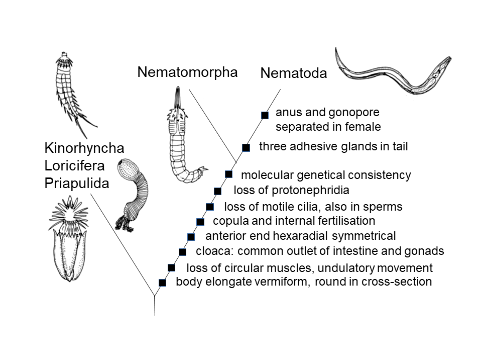
Several additional traits shared by Nematomorpha and Nematoda such as a terminal rather than subventral stoma, food intake by suction, a circum-pharyngeal central nervous system, a multilayered cuticle, and molting during ontogeny are broader apomorphies of Cycloneuralia and do not specifically support their sister-group relationship [19, 24]. Other Cycloneuralian taxa include Kinorhyncha, Loricifera, and Priapulida (Figure 1). The stem species of Cycloneuralia was likely small, inhabiting marine interstitial spaces of sediment, where its cuticle provided mechanical protection against shifting sand grains [24].
The cylindrical body plan and hexaradial anterior symmetry in the Nematomorpha-Nematoda lineage may represent adaptations to a benthic marine lifestyle [25]. The presence of adhesive glands, which anchor nematodes to substrates, further supports the idea that their ancestors lived in unstable sandy habitats. Since these caudal glands are unique to Nematoda within Cycloneuralia, they likely represent an evolutionary novelty [24]. There is no evidence that adhesive glands were present in the stem species of Nematoda-Nematomorpha and subsequently lost in Nematomorpha due to parasitic adaptation.
In this context, we reject the hypothesis that the three terminal lobes (without glands) in female Paragordius (Nematomorpha) are homologous to the tri-lobed caudal glands of nematodes, despite their similar dorsal and subventral orientation, which is considered plesiomorphic in nematodes. Additionally, because adhesive glands were absent in the Nematoda-Nematomorpha stem species, we also dismiss the idea that hexaradial anterior symmetry evolved in connection with a hemi-sessile lifestyle relying on secretions for substrate attachment [21]. Lastly, historical attempts to homologize nematode caudal glands with the foot glands of Rotifera or Gastrotricha are now considered obsolete [26, 27, 28, 29, 30]. These taxa belong to Lophotrochozoa, the sister group to Ecdysozoa (which includes Nematoda) [31], where adhesive glands primarily were absent.
Typically, three unicellular caudal glands are located in the anterior region of the nematode tail, arranged in tandem with slight overlap, likely due to spatial constraints. We can hypothesise that their arrangement follows a consistent sequence, allowing for the identification of homologous cell bodies (Figure 2). Following the nomenclature proposed by Coomans, et al. [32], we designate these as cg1 (anterior, usually dorsal), cg2 (middle), and cg3 (posterior).
![Figure 2: Caudal glands (cg) in tandem. A) Oncholaimus sp., precaudal gland cells left (l) or right (r) of intestine [115]; B) Synonema cosmopoliticum [44]; C) Araeolaimus filpjevi [231]; D) Thalassogenus tortuosus female, two sets of caudal glands [173]; arrows: extra gland cells.](/fulltextimages/13910/fig_2.png)
Structure and Functioning of Caudal Glands
Glandular Cells for Adhesion and Detachment
The structure of caudal glands has been studied in detail through light and electron microscopy in only a few species, including Chromadorina germanica (Chromadorida) [33], Gonionchus australis (Monhysterida) [34], Theristus caudasaliens (Monhysterida), and Perepsilonema sp. (Desmodorida) [35]. Each gland cell extends into a duct lined by a thin layer of cytoplasm [33] and is flanked by a circomyarian muscle cell. The ducts primarily open separately at the tail tip (Figure 3A-E; 5B), but in most taxa, they merge with those of other gland cells to form a common pore. The secretory cells are relatively large and exhibit features indicative of high metabolic activity. Each gland cell contains a prominent nucleus with multiple strands of chromatin and numerous dense secretory vesicles in the peripheral cytoplasm. Light microscopy of C. germanica revealed large, globular caudal gland cells with conspicuous nuclei. The presence of RNA was confirmed using acridine orange fluorescent staining. Ultrastructural analysis further supported the secretory function of these glands, showing an abundance of rough endoplasmic reticulum, mitochondria, and a well-developed Golgi complex. Additionally, microvilli containing ~20 nm particles were observed projecting into the gland cell lumen (or duct). These particles were proposed as precursors to secretory granules, which range from 0.5 to 2.0 µm in size; however, the chemical composition of the secretion remains unknown.
![Figure 3: Apical view of tail tip. The dorsal side to the top. A) _Dorylaimopsis variabilis_ with 3 openings, 1 dorsal [133]; B) _Leptonemella brevipharynx_ with 3 openings, 2 dorsal, and a small central pore [12]; C) _Pternepsilonema servaesae_ [133]; D) _Triepsilonema tripapillata_ [133]; E) _Epsilonema pustulatum_ [155]; F) _Zalonema ditlevseni_ with a single pore [232]; G-J) Two openings. G) _Laxus sakihariiae_, the dorsal opening larger [51]; H) _Eubostrichus hopperi_ [12]; J) _Bathyepsilonema lopheliae_ male with a medioventral pore and a curved slit-like dorsal opening [133].](/fulltextimages/13910/fig_3.png)
Figure 3: Apical view of tail tip. The dorsal side to the top. A) Dorylaimopsis variabilis with 3 openings, 1 dorsal [133]; B) Leptonemella brevipharynx with 3 openings, 2 dorsal, and a small central pore [12]; C) Pternepsilonema servaesae [133]; D) Triepsilonema tripapillata [133]; E) Epsilonema pustulatum [155]; F) Zalonema ditlevseni with a single pore [232]; G-J) Two openings. G) Laxus sakihariiae, the dorsal opening larger [51]; H) Eubostrichus hopperi [12]; J) Bathyepsilonema lopheliae male with a medioventral pore and a curved slit-like dorsal opening [133].
To enable temporary adhesion, nematode caudal gland cells rapidly release an adhesive secretion under nervous control. This secretion hardens immediately in water, allowing the nematode to adhere to various substrates. The attachment is strong yet reversible, enabling frequent detachment and reattachment of the tail tip within short time intervals. In an experiment with the monhysterid Sphaerolaimus gracilis, Turpeenniemi, et al. [36] demonstrated that the nematode could anchor itself to a sediment particle by secreting a cement-like substance from the caudal glands, forming a thread more than one body length long. The adhesion is so firm that detachment by mechanical means such as using a needle or pipette suction is mostly unsuccessful Schiemer, et al. [37, 38] suggesting a chemical component is required for releasing. The dual function of attachment and release is accomplished by a two-part glandular system, recorded in three species: Perepsilonema conifer, Sphaerolaimus gracilis, and Theristus caudasaliens [35, 36]. This system consists of two or three voluminous adhesive gland cells that secrete a rapidly hardening adhesive, along with approximately two smaller releasing gland cells thought to produce a “de- adhesive” substance that facilitates detachment by chemical means. Similar duo-gland adhesive systems are known from various interstitial meiofaunal taxa [35, 39]. We propose that this cooperative adhesive-releasing system, consisting of 3+2 glandular cells, is homologous across different nematode taxa and was likely present in the last common ancestor of Desmodorida (Perepsilonema) and Monhysterida (Sphaerolaimus, Theristus) (Figure 4, species X). It will be shown that this ancestral species had three separate glandular outlets.
![Figure 4: Phylogenetic diagram of Nematoda compiled from data in [5,9,10,16,17,81]. Paraphyletic taxa after Ahmed, et al. [17] in parentheses. The greyish circles mark points not resolved into dichotomies so far. Following our hypothesis that the stem species had three caudal glands with separate outlets, the lineages are marked where the three outlets must have joined to a single one and where caudal glands were completely reduced.](/fulltextimages/13910/fig_4.png)
Figure 4: Phylogenetic diagram of Nematoda compiled from data in [5, 9, 10, 16, 17, 81]. Paraphyletic taxa after Ahmed, et al. [17] in parentheses. The greyish circles mark points not resolved into dichotomies so far. Following our hypothesis that the stem species had three caudal glands with separate outlets, the lineages are marked where the three outlets must have joined to a single one and where caudal glands were completely reduced.
Hypotheses on the Existence of Releasing Cells and their Origin
Releasing gland cells may have been frequently overlooked in light microscopy studies, particularly when only three caudal gland cells were expected, or they may not have been resolvable in many cases. However, descriptions of several species suggest the presence of additional gland cells. For instance, two extra cells were illustrated in Belgopeltula belgica (Araeolaimida) [40], Monhystera (= Diplohystera) inflata [41], Linhomoeus parmacramphis (Monhysterida) [42], Plectus acuminatus (Plectida) [43], and Synonema cosmopoliticum (Desmodorida) [44] (Figure 2B).
Four to five glandular cells were documented in Caprionchulus diversipapillatus (Triplonchida) [45], while five gland cells were reported in Phanodermopsis nana (Enoplida) [46]. Chitwood described five uninucleate glands arranged in tandem anterior to the anus in Eurystomina americana (Enoplida) [47], likely comprising three caudal glands and two additional supplementary glands. Similarly, Aphanolaimus pseudoattentus (Plectida) exhibited two extra glandular cells extending dorsally from the intestine to nearly one tail length anterior to the cloaca, which may represent releasing gland cells [48].
Occasionally an additional cell was observed in Chromadorina germanica [33], positioned posterior to the main caudal gland cell but lacking a separate duct. Various authors have depicted additional gland-like cells in species across Araeolaimida, Chromadorida, Desmodorida, Desmoscolecida, Enoplida, Monhysterida, Mononchida, Plectida, and Triplonchida, often shading or greying them in illustrations to indicate structural or content differences (Table 1). These cells are typically found anteriorly adjacent or dorsal to the caudal gland cells, though some cases show a posterior or even ventral position (Figure 2C). Despite the lack of commentary by many authors, these observations warrant careful consideration. A targeted reexamination, guided by the hypothesis of additional releasing gland cells, is necessary. However, alternative functions for these adjacent cells should also be explored. In Plectus species, for example, these cells were interpreted as (pseudo)coelomocytes by Andrássy [49, 50], a view also proposed for Hemiplectus [6].
| species name | group | author |
|---|---|---|
| Brevitobrilus graciloides | Tobrilidae, Triplonchida | Abebe, et al. [234] |
| Epitobrilus setosus | Tobrilidae, Triplonchida | Abebe, et al. [234] |
| Triodontolaimus acutus | Triodontolaimidae, Triplonchida | Lorenzen [235] |
| Bathylaimus arthropappus | Tripyloididae, Enoplida | Wieser, et al. [111] |
| Tripyloides imitans | Tripyloididae, Enoplida | Wieser [134] |
| Synonchus dubius | Leptosomatidae, Enoplida | Wieser [42] |
| Phanoderma segmentum | Phanodermatidae, Enoplida | Murphy [236] |
| Iotonchus singaporensis | Iotonchidae, Mononchida | Ahmad, et al. [237] |
| Miconchus sp. | Iotonchidae, Mononchida | Ahmad, et al. [238] |
| Mylonchulus brachyuris | Mononchidae, Mononchida | Zell [239] |
| Chromadora macrolaima | Chromadoridae, Chromadorida | Steiner [116] |
| Chromadora micropapillata | Chromadoridae, Chromadorida | Wieser [240] |
| Chromadora quadrilineoides | Chromadoridae, Chromadorida | Chitwood [47] |
| Chromadorella filiformis (= C. filiformoides) | Chromadoridae, Chromadorida | Chitwood [47] |
| Chromadorina germanica (= C. minor), C. laeta | Chromadoridae, Chromadorida | Wieser [240] |
| Chromadorita mucrodonta | Chromadoridae, Chromadorida | Steiner [116] |
| Dichromadora amphidiscoides | Chromadoridae, Chromadorida | Kito [56] |
| Sabatiera abyssalis | Comesomatidae, Chromadorida | Timm [147] |
| Sabatieria longispinosa, S. pulchra | Comesomatidae, Chromadorida | Tchesunov [241] |
| Paracyatholamus lewisi | Cyatholaimidae, Chromadorida | Coomans, et al. [242] |
| Cobbionema brevispicula | Selachinematidae, Chromadorida | Ahmed, et al. [243] |
| Microlaimus porosus | Microlaimidae, Microlaimida | Miljutin, et al. [244] |
| Synonema braziliense | Aponchiidae, Desmodorida | Wieser [62] |
| Spirinia parasitifera | Desmodoridae, Desmodorida | Vincx, et al. [245] |
| Protricoma squamosa | Desmoscolecidae, Desmoscolecida | Decraemer [246] |
| Tricoma similis | Desmoscolecidae, Desmoscolecida | Decraemer [247] |
| Linhomoeus buculentus | Linhomoeidae, Monhysterida | Wieser [134] |
| Linhomoeus elongatus | Linhomoeidae, Monhysterida | Steiner [116] |
| Linhomoeus hirsutus | Linhomoeidae, Monhysterida | Wieser [62] |
| Linhomoeus ponticus | Linhomoeidae, Monhysterida | Schneider [249] |
| Monhystera rolandi | Monhysteridae, Monhysterida | Khan, et al. [250] |
| Araeolaimus filipjevi | Diplopeltidae, Araeolaimida | De Coninck et al. [231] |
| Diplopeltula sundensis | Diplopeltidae, Araeolaimida | Jensen [251] |
| Intasia monohystera | Diplopeltidae, Araeolaimida | Tchesunov, et al. [252] |
| Aphanolaimus aquaticus | Aphanolaimidae, Plectida | Meyl [253] |
| Dagda bipapillata | Camacolaimidae, Plectida | Holovachov, et al. [221] |
| Stephanolaimus elegans | Camacolaimidae, Plectida | Holovachov, et al. [221] |
| Anaplectus granulosus | Plectidae, Plectida | Holovachov, et al. [254] |
| Arctiplectus alaskanus | Plectidae, Plectida | Andrássy [50] |
| Ceratoplectus armatus | Plectidae, Plectida | Andrássy [43] |
| Chiloplectus andrassyi | Plectidae, Plectida | Andrássy [43] |
| Chiloplectus loricatus | Plectidae, Plectida | Andrássy [43] |
| Hemiplectus muscorum | Plectidae, Plectida | Holovachov, et al. [6] |
| Perioplectus secundus | Plectidae, Plectida | Andrássy [50] |
| Plectus antarcticus, P. belgicae, P. frigophilus, P. insolens, P. meridianus, P. tolerans | Plectidae, Plectida | Andrássy [49] |
| Plectus auracanorum | Plectidae, Plectida | Andrássy [256] |
| Plectus australis, P. intermedius, P. pusteri, P. zelli | Plectidae, Plectida | Zell [255] |
| Plectus cirratus, P. palustris, P. silvaticus, P. tenuis | Plectidae, Plectida | Andrássy [43] |
| P. communis, P. elegans, P. montanus, P. rotundilabiatus, P. similis, P. thornei | Plectidae, Plectida | Zell [255] |
| Plectus infundibulifer, P. parietinus | Plectidae, Plectida | Andrássy [43] |
| Plectus murrayi | Plectidae, Plectida | Zell [255] |
| Plectus parietinus | Plectidae, Plectida | Steiner [257] |
| Plectus sambesii | Plectidae, Plectida | Zell [258] |
| Plectus spicacaudatus | Plectidae, Plectida | Andrássy [50] |
| Tylocephalus cornutus | Plectidae, Plectida | Zell [259] |
Table 1: Existence of additional releasing cell(s) or pseudocoelomocyte(s).
If only three caudal gland cells exist without additional releasing cells, this does not necessarily imply that nematode detachment is mechanically driven by muscle action. Besides the previously discussed system of three adhesive and two releasing cells, an alternative duo-gland system could exist, wherein all three gland cells perform either adhesion or release functions. This possibility is suggested by the presence of two distinct glandular outlets in some species, such as Laxus sakihariiae (Desmodoridae), where one of the pores is dorsally positioned and slightly larger (Figure
3G) [51]. A stronger indication of functional differentiation among the three gland cells comes from observations of secretory vesicle differences. In Gonionchus australis, for example, one of the three gland cells contained secretory vesicles distinct from those in the other two [34], possibly representing a releasing function.
Similarly, in Paramphimonhystrella species (Xyalidae), the anterior-most caudal gland cell (cg1) is particularly voluminous [52], with structural and staining differences also noted in Eumonhystera filiformis [53], Gammarinema gammari and G. ligiae [54, 55], Halomonhystera (= Monhystera) cameroni [2], Monhystera refringens [56], M. disjuncta and Odontobius ceti [55], and possibly Chromadorina germanica [33] and Daptonema conicum [57].
Notably, in Cryonema crassum and C. tenue, it is the middle gland cell (cg2) that differs in secretion content [55]. Except for Chromadorina, all these species belong to the closely related families Monhysteridae or Xyalidae.
Despite its location in the thickest part of the tail, cg1 is often the smallest of the three gland cells, as observed in numerous monhysterids, including Daptonema hirsutum [57], Geomonhystera glaciei [58], Halomonhystera cameroni [2], H. parasitica [59], Monhystera floreanae and M. somereni [60], M. tanae [61], Paramonohystera proteus [62], Theristus caudasaliens [35], T. polychaetophilus [63], T. pratti [64], T. denticulatus and T. roscoffiensis [57].
This size disparity suggests a functional difference between cg1 and the other two cells. This pattern provides insight into species that possess only two caudal gland cells (Table 4), where cg1 appears to be entirely reduced. In such cases, cg1 may not have been responsible for releasing substances, and the presence of additional, undiscovered releasing cells similar to those found in Sphaerolaimus gracilis remains a possibility. This species possesses only two caudal glands in contrast to other species of Sphaerolaimus [65], raising the possibility that both caudal glands perform distinct functions, with one producing the releasing substance. How attachment and release occur in species described with only a single functional caudal gland cell, remains an open question. This has been reported in several Chromadorida species, such as Chromadorita tenuis, C. tentabunda, and Graphonema amokuroides [38], as well as in Cryptonchidae (Mononchida), including Cryptonchus nudus [66] and C. tristis [67]. Reports of species with only one gland, including members of Desmodorida (Draconema, Epsilonema, Notochaetosoma, Prochaetosoma), Desmoscolecida (Desmoscolex), and Triplonchida (Neotobrilus), require re-examination.
From these observations, we infer the existence of two distinct adhesive systems in nematodes. However, a key evolutionary question remains: which system is ancestral, and how and in which lineage did the transition between systems occur? Due to gaps in available data, any evolutionary hypothesis remains speculative, but these uncertainties should inspire further investigation. Since no additional releasing cells have been reported in species of Enoplida or Chromadorida, we propose that the ancestral nematode had three caudal glands, likely differentiated into two adhesive cells and one releasing cell. Each gland cell probably had its own duct and separate outlet at the tail tip. Arguments supporting the ancestral presence of three separate outlets will be discussed later. We hypothesise that the dorsal opening was associated with cg1, functioning as the releasing gland, while the two adhesive glands opened subventrally. The emergence of two additional releasing cells would thus represent a novel trait that may have originated in the stem ancestor of Desmodorida and Monhysterida (Figure 4, species X) or evolved independently in both clades.
The positioning of the two additional releasing glands suggests they may have originated through duplication of the ancestral anterior releasing cell. These new glands could have opened into a central pore, as seen in Leptonemella brevipharynx (Desmodorida) (Figure 3B) [12] and possibly Desmoscolex (Desmolorenzenia) gourbaultae (Desmoscolecida) [68]. Alternatively, they may have joined the dorsal outlet, contrasting with lineages where two subventral outlets presumably fused. This fusion could explain the glandular arrangement observed in Laxus sakihariiae (Figure 3G). The emergence of two additional releasing cells may have altered the function of the primary releasing cell, either shifting it toward adhesive secretion or leading to its reduction. While this hypothesis appears promising, new ultrastructural investigations are needed to critically assess the many assumptions made in this context.
Biological Role of Caudal Glands
Caudal glands play a crucial role in the survival and feeding behavior of aquatic nematodes, enabling them to anchor their tail tips temporarily to the substrate. This adhesion helps them resist water currents and turbulence, allowing specialized species to inhabit dynamic environments such as shifting sands on shores, riverbeds, and even torrents without being swept away. In more stable substrates like muddy sand, some nematodes adopt a hemi-sessile lifestyle. For instance, the long-tailed enoplid Trefusia spp. remains anchored by its tail tip while making exploratory movements in various directions. When disturbed, it can rapidly retreat, coiling its flagelliform tail or even its entire body into compact spirals [37]. Similarly, Chromadorita tentabunda maintains near-permanent attachment to the substrate via its tail while conducting intensive searching movements with its anterior end [38].
In terrestrial environments, adhesive glands may seem redundant. However, they persist in certain soil-dwelling adenophorean lineages, despite no longer being essential for locomotion [69]. The function of caudal glands in terrestrial nematodes such as those in Chromadorida, Monhysterida, Mononchida, Plectida, and Triplonchida remains uncertain. One untested hypothesis suggests that these glands may help prevent nematodes from being washed into deeper soil layers during heavy rainfall. Additionally, species with vestigial caudal glands lacking external outlets may still utilize them for unknown internal physiological functions.
In marine environments, caudal glands are integral to the distinctive movements of Draconematidae and Epsilonematidae. These nematodes exhibit a looper-like crawling behavior on marine plants, alternating between attachment and release of their anterior and posterior ends via caudal gland secretions [70]. A peculiar hopping locomotion is observed in Theristus species (Xyalidae) and Innocuonema tentabunda (= Chromadorita tentabunda) (Chromadoridae), which use their leftward-bent tails to adhere to sand grains. By straightening and curling the posterior part of their bodies, they propel themselves forward in a jumping motion, sometimes alternating attachment between the tail and the anterior end [71]. Interestingly, Innocuonema tentabunda, despite having only a single caudal gland, performs this movement in the same manner as related species with three glands [38]. Similarly, in Daptonema setosum (Xyalidae), caudal glands assist in locomotion [72].
Observations dating back to Cobb describe Chromadorina germanica (= C. minor, Chromadoridae) using an unusual mode of movement [73]. This species anchors itself by suctioning its mouth onto glass slides, then loops its body by bringing its tail close to its head and securing a hold via the caudal spinneret. It then advances by releasing its head, executing a movement similar to the inching motion of geometrid moth larvae.
These diverse adaptations underscore the multifunctionality of caudal glands in nematodes, aiding both adhesion and locomotion in aquatic and terrestrial environments. However, the precise function of these glands in certain soil-dwelling species remains an open question, meriting further investigation.
Caudal glands serve additional functions in certain marine nematode lineages beyond adhesion and locomotion. In some species, these glands secrete mucus to construct microtubes, providing a protective habitat within soft sediments [74]. The epizoic monhysterid Gammarinema utilizes its caudal gland secretions to assemble clumps of detritus and feces, creating a substrate on its crustacean host for egg deposition [75].
An intriguing foraging-related function of caudal glands was proposed in the “mucus-trap hypothesis” [76]. The authors suggested that slimy secretions from caudal glands along with those from the pharyngeal, ventral (renette), and epidermal glands can trap detritus particles, microorganisms, and macromolecules, which are subsequently ingested by the nematode. A remarkable example in this context is that Schneider (p. 55) documented an enoplid producing a several-millimeter-long thread of secretion from its “silk glands” on a glass slide [77].
Another significant role of caudal gland secretions may be in “enzyme sharing” between nematodes and microbes, facilitating the degradation of organic detritus. These secretions appear to agglutinate detritus into mucus- laden threads [78], which may later be decomposed by microbial activity. In Desmodora schulzi, Riemann [79] frequently observed sediment particles aggregating on the ventral side of the female’s body between the vulva and anus forming a ‘micro-garden’ of coccoid, filamentous, and septate microorganisms, likely promoted by caudal gland secretions.
Some members of Anoplostomatidae_,_ Oncholaimidae, and Comesomatidae have been reported to foster microbial growth around tail-secreted threads, potentially contributing to cellulose degradation. Their role in detritus processing is further highlighted by their apparent inability or reluctance to graze on the microorganisms responsible for cellulose breakdown [80]. These diverse functions underscore the multifaceted role of caudal glands in nematodes, extending beyond adhesion and locomotion to include habitat construction, foraging, and microbial interactions that may influence nutrient cycling in sedimentary environments.
The Stem Species of Nematodes Had Three Caudal Gland Cells with Separate Openings
The Discovery of Separate Gland Outlets
It can be assumed that three unicellular caudal glands were already present in the stem species of Nematoda [22]. These glands may have evolved as specialised derivatives of distal epidermal gland cells, arranged terminally in a triplet as a functional unit. As reasoned below, this set of large glands likely consisted of one dorsal cell and two subventral cells.
Their pyriform or ellipsoidal bodies are confined to the upper part of the tail, each containing a large nucleus with a prominent nucleolus. Their elongated ducts open terminally, reinforcing a triradiate organization at the posterior end of the nematode’s body similar to the anterior end, where bilateral symmetry is also overridden by triradiate structures. Contrary to the general view (eg. [22]), we agree with Lorenzen [82], that each gland cell originally had its own separate outlet rather than a common pore (Figure 5B).
![Figure 5: A) Three caudal glands with one terminal opening and spinneret; B) three separate outlets (exemplified by Diplopeltis incisus); C) three-pointed tail in a Secernentea lacking caudal glands (exemplified by Molinostrongylus). Sketches by Osche [107], newly arranged.](/fulltextimages/13910/fig_5.png)
The first documented case of separate glandular outlets was reported in Pseudodilaimus pilosus [83], which was described as having a truncated female tail where one gland opens dorsally and two open ventrally or subventrally (Figure 10D). Though the description of the species is quite well, it has never been found again, was classified as incertae sedis within the “Adenophorea”, and its existence of separate gland outlets was never cited. It happened similarly to Schulz [84], who observed a thin glandular duct opening laterally near the tip of the swollen tail in Sphaerolaimus makrolasius and S. sabulosus (Monhysterida), instead of a terminal opening, and hypothesised that there were three such ducts corresponding to three gland cells. This hypothesis was later supported by findings in S. gracilis and S. occidentalis, where three distinct glandular outlets were confirmed [85].
The hypothesis stating that separate caudal gland openings one dorsal and two subventral represent a plesiomorphic condition in Nematoda, was first proposed by Osche [86]. He observed remnants of a “triply-pointed” tail in a high percentage of females in a population of Pellioditis (= Rhabditis) papillosa, which he interpreted as an atavistic trait. Additionally, he noted similar tail structures in various nematode lineages, particularly among Secernentean parasites, where the tail exhibited three distinct cone-like protuberances arranged in the same way (Figure 5C). Osche suggested that this triply-pointed tail persisted as a cryptic feature of nematode body architecture for a prolonged evolutionary period, resurfacing repeatedly, particularly within Secernentea [87].
Osche further pointed out the occurrence of “archaic” three-pointed tails in females of the Strongylacanthinae (Trichostrongylidae) [88], implying that this trait may have been present in the stem species of Secernentea but was subsequently lost multiple times at the phenotypic level. However, we reject the idea of phenotypic continuity due to the improbability of such frequent reductions across nearly all lineages of Strongylacanthinae_._ Instead, we infer the persistence of the underlying genetic information for a three-pronged tail structure, followed by parallel phenotypic reversals driven by the same gene complexes. Although gland vestiges were never observed within the three tail cusps, their positional correspondence with the glands of “Adenophorea” is striking. In this context, Osche [86] referenced an earlier observation by Gerlach [89], who reported that two species of Diplopeltidae possessed three caudal glands with separate openings one dorsal and two subventral (Figure 5B).
Separate Outlets is a Plesiomorphic Character
Subsequent studies, influenced by Osche’s hypothesis, carefully examined gland termination and confirmed separate glandular outlets in approximately 77 species (4 questionable) across 39 genera (3 questionable) spanning Microlaimida, Desmodorida, Ceramonematoidea, Desmoscolecida, Monhysterida, and Araeolaimida, with possible occurrences in Enoplida (Table 2).
This trait is considered homologous across these taxa. In a few lineages where the dorsal gland was completely reduced (Daptonema trabeculosum, Echinotheristus teutonicus, Paramonohystera concinna, Pseudechinotheristus nudus, and likely Eubostrichus hopperi, Laxus spp., and Rhynchonema spp.), the two remaining gland outlets shifted laterally and slightly anterior to the tail tip [82]. This modification appears to have occurred independently multiple times.
| species name | group | author(s) |
|---|---|---|
| Pseudodilaimus pilosus (?) | Enoplida incertae sedis | Kreis [83] |
| Ixonema deleyi | Microlaimidae, Microlaimida | Muthumbi, et al. [153] |
| Ixonema sordidum | Microlaimidae, Microlaimida | Lorenzen [98] |
| Ixonema sp. | Microlaimidae, Microlaimida | Raes, et al. [133] |
| Desmodora bilacinia (possibly 2, dorsal/ ventral) | Desmodoridae, Desmodorida | Leduc, et al. [129] |
| Akanthepsilonema sinecornibus | Epsilonematidae, Desmodorida | Raes, et al. [133] |
| Bathyepsilonema lopheliae male | Epsilonematidae, Desmodorida | Raes, et al. [133] |
| Epsilonema multispiralum | Epsilonematidae, Desmodorida | Raes, et al. [132] |
| Epsilonema pustulatum | Epsilonematidae, Desmodorida | Karssen, et al. [155] |
| Triepsilonema tripapillata | Epsilonematidae, Desmodorida | Decraemer [100] |
| Eubostrichus hopperi (2, dorsal/ventral) | Stilbonematinae, Desmodorida | Armenteros, et al. [12] |
| Eubostrichus topiarius | Stilbonematinae, Desmodorida | Berger, et al. [260] |
| Laxus parvum (2, dorsal/ventral) | Stilbonematinae, Desmodorida | Armenteros, et al. [12] |
| Laxus sakihariiae (2, dorsal/ventral) | Stilbonematinae, Desmodorida | Leduc, et al. [51] |
| Leptonemella brevipharynx | Stilbonematinae, Desmodorida | Armenteros, et al. [12] |
| Leptonemella vestari | Stilbonematinae, Desmodorida | Hoschitz, et al. [261] |
| Robbea hypermnestra | Stilbonematinae, Desmodorida | Ott, et al. [152] |
| Diplopeltoides asetosus, D. bulbosus, D. grandis, D. linkei, D. longicaudatus, D. nudus, D. pumilus, D. suecicus | Ceramonematoidea | Holovachov, et al. [262] |
| Diplopeltoides axayacatli, D. santaclarae, D. paramastigia | Ceramonematoidea | Holovachov, et al. [263] |
| Diplopeltoides mastigia | Ceramonematoidea | Tchesunov [264] |
| Desmoscolex gourbaultae (not quite sure) | Desmoscolecidae, Desmoscolecida | Decraemer [68] |
| Tricoma tripapillata | Desmoscolecidae, Desmoscolecida | Soetaert [265] |
| Desmolaimus zeelandicus (?) | Linhomoeidae, Monhysterida | Meldal [81] |
| Metasphaerolaimus crassicauda, M. hamatus | Sphaerolaimidae, Monhysterida | Jensen [266] |
| Sphaerolaimus gracilis (2) | Sphaerolaimidae, Monhysterida | Turpeenniemi, et al. [36] |
| Sphaerolaimus occidentalis (2) | Sphaerolaimidae, Monhysterida | Turpeenniemi [85] |
| “several species” | Sphaerolaimidae, Monhysterida | Lorenzen [82] |
| Amphimonhystera anechma | Xyalidae, Monhysterida | Lorenzen [57] |
| Amphimonhystrella bullacauda | Xyalidae, Monhysterida | Tchesunov, et al. [267] |
| Cobbia sp. (presumably) | Xyalidae, Monhysterida | Lorenzen [57] |
| Daptonema conicum, D. fistulatum, D. hirsutum, D. trabeculosum (2) | Xyalidae, Monhysterida | Lorenzen [57] |
| Daptonema normandicum, D. oxycerca | Xyalidae, Monhysterida | Blome [65] |
| Echinotheristus cimbricus (2), E. teutonicus (2) | Xyalidae, Monhysterida | von Thun, et al. [101] |
| Elzalia spp. | Xyalidae, Monhysterida | Hope, et al. [268] |
| Gonionchus inaequalis, G. sensibilis | Xyalidae, Monhysterida | Lorenzen [57] |
| Metadesmolaimus gelana | Xyalidae, Monhysterida | Lorenzen [57] |
| Metadesmolaimus varians | Xyalidae, Monhysterida | Lorenzen [93] |
| Paramonohystera (P.) concinna (2), P. (P.) levicula | Xyalidae, Monhysterida | Lorenzen [57] |
| Pseudechinotheristus nudus (2) | Xyalidae, Monhysterida | Blome [269] |
| Rhynchonema lyngei (2), R. separatum (2) | Xyalidae, Monhysterida | Lorenzen [102] |
| Steineria pilosa | Xyalidae, Monhysterida | Lorenzen [270] |
| Stylotheristus mutilus | Xyalidae, Monhysterida | Lorenzen [57] |
| Theristus (T.) balticus, T. (T.) bastiani, T. (T.) denticulatus, T. (T.) roscoffiensis | Xyalidae, Monhysterida | Lorenzen [57] |
| Theristus biarcospiculoides | Xyalidae, Monhysterida | Blome [65] |
| Theristus caudasaliens (2) | Xyalidae, Monhysterida | Adams, et al. [35] |
| Valvaelaimus maior | Xyalidae, Monhysterida | Lorenzen [57] |
| Xyala riemanni | Xyalidae, Monhysterida | Lorenzen [57] |
| Dorylaimopsis variabilis | Comesomatidae, Araeolaimida | Muthumbi, et al. [154] |
| Diplopeltula breviceps | Diplopeltidae, Araeolaimida | Gerlach [89] |
| Mudwigglus nellyae, M. patumuka | Diplopeltidae, Araeolaimida | Leduc [303] |
| Neodiplopeltula barentsi (= Diplopeltula cuspidiboja) | Diplopeltidae, Araeolaimida | Leduc, et al. [148] |
| Neodiplopeltula bathmanni, N. onusta | Diplopeltidae, Araeolaimida | Holovachov, et al. [271] |
| Neodiplopeltula (Diplopeltis) incisa | Diplopeltidae, Araeolaimida | Gerlach [89] |
| Pararaeolaimus nudus | Diplopeltidae, Araeolaimida | Lorenzen, et al. [99] |
| Aegialoalaimus elegans | incertae sedis, Araeolaimida | Tchesunov [264] |
| Aegialoalaimus setosus | incertae sedis, Araeolaimida | Holovachov pers. com. |
Table 2: Three (or two) separate outlets of caudal glands.
The most parsimonious interpretation of these findings is that the presence of three separate gland openings represents a plesiomorphic trait within nematodes. Thus, the last common ancestor of Microlaimida, Desmodorida, and Secernentea likely possessed distinct caudal gland outlets, one dorsal and two subventral (Figure 4, species X). This same ancestor is reconstructed to have had a dual gland system consisting of three adhesive and two release gland cells, though it remains uncertain how each gland type corresponded to the three openings. As explored in the next section, the transition to a single common opening likely occurred multiple times through gradual tail-tip transformations and remodelling of the original three outlets.
What type of glandular openings might have been present in the outgroup of the clade containing stem species X? So far, no separate glandular outlets have been observed in the early-diverging lineages of the nematode tree. The only documented case was Pseudodilaimus pilosus [83], mentioned earlier (Figure 10D), which might be an artifact, but no similar structures have been reported since.1 Nevertheless, from a functional perspective, it can If it turns out to be right, as suggested, that the Muspiceida belongs to the Trichinellida clade [96], they provide an example of separate openings of the two caudal glands in one of the early nematode lineages ("Adenophorea"). However, presumably, this taxon belongs to Secernentea and more precise ly to the Spiroascarida [97], while the paired glands represent phasmids, which will be discussed in the chapter XIV on phasmids.
be argued that even the stem species of Nematoda likely possessed separate gland outlets. This reasoning stems from the improbability of a functional transition from a common opening potentially involving a complex spinneret apparatus (see below) and possibly a shared ampulla to three entirely separate outlets. Such a drastic shift in glandular architecture seems less parsimonious than the alternative scenario: the stepwise evolution of a common opening via the progressive invagination of the tail end, gradually consolidating the three separate outlets. Given the multiple independent occurrences of a common opening across nematode lineages (as explored in the section after the next), this evolutionary trajectory appears far more plausible.
Historical Remnants of Former Three Outlets
Caution is advised when interpreting caudal gland data reported in taxonomic descriptions, certain details have been elucidated. Cobb [90] reported that in various nematodes including Synodontium fecundum (Araeolaimida), Mesonchium poriferum (Chromadorida), Chromaspirina (= Bolbolaimus) pellucida and Synonema braziliense (Desmodorida), Tripylium carcinicola (Monhysterida), and Acontiolaimus (= Digitonchus) uniformis (Plectida) the caudal glands discharge through separate ducts. In close relatives of these species, three glandular outlets are frequently observed. Notably, in Synodontium, “the ducts of the caudal glands are separate, practically to the spinneret pore” (p. 280), indicating that while three ducts remain distinct for most of their length, they ultimately converge at a single common spinneret rather than terminating in three separate pores at the tail tip (Figure 10F). Similarly, in Oxyonchus spp. (Enoplida), “the caudal glands open by three ducts through a spinneret” [91]. Deontolaimus papillatus (Plectida) (Figure 10C) also possesses a common spinneret [92], contradicting Lorenzen’s [93] assumption based on Meyl’s [94] depiction of seemingly three terminal tubes that it had separate outlets.
However, it is of much interest that in some cases, the three gland ducts expand into three distinct ampullae before ultimately merging into a single terminal outlet. This structural organisation may be a remnant of an ancestral condition in which three separate openings were present in the nematode body plan. Such configurations have been observed in Cynura uniformis (Araeolaimida), Gammanema ferox (Chromadorida), Synonema braziliense (Desmodorida), Chromaspirina (= Bolbolaimus) pellucida (Desmodorida), and Trissonchulus oceanus (Enoplida) (all reported in [90]). Additionally, similar glandular arrangements have been noted in monhysterids such as Gammarinema gammari [55], Halomonhystera cameroni [2], Monhystrium inquilinus [55], M. wilsoni [90], and Theristus meyli [95].
A particularly intriguing case is Phanoderma (= Cophonchus) ocellatum (Enoplida), where it appears that one of the glands “the one on the ventral side” leads to a separate ampulla, while the other two ducts run parallel and seem to join within a single, much larger ampulla [90]. Given the structural diversity of glandular arrangements across taxa, a more detailed investigation would likely reveal additional examples of this intermediate condition. However, most Adenophorean taxa appear to have a single common ampulla, reinforcing the idea that the fusion of glandular ducts into a single opening occurred repeatedly and independently in nematode evolution.
A Cryptic Base for the Three-Pointed Tail Tip
In Ixonema sordidum and Pararaeolaimus nudus, the tail end features three protuberances, each serving as a separate outlet for the three caudal glands [98, 99]. Similarly, in Triepsilonema tripapillata, the three glandular outlets are positioned on prominent caudal papillae (Figures 3D & 10H) [100]. In contrast, Echinotheristus cimbricus and E. teutonicus possess only two caudal glands, which open separately on distinct protuberances or tubercles [101]. A similar arrangement has been observed in species of Rhynchonema [102].
Interestingly, in unrelated lineages of Secernentea where caudal glands are entirely absent prominent projections or cusps often appear dorsally and/or subventrally at the tail end, either as species-specific traits or as features of a particular developmental stage or sex (Figure 5C). For example, in the diagnosis of Histiostrongylus (Strongyloidea) (Figure 10E), Yorke, et al. [103] (p. 132) noted: “the posterior extremity of the female is provided with three strong spines, one dorsal and two subventral, between which the atrophied tail is found”.
This suggests that once caudal glands are lost, the tail shape is no longer constrained by the functional necessity of terminal glandular openings, potentially allowing the reactivation of an underlying developmental program for such protuberances. Slight dorsal and subventral protrusions could then be selectively enhanced, especially if they contribute to anchoring the nematode to its host. This may partly explain the recurrent evolution of cusps or mucros at the tail end in many unrelated, predominantly zooparasitic nematodes of Secernentea, as well as in multiple independent lineages of Strongylina parasitising bats (Chiroptera) [88]. However, the potential selective advantage of these structures remains unknown.
If a three-pointed tail is a cryptic feature of the nematode bauplan rather than being exclusive to Secernentea its occasional reappearance should also be expected in certain taxa of “Adenophorea” where caudal glands have been lost. However, such occurrences appear to be rare. In species of Lenonchium (Dorylaimida), both sexes exhibit three or four finger-like projections at the tip of their elongated tails (Figure 10A) [104, 105].
An exceptional case was reported in a female Benthimermis sp., which displayed three terminal lobes in dorsal and subventral positions [106]. Osche also cited Chronogaster (Plectida) as an example [107], but subsequent observations indicate that species in this genus primarily possess hook-like spines or other distinct mucro shapes at the tail terminus [108]. In one peculiar case, a male Trefusialaimus monorchis (Enoplida) (Figure 10B) was found with three cusps at the terminal end of its cylindrical tail. However, rather than representing an ancestral trait, these structures were suspected to result from wound healing [109].
Towards A Common Outlet and A Spinneret Apparatus
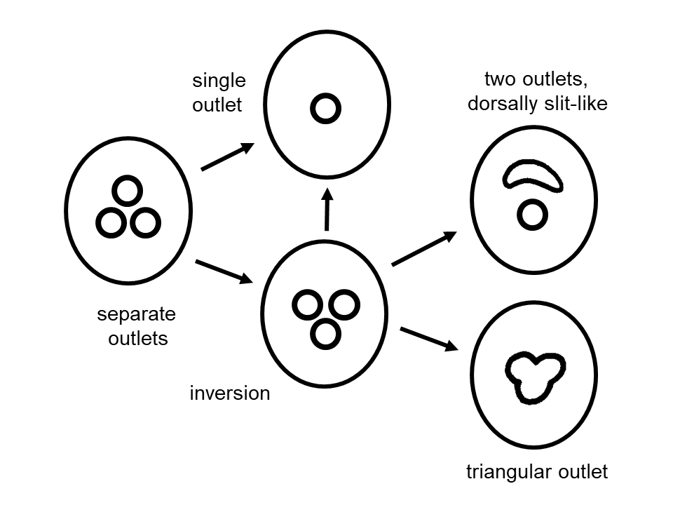
We hypothesise that three caudal glands with three separate terminal openings represent the ancestral state in nematodes, and a common exit realized by fusion is apomorphic. This hypothesis is supported by the presence of separate openings in multiple species within the clade that includes species X as an ancestor (Figure 4). Additionally, some taxa with a common opening retained three separate ampullae. In contrast, others rarely exhibited remnants of a three-pointed tail tip.
A unique relic of this ancestral condition is found in Pternepsilonema servaesae, where a triradial tube with a triangular aperture outlines three duct orifices (Figure 3C) [110]. In contrast, nematodes with a single outlet typically possess a cuticularized tube with a circular cross-section.
Similarly, a three-lobed spinneret is observed in some Araeolaimida, such as Synodontium fecundum (Figure 10F) [90] and Axonolaimus hexapilus [111], while a tripartite supporting structure is present in the mononchid Oionchus obtusicaudatus [112].
A functional argument against the presence of a common exit in the nematode stem species is the improbability of losing a complex and highly efficient (effective) spinneret apparatus while gland cells remained ecologically functional. A possible intermediate stage in this evolutionary transition would involve the retention of three separate ampullae, with early signs of fusion evident in the structure of the opening. This suggests a gradual transformation from three distinct gland ducts, ampullae, and openings to a configuration with at least one common ampulla, a valve, and a joint opening (Figure 6).
If this reasoning is correct, then the common opening seen in the vast majority of adenophorean species is not an ancestral trait but rather a unique feature that evolved multiple times independently. This pattern of convergent evolution is reflected in the phylogenetic tree (Figure 4) and will be further discussed in the next section.
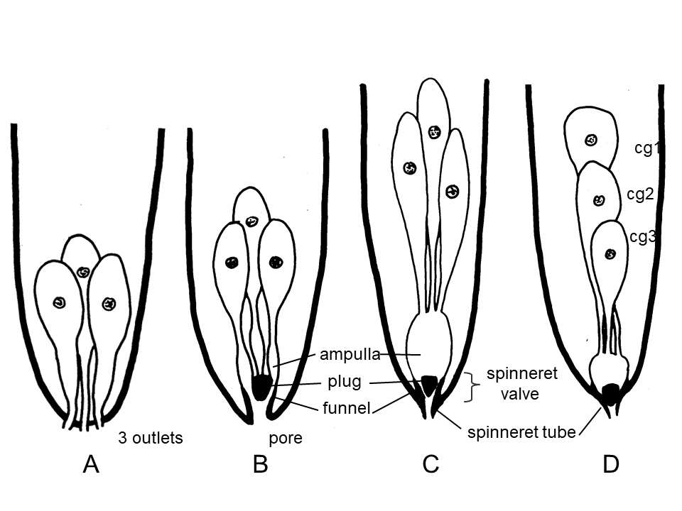
Figure 6: Different characteristics of the caudal glands complex and terminology. A) Beginning with three separate openings; B) in a second step with a common pore and a valve represented by a cuticularised plug in a funnel, each gland with an ampulla; C) the separate ampullae are joined to a common ampulla and the simple pore is developed to a cuticular spinneret tube; gland cells in tandem, numbered from anterior to posterior; A-B) gland cells grouped.
The common opening is typically part of a functional complex that Cobb first referred to as a “spinneret” [113], drawing an analogy to the silk-spinning organs of spiders. In Mononchulus ventralis, M. nodicaudatus and Mononchus lacustris [114, 115]), Cobb described a sophisticated apparatus associated with this structure [113]. A similar mechanism was noted in Klugea (= Enchelidium) polaris (Enoplida) [116], although without illustration. This apparatus consists of a conical plug housed within a cuticular funnel (Figure 6), functionally resembling a needle valve that finely regulates secretion flow. Small retractor muscles open a ring-shaped passage around the plug, which is always positioned obliquely to the dorsal body wall, allowing secretions to be released. The valve then closes due to the turgor pressure of the body fluid [33], enabling secretion to be controlled under neural regulation. The term “spinneret” is often applied to a simple terminal opening (pore), but more commonly, it refers to a mucro-like offset terminal tube (historically termed an “adhesive tube”), whose length and shape are often diagnostic in species identification (e.g., in Monhystera [117]). However, inconsistencies exist: some authors refer to the valvular apparatus as the “spinneret” [115, 116, 117, 118, 119], others use the term to describe the apparatus plus the terminal tube [55], and some remain ambiguous in their usage [120, 121, 122].
We emphasize the importance of distinguishing between a “spinneret valve” and a “spinneret” (Figure 6). The term “spinneret” should be used as a general descriptor for the common outflow of the caudal glands, given its widespread usage in species descriptions. Based on structural differences, we propose the following terminology: (1) a simple opening, as depicted in Cobb’s illustrations, should be referred to as a “pore” or “spinneret pore”; (2) a cuticular tube should be designated as a “spinneret tube” or “tubular spinneret”; and (3) specialized structures (valves) should be described more precisely. As these structures have evolved multiple times independently, these terms serve as functional descriptors within our evolutionary framework.
In many lineages, the separate ducts of the glands, which form part of the gland complex, converge into a common ampulla for secretion storage, enabling immediate release when needed. During evolution, a distinct ampulla or expanded collecting duct likely emerged from the fusion of three ducts or from separate ampullae, as observed in Monhystera species [61]. A prominent ampulla is documented across diverse lineages, including Mononchida, Triplonchida, Enoplida, Chromadorida, Desmodorida, Monhysterida, and Araeolaimida, based on various studies. However, estimating the frequency of convergent evolution for this trait is challenging, as it is rarely considered in species descriptions. Notably, an ampulla is absent in Chromadorina and Perepsilonema [33, 35].
According to our hypothesis, the fusion of three ducts into a single ampulla and outlet, along with the formation of a valve or “caudal cone,” likely occurred simultaneously. Malakhov (p. 109) described the cuticular tail cone as forming via the submersion of a cuticle segment located between gland pores [123]. Although comparative studies on this topic are lacking, structural variations of the apparatus among unrelated taxa are expected. Pternepsilonema servaesae, for instance, possesses a triangular aperture, which may represent a transitional stage within the Epsilonematidae family. Gerlach described three cuticular components (one dorsal, two subventral) forming the outlet in Cynura uniformis (Leptolaimidae) [124]. Similarly, Kreis noted three sclerotized structures laterally positioned around the outlet in Symplocostoma papillatum (Enchelidiidae), however one ventral and two subdorsal [125].
Information on the internal sclerotized valve structure across taxa remains limited, and structural differences among lineages such as Mononchida, Chromadorida, and Monhysterida are likely (for monhysterids, see [55]). The homology of the spinneret apparatus in these taxa was questioned [33]. However, at present, we assume that these species exhibit structural similarity with only minor variations, such as a needle-shaped versus conoid spinneret plug, and differences in the number of retractor muscles ranging from one [33] to two [126] or several [127]. Wilsonematinae species lack needle valves [128]. The closure mechanism in species with separate outlets and no valves remains unknown [35, 36].
Multiple Transformations to A Single Opening
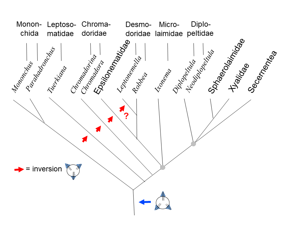
It is highly probable that the stem species of Nematoda possessed three distinct apertures for the caudal glands, a trait that has been retained in many descendants of species X (Figure 4). However, most Adenophorean taxa share a common outlet, suggesting that multiple lineages independently converged on this trait. This study aims to determine where these transformations occurred in phylogeny (see tree for comparison; Figure 4).
The absence of distinct gland openings in all early- branching species of “Adenophorea” supports the hypothesis that a common outlet evolved independently in Chromadorida, Enoplida/Triplonchida, and the clade encompassing Mononchus and Trichinella (Figure 4). In closely related species of the clade defined by the stem species X both separate and common gland openings are present. Further investigation is required to clarify evolutionary transitions within these taxa (Figure 7).
Microlaimida Among Microlaimidae, only Ixonema exhibits separate gland outlets. Therefore, we infer that the stem species of Microlaimidae possessed this trait, with at least one subsequent transformation to a common outlet within this family taxon.
Aponchiidae (Aponchium, Synonema), Monoposthiidae (Monoposthia, Monoposthioides, Nudora, Rhinema), and Richtersiidae (Desmotersia, Richtersia) all possess a spinneret.
This suggests that the stem species of Microlaimida originally had separate gland openings, which were independently modified into a common pore in at least two lineages.
Desmodorida Desmodoridae: The condition in Desmodora bilacinia remains uncertain, as there appear to be two superimposed openings (Figure 8E). Aside from this ambiguous case and the Stilbonematinae subfamily, all Desmodoridae exhibit a common outlet with a spinneret [129].
Stilbonematinae: Since multiple species in this subfamily retain separate apertures, it is likely that both the stem species of Stilbonematinae and the broader Desmodoridae initially had separate gland openings.
While Adelphos may possess a single terminal pore [130], historical claims of a “spinneret present” in Catanema and Stilbonema [120] require verification.
Eubostrichus includes species with both separate gland openings (Figure 3H) and a single spinneret [131], indicating at least two independent transitions to a common outlet within Stilbonematinae.
At least a third independent transformation must be postulated for the remaining Desmodoridae. • All species of Draconematidae have a single outlet for the caudal glands and usually a spinneret, which is interpreted as an apomorphic trait.
• In Epsilonematidae, some genera exhibit three separate outlets (Akantepsilonema, Epsilonema, Triepsilonema: Figures 3D,3E), while others have a single outlet (Glochinema, Metepsilonema, Perepsilonema). In Bathyepsilonema, there is one pore and a slit in males (Figure 3J) and a single pore in females [132, 133], which may indicate the fusion of two pores. The presence and structure of pores in Polkepsilonema and Pternepsilonema remain ambiguous [110]. It is likely that the stem species of Epsilonematidae had three separate gland openings, with at least one independent transformation to a joint opening within this group. Similarly, at least five independent transformations to a common duct for caudal glands must be assumed within Desmodorida.
Ceramonematoidea Unlike other taxa within Ceramonematoidea, species of Diplopeltoides possess separate gland openings. This suggests that the transition from separate openings in the stem species to a single joint pore occurred at least once within this group.
Desmoscolecida The only species of Desmoscolecida known to have separate outlets is Tricoma tripapillata, though they may also be present in Desmoscolex gourbaultae [68]. Since a terminal spinneret is present in other species of Tricoma, it suggests that the separate outlets reconstructed for the stem species of Desmoscolecidae were modified into a terminal spinneret at least twice independently within this group.
Monhysterida • Linhomoeidae: All species possess a spinneret. Meldal reported separate openings in Desmolaimus zeelandicus [81], but other authors have described only a single terminal pore in Desmolaimus species [84, 134]. Therefore, we question the validity of Meldal’s observation. At least one transition to a single duct must be assumed here.
• Sphaerolaimidae: All species should possess three separate outlets. However, there are reports of a spinneret in Doliolaimus agilis [135] and various Sphaerolaimus species [62, 134, 136].
• Monhysteridae: All taxa have a common opening for the glands, which is considered an apomorphy supporting the monophyly of the group [82].
• Xyalidae: The stem species of Xyalidae undoubtedly had separate outlets for the three caudal glands, a feature retained in most genera (Amphimonhystera, Daptonema, Echinotheristus, Elzalia, Gonionchus, Metadesmolaimus, Pseudechinotheristus, Stylotheristus, Valvaelaimus). However, some genera contain both species with three outlets (references in Table 2) and a single pore or spinneret, such as Paramonhystera [56], Rhynchonema [137], Steineria [138, 139], Theristus [140, 141, 142], and Xyala [143, 144]. Other genera consistently exhibit a common outlet (Capsula, Dactylaimoides, Hofmaenneria, Lamyronema, Promonhystera, Sphaerotheristus). The character state in Cobbia remains uncertain [57, 144]. If these character states are correctly assessed, approximately eleven independent transitions to a joint outlet happened within Xyalidae.
Thus, within Monhysterida, there have been approximately thirteen transitions to a common pore, one each in the lineages leading to Linhomoeidae and Monhysteridae, and eleven transformations within Xyalidae.
Araeolaimida • Comesomatidae: Three gland pores (one dorsal, two subventral) have been reported only for Dorylaimopsis variabilis (Figure 3A), distinguishing it from other, spinneret-bearing Dorylaimopsis species [145, 146] and all other genera within the family. This suggests that a transformation to a common duct occurred at least twice within Comesomatidae.
• Diplopeltidae: Caudal glands open via three separate pores in Diplopeltula, Mudwigglus, Neodiplopeltula, and Pararaeolaimus nudus, whereas other species of Pararaeolaimus [147, 148] and all other genera possess a spinneret. Notably, caudal glands are absent in Edalonema. These observations indicate that Diplopeltidae inherited separate openings from its stem species, with at least two independent transitions to a common spinneret within this group.
Aegialoalaimus: At least two species of Aegialoalaimus possess three separate openings (Table 2), while others exhibit a common pore [16]. It is hypothesised that fusion occurred only once within Aegialoalaimus. An intriguing observation by Tchesunov suggests that in A. elegans, adults exhibit three separate outlets, whereas juveniles appear to have a common pore [149]. However, before proposing that a common pore represents an ancestral state in juveniles, potentially challenging our hypothesis of separate pores as plesiomorphic, further confirmation of character states in both developmental stages is needed. Consequently, caudal glands have independently evolved to discharge via a common opening at least five times in the lineage of Araeolaimida.
Plectida The timing of the evolution of a single duct in Plectida remains uncertain. In the Secernentea/Teratocephalida lineage, caudal glands were completely reduced. It is assumed that the three-pointed tails found within Secernentea represent a plesiomorphic condition corresponding to the existence of separate outlets. This implies that a joint outlet was a derived trait, emerging either in the lineage to Plectida or in the sister lineage to Metateratocephalidae (Figure 4), suggesting at least one such transformation. However, the exact point of this transition remains undetermined (Figure 4).
![Figure 9: Phylogenetic diagram of Enoplida appraising the data in [11,13,17]. Paraphyletic taxa after Ahmed, et al. [17] in parentheses. The greyish circles mark points not resolved into dichotomies so far.](/fulltextimages/13910/fig_9.png)
In conclusion, in the clade derived from stem species X, caudal glands evolved to open via a single pore at least 29 times independently (Figure 4). Additionally, as noted earlier, three similar transformations could be inferred within the early lineage of “Adenophorea” (Figure 4). Thus, it can be hypothesised that the single caudal gland outlet evolved independently about 32 times in Nematoda. This exceptionally frequent parallel evolution suggests a selective advantage for a single outlet, although its functional benefits remain unclear. A newly developed valve may regulate secretion flow precisely, but the challenge of coordinating the discharge of secretions from both adhesive gland cells and releasing gland cells through the same pore requires further investigation. The convergent evolution of joint pores suggests that spinneret structures might differ across lineages, of which three-lobed spinnerets give examples. A comparative SEM study could provide valuable insights into their structural diversity and functional implications.
Dorsoventral Positioning of Caudal Glands
Two distinct orientations of caudal glands can be observed within Nematoda. The most common configuration consists of an anteriorly positioned dorsal gland and two ventrally located caudal glands or their ducts. However, the exact positions of these glands have seldom been described in the literature, with exceptions such as Mononchus italicus [150] or illustrated cases like the mononchid Parahadronchus shakili [151] and the monhysterid Theristus caudasaliens [35].
![Figure 10: Structure of tail terminus in different nematode taxa. A) Lenonchium fimbricaudatum [104]; B) Trefusialaimus monorchis [109]; C) Deontolaimus papillatus; D) Pseudodilaimus pilosus [83]; E) Histiostrongylus spineus [233]; F) Synodontium fecundum [90]; G) Caribplectus magdalenae [201]; H) Triepsilonema tripapillata [133].](/fulltextimages/13910/fig_10.png)
Instances where glands open through three separate apertures at the tail tip show a consistent pattern: one dorsal and two subventral openings [82, 83, 89]. Examples include Robbea hypermnestra [152] in Desmodoridae, Ixonema deleyi [153] in Microlaimidae, and Dorylaimopsis variabilis [154] in Araeolaimida. A similar arrangement is found in glandless Secernentea, where three-pointed tails also feature one dorsal and two subventral protrusions.
An unusual deviation from this pattern was reported by Lippens (p. 182) in Chromadorina germanica and Chromadora nudicapitata (Chromadoridae), where “the duct of the anterior gland cell is located ventrally... [and] the ducts of the two posterior cells assume a dorso-sublateral position” [33]. A similar reversed orientation was noted in Tuerkiana (=Thoracostoma) strasseni (Leptosomatidae, Enoplida), where Türk (p. 308) observed a ventromedial gland duct alongside two subdorsal ducts [1].
This orientation is also seen in Epsilonematidae (Desmodorida), as reported for Akanthepsilonema sinecornibus, Epsilonema pustulatum, and Triepsilonema tripapillata, which possess one medioventral and two subdorsal outlets [155]. Furthermore, SEM imaging of Leptonemella brevipharynx (Desmodoridae) reveals two laterodorsal outlets and one ventral opening (Figure 3B).
Despite these observations, none of the cited authors have addressed the significance of these variations, and to our knowledge, this discrepancy has not been discussed in the literature. One possible explanation could be situs inversus, as observed in the triradiate stoma of five species within “Rhabditidae” by Osche [156], suggesting that similar developmental modifications may have influenced the caudal gland arrangement in these nematode groups.
The inverse orientation of the three duct orifices in Pternepsilonema servaesae [110] may also be indicative of a corresponding inversion in caudal gland positioning. In this context, the sclerotizations observed at the outlet of Symplocostoma papillatum (Enoplida) by Kreis [125] may be relevant. Similarly, in Phanoderma ocellatum (Enoplida), the orientation of the caudal glands might resemble that of Chromadoridae [90]. However, these interpretations require confirmation, as the available data remain too imprecise for meaningful phylogenetic discussion.
Based on the available evidence, an apomorphic inverse orientation of caudal glands appears to have evolved independently in at least four distantly related lineages: Leptosomatidae, Chromadoridae, Epsilonematidae, and likely within Desmodoridae. Some of the inferred transformations of gland positions and their respective openings are outlined in Figure 7.
Location and Arrangement of Caudal Gland Cells, Especially in Enoplids
Caudal glands in nematodes typically follow one of two main arrangements: a tandem sequence (cg1-cg3) (Figure 6D) or a grouped configuration, where cg1 is positioned anteriorly and cg2 and cg3 lie side by side at the same level (Figure 6B, C).
In some cases, these differences serve as taxonomic markers for distinguishing closely related species. However, no clear pattern has emerged in the distribution of these arrangements, suggesting that both configurations have evolved multiple times in different directions. It is plausible to assume that the grouped arrangement was ancestral in nematodes [21, 115].
Based on available data, caudal glands in nematodes can be categorized into two primary spatial configurations: incaudal and precaudal.
Incaudal: The glands are predominantly confined to the tail, with cg1 usually reaching the level of the anus/cloaca or extending slightly beyond (Figure 2B, C). In some cases, cg1 and even cg2 may extend anterodorsally beyond the rectum, occasionally reaching the end of the intestine. This condition is termed “suprarectal.” Meanwhile, cg3 remains entirely within the tail.
Precaudal: All three gland cells, including their nuclei, are located anterior to the anus/cloaca, typically arranged sequentially without overlap. They are aligned parallel to the intestine and often the gonads (Figure 2A). This precaudal positioning allows the gland cells to enlarge and potentially produce greater secretions.
Even within the incaudal configuration, the extension of cg1 (and sometimes cg2) anterior to the anus/cloaca likely serves to increase secretion output based on species-specific ecological demands. In some cases, this extension is sexually dimorphic being more pronounced in females than in males. A possible explanation is that, in males, the anterior expansion of caudal glands may be constrained by the presence of copulatory structures such as spicules, the gubernaculum, and associated muscles. However, exceptions exist: in Enoplus [23] and Synonchus fasciculatus [73], caudal glands extend further anteriorly in males, while remaining within the tail in females. Similarly, in Graphonema antarcticum (Chromadoridae), all caudal gland cells are located anterior to the cloaca in males, whereas only one gland is preanal in females [157].
In contrast, taxa with gland bodies located far precaudally are particularly notable and occur frequently in Enoplida. The mixed distribution of incaudal and precaudal gland positioning within this group (or clade) allows for multiple evolutionary interpretations: (1) caudal glands were originally confined to the tail, with precaudal displacement occurring repeatedly in parallel; (2) a single primary shift to a precaudal position occurred, with occasional reversions; or (3) continuous back-and-forth displacements happened throughout evolutionary history. Lorenzen proposed an explanation based on the concept of “underlying apomorphy” [23, 158] suggesting that a genetic predisposition for precaudal displacement was inherited from a common ancestor with incaudal glands. According to this hypothesis, minor transformations could repeatedly lead to an anterior shift. We agree with Lorenzen that the precaudal gland position in Enoplida is homologous in origin. However, distinguishing between evolutionary scenarios (1) and (2) remains challenging.
To explore this question, we consider the possibility of independent precaudal shifts. If unrelated taxa exhibit differences in gland positioning relative to the intestine and rectum, this could support the hypothesis of multiple independent shifts. Unfortunately, data to test this hypothesis are scarce.
Based on our observations, we propose a positional formula for precaudal gland cell arrangement - left (l) or right (r) of the intestine - in the sequence cg1-cg3: Phanoderma tuberculatum [21]: <l-l-l> Oncholaimus sp. [21], see Fig. 2A: <l-r-l> Viscosia viscosia [159]: <r-l-l> Eurystomina terricola [160]: <r-l-r> Oncholaimus jessicae [32]: <r-l-r>, but some specimens exhibit <l-r-l>.
This pattern highlights variation in gland positioning, which could provide further insights into the evolution of caudal gland arrangements in nematodes.
In Triplonchida, the supposed sister group of Enoplida, the three caudal glands are invariably incaudal, which is likely plesiomorphic, suggesting that the ancestral condition in the Enoplida lineage was also incaudal. Consequently, within Enoplida, the incaudal condition is presumed always to be the ancestral state, ruling out any reversals from a precaudal site back to the original configuration. While the exact evolutionary relationships within Enoplida remain uncertain (Figure 9), the potential positioning of caudal glands in well-supported monophyletic subgroups of Enoplida can be outlined. Additionally, it is assumed that most of the genera listed below are monophyletic in this preliminary analysis. The data sources include various species descriptions, with references provided only in exceptional cases. Estimating the number of parallel evolutionary events is challenging, particularly given the lack of robust phylogenetic trees. However, it is hoped that studies such as the present one will encourage further research on this subject.
Campydoroidea: All taxa (Campydoroides, Udonchus, Rhabdolaimus, Rogerus, and most species of Syringolaimus) possessing caudal glands exhibit an incaudal arrangement.
Alaimidae: In the stem species of Alaimus and Amphidelus, caudal glands were lost. However, their ancestral lineage likely possessed incaudal glands.
Ironidae: In most genera (Dolicholaimus, Ironus, Syringolaimus, Trissonchulus), the caudal glands remain within the tail or extend suprarectally. However, the precaudal positioning of glands in Conilia, T. jungi within Thalassironus, and possibly in Ironella suggests at least three independent gland dislocations within Ironidae.
Trefusiidae: In genera such as Africanema, Rhabdocoma, Trefusia, Tripylina, and Trischistoma, caudal glands are always in the tail, rarely extending suprarectally.
Tripyloididae: In Bathylaimus, Paratripyloides, and Tripyloides, caudal glands are consistently confined to the tail.
Oxystominidae: This group may be paraphyletic. The plesiomorphic incaudal condition is observed in Halalaimus, certain species of Litinium (e.g., L. bananum), Nemanema (N. rectoresectum) and Thalassoalaimus (T. longicaudatus, T. montemari). However, Oxystomina (O. astridae, O. clavicauda, O. exilis) and most species of Litinium, Nemanema, Paroxystomina, and Thalassoalaimus exhibit a suprarectal gland position. Thus, at least five independent precaudal gland shifts are expected within Oxystominidae.
Rhaptothyreidae: In Rhaptothyreus, the caudal glands remain incaudal.
Enchelidiidae: Plesiomorphic caudal glands are incaudal or suprarectal in Belbolla heptabulba, species of Ledovitia, Eurystomina (E. bilineata, E. chilensis, E. vincxae), and Polygastrophora (P. hexabulba, P. septembulba). However, they are often arranged in tandem anterior to the anus/cloaca or at a much greater distance in Calyptronema (= Catalaimus), Ditlevsenella, Illium, Symplocostoma, Symplocostomella, Thoonchus, and Belbolla sundaensis, as well as in most species of Eurystomina and Polygastrophora. In Bernardius lineatus, a transitional state is observed, with two caudal glands extending up to two spicule lengths anterior to the cloaca, while cg3 terminates at the cloacal level [161]. Within Enchelidiidae, at least eight to nine independent gland displacements have occurred. However, the loss of caudal glands in Abelbolla, Bathyeurystomina, Megeurystomina, and Pareurystomina cannot be precisely traced to an incaudal or precaudal origin.
Oncholaimidae: This group appears to be paraphyletic. The presumed plesiomorphic incaudal and possibly suprarectal gland positions are seen in Pelagonemella, Thalassogenus, and certain species of Adoncholaimus, Oncholaimus, and Viscosia. However, most species in these genera exhibit the apomorphic precaudal condition. Transitional states are observed in Dioncholaimus brevicavatus and Metoncholaimus isopapillatus, where two caudal gland cells are precaudal while one remains incaudal [162, 163]. In other species of Metoncholaimus and other genera (Anoncholaimus, Krampia, Metoncholaimoides, Meyersia, Mononcholaimus, Oncholaimellus, Pelagonema, Pontonema, Prooncholaimus, Pseudoncholaimus, Pseudopelagonema, and Vasculonema), the precaudal condition is well established. A cautious estimation suggests that the precaudal gland position evolved independently at least 16 times in Oncholaimidae.
Anoplostomatidae: In Anoplostoma and Chaetonema, the caudal glands are precaudal, suggesting an apomorphic state for the family.
Anticomidae: The caudal glands are incaudal to suprarectal in Anticoma, Anticomopsis, and Paranticoma.
Leptosomatidae: This possibly paraphyletic group shows incaudal to suprarectal glands in Leptosomatum caecum, L. groenlandicum, Orthophallonema, Paratuerkiana, Pseudocella, Synonchus, and Tuerkiana. However, precaudal glands are found in Bongersia, Corythostoma, Cylicolaimus, Deontostoma, Leptosomella, Leptosomatides, Paraleptosomatides, Thoracostoma, and certain Leptosomatum species. Due to limited data and uncertain phylogenies, nine precaudal shifts are estimated within Leptosomatidae.
Thoracostomopsidae: Many taxa exhibit incaudal or suprarectal glands, including Fenestrolaimus, Filipjevia, Metenoploides, Parenoplus, Thoracostomopsis galeata, and several species of Enoploides, Enoplolaimus, Epacanthion, Mesacanthion, Oxyonchus, Paramesacanthion, Saveljevia, and Trileptium. However, approximately 13 independent precaudal transitions have occurred (in Africanthion, Fleuronema, Mesacanthoides, Okranema, Enoplolaimus pectinatus, Epacanthion, Oxyonchus dentatus, Paramesacanthion tricuspis, Saveljevia kolaensis, Thoracostomopsis barbata, Trileptium sp. and within Enoploides and Mesacanthion).
Phanodermatidae: Caudal glands are generally incaudal or suprarectal. However, in Phanoderma campbelli and Paraphanoderma robynae, they are precaudal, indicating two independent shifts.
Enoplidae: In Enoplus, caudal glands extend precaudally in males, whereas they remain incaudal to suprarectal in females.
Overall, caudal glands remain largely restricted to the tail in six of the 15 groups analyzed, supporting their plesiomorphic nature. However, gland migration anterior to the anus/cloaca has occurred approximately 58 times independently. This high frequency suggests that gland dislocation is genetically predisposed and phenotypically adaptable, reinforcing the concept of underlying apomorphy within Enoplida.
In the outgroup of Enoplida the caudal glands are located in the anterior part of the tail, but frequently cg1 and sometimes also cg2 extend suprarectally near the end of the intestine while cg3 still remains entirely incaudal. Possibly there are few exceptions of species of Araeolaimida, Chromadorida, Desmodorida and Monhysterida (Table 3) where all gland bodies are situated suprarectally anterior to anus/cloaca.
In Onyx adenophorus (Desmodoridae) they are located considerably anterior similar to some enoplids [164]. In comparison with their closely related taxa, the species in Table 3 demonstrate continuity between both conditions (incaudal to suprarectal and precaudal), which are defined with respect to the location of the posterior-most gland cell (more precisely: the cell portion of cg3 containing the nucleus) posterior versus anterior to anus/cloaca. In Diplopeltula striolata (Araeolaimida) two gland cells extend far anterior but cg3 just reach the cloaca [165].
| species name | group | author(s) |
|---|---|---|
| Steineridora (= Spilophora) loricata | Chromadoridae, Chromadorida | Steiner [116] |
| Onyx adenophorus | Desmodoridae, Desmodorida | Blome, et al. [164] |
| Xenonema obesum | Desmodoridae, Desmodorida | Cobb [90] |
| Dracograllus filipjevi | Draconematidae, Desmodorida | Rho, et al. [272] |
| Draconema cephalatum | Draconematidae, Desmodorida | Steiner [21] |
| Draconema micoletzkyi | Draconematidae, Desmodorida | Kreis [83] |
| Perepsilonema conifera | Epsilonematidae, Desmodorida | Adams, et al. [35] |
| Polkepsilonema mombasae | Epsilonematidae, Desmodorida | Verschelde, et al. [110] |
| Desmolaimus zeelandicus | Linhomoeidae, Monhysterida | Meldal [81] |
| Thalassomonhystera diplops | Monhysteridae, Monhysterida | Cobb [73] |
| Diplopeltula striolata | Diplopeltidae, Araeolaimida | Blome, et al. [164] |
Table 3: All caudal glands anterior to anus/cloaca in the outgroup of Enoplida.
Change in the Number of Caudal Glands
Additions As discussed earlier, a special cell associated with the three gland cells could either be a releasing cell or a pseudocoelomocyte. However, in many cases, additional cells cannot be distinguished from caudal glands. In such instances, the multiplication of gland cells may have occurred, resulting in a total of four to six cells.
Four gland-like cells have been reported in the Enoplida species Bathylaimus arthropappus and Tripyloides imitans, as well as in Linhomoeus spp. (Monhysterida), Aphanolaimus aquaticus, and various Plectus species (Plectida) (Table 1). In Steineridora (= Spilophora) loricata (Chromadoridae), Steiner depicted four gland cells located precaudally on the left side, except for the third gland, which was positioned to the right of the intestine [116].
In the section on releasing glands, two supernumerary gland cells were noted in Caprionchulus, Eurystomina, and Phanodermopsis. Additionally, a series of unidentified cells potentially gland cells or pseudocoelomocytes have been documented in the Mononchida species Miconchus studeri [166] and Mylonchulus obtusicaudatus [167], as well as in Triplonchida species such as Eutobrilus [168, 169] and Tobriloides [170, 171]. Similar features were observed in Anticyclus exilis (Monhysterida) [90].
Notably, species of Thalassogenus (Oncholaimidae) exhibit six caudal glands arranged in two sets of three in tandem (Figure 2D), each with a separate duct leading to a large common ampulla [172, 173]. This configuration suggests a duplication of the entire gland complex in the stem species of Thalassogenus.
Reduction in the number of gland cells While increases in gland cell numbers are rare, reductions have occurred many times independently. In several lineages, one gland has been lost, leaving only two gland cells. This phenomenon has been documented in approximately 40 species across Chromadorida, Desmodorida, Enoplida, Monhysterida, Mononchida, and Triplonchida (Table 4), where closely related species typically possess three glands.
If these reports are accurate, the complete reduction of one gland cell would have occurred at least 24-27 times independently, though it is likely to have happened even more frequently.
| species name | group | author(s) |
|---|---|---|
| Pandolaimus spp. | Pandolaimidae, Triplonchida | Lorenzen [23] |
| Rhabdodemania major | Rhabdodemaniidae, Triplonchida | Naveen, et al. [273] |
| Rhabdodemania minor | Rhabdodemaniidae, Triplonchida | Murphy, et al. [274] |
| Anticoma arctica | Anticomidae, Enoplida | Steiner [116] |
| Trileptium sp. | Thoracostomopsidae, Enoplida | Smol, et al. [195] |
| Miconchus japonicus | Anatonchidae, Mononchida | Ahmad, et al. [238] |
| Cryptonchus sp. | Cryptonchidae, Mononchida | Lorenzen [23] |
| Prionchulus japonicus, P. punctatus | Mononchidae, Mononchida | Ahmad, et al. [238] |
| Chromadorita heterophya | Chromadoridae, Chromadorida | Allgén [275] |
| Dichromadora geophila | Chromadoridae, Chromadorida | Schiemer, et al. [38] |
| Latronema orcinum | Selachinematidae, Chromadorida | Blome [276] |
| Eubostrichus hopperi (?) | Stilbonematinae, Desmodorida | Armenteros, et al. [12] |
| Laxus parvum (?) | Stilbonematinae, Desmodorida | Armenteros, et al. [12] |
| Monhystrella hastata (?) | Monhysteridae, Monhysterida | Abebe, et al. [60] |
| Monhystrium transitans | Monhysteridae, Monhysterida | Cobb [90] |
| Monhystrium aff. transitans | Monhysteridae, Monhysterida | Riemann [30] |
| Thalassomonhystera bathislandica | Monhysteridae, Monhysterida | Riemann [277] |
| Thalassomonhystera oxycephalata | Monhysteridae, Monhysterida | Tchesunov, et al. [267] |
| Tripylium carcinicola | Monhysteridae, Monhysterida | Riemann [30] |
| Astomonema otti | Siphonolaimidae, Monhysterida | Vidakovic, et al. [278] |
| Sphaerolaimus gracilis | Sphaerolaimidae, Monhysterida | Turpeenniemi, et al. [36] |
| Sphaerolaimus macrocirculus | Sphaerolaimidae, Monhysterida | Rosli, et al. [279] |
| Sphaerolaimus occidentalis | Sphaerolaimidae, Monhysterida | Turpeenniemi [85] |
| Subsphaerolaimus brevicauda | Sphaerolaimidae, Monhysterida | Jensen [266] |
| Daptonema trabeculosum | Xyalidae, Monhysterida | Lorenzen [57] |
| Echinotheristus cimbricus, E. teutonicus | Xyalidae, Monhysterida | von Thun, et al. [101] |
| Gnomoxyala spina | Xyalidae, Monhysterida | Lorenzen [57] |
| Pseudechinotheristus nudus | Xyalidae, Monhysterida | Blome [269] |
| Amphimonhystera marisalbi | Xyalidae, Monhysterida | Tchesunov, et al. [280] |
| Paramonohystera concinna | Xyalidae, Monhysterida | Lorenzen [57] |
| Rhynchonema amakusanum | Xyalidae, Monhysterida | Bezerra, et al. [137] |
| Rhynchonema fossum, R. lyngei, R. megamphidum, R. ornatum, R. semiserratum, R. separatum, R. tremendum | Xyalidae, Monhysterida | Lorenzen [102] |
| Theristus acer | Xyalidae, Monhysterida | Lorenzen [57] |
| Theristus flevensis, T. pertenuis | Xyalidae, Monhysterida | Lorenzen [57] |
Table 4: Only two caudal glands stated.
The literature contains depictions of only two caudal glands in approximately 100 additional species across 22 family-level taxa, though without explicit mention of the observed number. This raises uncertainty as to whether these species truly possess only two glands or whether some illustrations were merely schematic, omitting a third gland. Cases were excluded where only two glands were depicted in illustrations but three were stated in the text. Accurately determining the number of glands is challenging, as authors sometimes correctly note that only two caudal glands were “visible.” In some species, two posterior gland cells may be positioned parallel, appearing as a single structure in lateral view. Additionally, one gland may be obscured and thus overlooked, as mentioned for various species of Monhysterida [169].
In Monhysterida, it is evident that the most anterior and presumably always dorsal gland cell (cg1) has been reduced in size. In other groups, we have not attempted a comparative analysis with closely related species possessing three gland cells to determine whether a different gland was lost in certain species. This would be particularly relevant in Chromadorida species, where the glands have an inverse orientation.
The reduction to a single gland cell, as discussed in the section on caudal gland function, has occurred more than five times independently. There is no indication that this reduction happened in a stepwise manner, with an intermediate stage of two retained cells. Moreover, the exact reasons for the complete loss of one or two gland cells remain unclear.
Processes of Reduction of Caudal Glands
Degenerative evolution leading to the loss of caudal glands has been observed across multiple taxa. In various populations of Plectus species, researchers have identified aberrant specimens lacking caudal glands and a spinneret [108]. Similar observations have been made in Chiloplectus andrassyi [174].
Within Mononchida, the complete reduction of caudal glands has occurred across numerous lineages, with several indications of ongoing degeneration. For instance, species of Clarkus, Coomansus, and Prionchulus exhibit vestigial tail glands that lack an orifice [175]. Similar conditions have been reported in species of Crassibucca, Iotonchus, Jensenonchus, and Mulveyellus [176]. In species descriptions, terms such as “caudal glands weakly developed” or “reduced” are frequently used, but these should be qualified with descriptors like “partially” or “entirely.” In some cases, these terms indicate a process of gradual reduction. Table 5 presents 38 such instances. Transitional stages are evident in species where caudal glands are mostly absent or vestigial in only a few specimens, such as Clarkus papillatus [177] and Margaronchulus adenticulatus [178].
| species | caudal glands | terminal opening | author(s) |
|---|---|---|---|
| Clarkus enigmatus | indistinct | lacking | Ahmad, et al. [238] |
| Clarkus papillatus | absent, rudimentary in one specimen | absent | Tahseen, et al. [177] |
| Clarkus parvus | rudimentary | absent | Rizvi [282] |
| Clarkus pulcherrimus | rudimentary | absent | Andrássy [178] |
| Clarkus sheri | weakly developed | lacking | Khan, et al. [283] |
| Comiconchus trionchus | rudimentary | absent | Loof [284] |
| Coomansus garhwalicus, C. minor | rudimentary | absent | Rizvi [282] |
| Coomansus intestinus, C. silvius, C. splendidus | reduced | absent | Andrássy [285] |
| Coomansus magellanicus, C. meridionalis | absent in female, reduced in male | absent in female, present in male | Guirado, et al. [179] |
| Coomansus (= Mononchus) mesadenus | present, reduced | present in male, not in female | Clark [182] |
| Granonchulus decurrens | reduced | absent | Andrássy [286] |
| Iotonchus arcuatus | very weakly developed | absent | Ahmad, et al. [238] |
| Iotonchus cuticaudatus | present | present in female, absent in male | Jana, et al. [181] |
| Iotonchus darreni, I. singaporensis | weakly developed | weakly developed | Ahmad, et al. [237] |
| Iotonchus indicus | poorly developed | zpresent | Rizvi [282] |
| Iotonchus maragnus, l. parazschokkei | present | absent | Clark [287] |
| Iotonchus sphagni | rudimentary or absent | absent | Loof [284] |
| lotonchus stockdilli | non-functional | not distinct | Yeates [288] |
Table 5: Remnants of caudal glands or associated structures in Mononchida.
- Jensenonchus sp. rudimentary absent
- Andrássy [176]
- Margaronchulus adenticulatus rudimentary mostly absent, weak if
- Miconchus japonicus poorly developed absent
- Ahmad, et al. [238]
- Mononchus papillatus present absent
- Clark [290]
- Mylonchulus caespitosus inconspicuous absent
- Andrássy [286]
- Mylonchulus psammophilus apparently vestigial absent
- Yeates [291]
- Mylonchulus striatus obscure rudimentary de Bruin [292]
- Parahadronchus egregius reduced lacking
- Andrássy [176]
- Prionchulus iranicus present absent
- Farahmand, et al.
- Prionchulus japonicus weakly developed lacking
- Ahmad, et al. [238]
- Prionchulus koriensis present absent
- Zakaullah, et al. [294]
- Prionchulus mordax reduced absent
- Andrássy [285]
- Prionchulus muscorum rudimentary or absent absent
- Tahseen, et al. [177]
- Sporonchulus ibitensis weakly developed narrow, inconspicuous
- Tahseen, et al. [177]
Table 6: Remnants of caudal glands or associated structures in Mononchida.
Of particular interest is the occurrence of sexual dimorphism, where caudal glands and their outlets are absent in females but present (albeit reduced) in males, as seen in Coomansus magellanicus and C. meridionalis [179]. The reverse situation is apparently observed in Prionchulus longus [180]. In other species, glands exist in both sexes, but the outlet is absent in one, such as in Iotonchus cuticaudatus (pore absent in males [181]) and Coomansus (= Mononchus) mesadenus (pore absent in females [182]). Ontogenetic differences have also been noted, with caudal glands being distinct in juveniles but obscure in adults, as reported in Anatonchus tridentatus and Miconchus digiturus [167, 183]. These observations suggest that, once adhesive secretion became unnecessary in evolutionary history, the initial step in reduction involved the closure of the terminal opening. Some instances of caudal gland cells persisting without an outlet suggest they may have acquired an accessory internal function. This is also observed in Dorylaimida, where remnants of caudal glands were reported in Lenonchium fimbricaudatum (Fig. 10A) [104].
The degeneration of caudal glands has also been documented in marine Enoplida. In Oncholaimidae, Kreis reported rudimentary caudal glands in Pseudoncholaimus elegans, while in Pontonema (= Paroncholaimus) ocellatum, females lacked caudal glands altogether, whereas males retained a single precloacal gland cell [162]. In Nemacoma (= Acoma) borealis (Oxystominidae), Steiner described a small residual bubble-shaped structure without a terminal opening [116]. Among Ironidae, Trissonchulus baldwini possesses weakly developed or rudimentary caudal glands and lacks a spinneret [184], whereas Parironus keiensis exhibits “vestigial or absent” caudal glands [185].
In Triplonchida (Onchulinae), several species exhibit caudal gland cells without a spinneret, including Onchulus flagellatus [171] and Caprionchulus diversipapillatus, where four to five tandem glandular cells persist without a spinneret, though remnants of a tubule are sometimes observed [45]. Similarly, in Monhystera cartaromanae (Monhysterida), caudal glands are in the process of degeneration, with no indication of a spinneret [186]. A comparable condition is found in Mediolaimus obtusicaudatus (Araeolaimida), where the spinneret is absent and caudal glands appear obscure [187], as well as in Leptolaimoides filicaudatus (Plectida) [188].
Unlike in Teratocephalida and Secernentea, vestigial caudal glands appear to be recapitulated in early stages of zooparasitic Benthimermithida and Marimermithida, which usually lack such glands. In Benthimermitidae, poorly developed caudal glands and a terminal pore are present in early juveniles of Trophomera conchicola [8], while in parasitic juveniles of Trophomera granovitchi, a “very thin terminal duct, probably a rudiment of a spinneret” has been observed [189].
Similarly, in Marimermithida, juveniles possess three caudal glands posterior to the anus and a terminal pore, whereas adults are entirely devoid of these structures [190]. The function of these vestigial glands in juveniles remains unknown.
Entire Loss of Caudal Glands
In terrestrial and zooparasitic nematodes, “there is no need for such glutinous glands” [191], leading to their loss in many lineages. Among parasitic groups within “Adenophorea,” this occurred independently in Dioctophymatida/Trichinellida and Mermithida. However, an exception exists in Domorganus (Ohridiidae, Plectida), a parasite in annelid intestines, which retains three caudal glands and a spinneret, likely for attachment to the host’s intestinal wall [192]. The parasitic Benthimermithida and Marimermithida exhibit vestigial caudal glands in juveniles. Their absence in zooparasitic Secernentea is attributed to loss in the stem species of Teratocephalida/Secernentea, which likely inhabited terrestrial environments.
Throughout evolution, many primarily marine nematode lineages transitioned to terrestrial habitats via moist soil. During this ecological adaptation, caudal glands frequently disappeared as they were no longer selectively advantageous. The presence and reduction of caudal glands across different groups, as inferred from phylogenetic relationships (Fig. 4), are discussed below.
Triplonchida In a clade including Bastianiidae, Diphtherophoridae, Odontolaimus, Prismatolaimus, and Trichodoridae, caudal glands are absent [193], despite contrary claims in older literature regarding Bastiania and Prismatolaimus. Nearly all species in this clade inhabit soil, indicating that caudal glands were lost in its stem species in adaptation to terrestrial life. Conversely, the predominantly aquatic sister group retained these glands.
The Onchulidae lineage also supports this trend. While its stem species likely lived in moist soil, caudal glands were retained in Tobriloides/Tobrilonchus and were undergoing degeneration in Caprionchulus and Onchulus. In the Onchulinae lineage (e.g., Kinonchulus, Limonchulus, Stenonchulus, Pseudonchulus inermis), complete gland loss occurred [194]. The presence of vestigial glands in Caprionchulus diversipapillatus, which presumably secondarily invaded freshwater, suggests no reversal to functional glands.
Enoplida The transition to terrestrial habitats within Enoplida led to caudal gland loss in the Alaimina lineage (Alaimus, Amphidelus, etc.). Similar reductions occurred in terrestrial species like Arenasoma terricola (Tripyloididae) and Campydora demonstrans [195]. Even within marine environments, some species lost caudal glands (Table 6).
| species | group | habitat | author(s) |
|---|---|---|---|
| Trissonchulus quinquepapillatus | Ironidae, Enoplida | dune sand | Yeates [291] |
| Platycoma cephalata (?) | Leptosomatidae, Enoplida | marine | Cobb [73] |
| Proplatycoma (= Platycoma) sudafricana | Leptosomatidae, Enoplida | marine | Inglis [295] |
| Proplatycoma tsukubae | Leptosomatidae. Enoplida | marine | Shimada, et al. [296] |
| Filoncholaimus prolatus | Oncholaimidae, Enoplida | marine | Neres [297] |
| Oncholaimus rapax | Oncholaimidae, Enoplida | marine | Kreis [162] |
| Phylloncholaimus immanis | Oncholaimidae, Enoplida | marine | Smol, et al. [195] |
| Halalaimus (= Halalaimoides) acuminatus | Oxystominidae, Enoplida | marine | Wieser [298] |
| Klugea morchella | Phanodermatidae, Enoplida | marine, soft bottom | Wieser [298] |
| Rhabdocoma (= Cytolaimium) obtusicaudata | Trefusiidae, Enoplida | marine | Chitwood [299] |
| Cryptonchus (= Gymnolaimus) exilis | Cryptonchidae, Mononchida | soil | Cobb [66] |
| Anticyathus tenuicaudatus | Linhomoeidae, Monhysterida | marine, sand | Cobb [90] |
| Didelta maculatum | Linhomoeidae, Monhysterida | marine, sea-grass | Cobb [90] |
| Zanema acanthurum | Linhomoeidae, Monhysterida | marine, mud, tide pool | Cobb [90] |
| Monhystera dintheriana | Monhysteridae, Monhysterida | soil | Schneider [249] |
| Siphonolaimus (= Chromagaster) nigricans, S. (= C.) purpureus | Siphonolaimidae, Monhysterida | marine, sand | Cobb [73] |
- Solenolaimus obtusus
- Siphonolaimidae, Monhysterida marine, sand
- Cobb [73]
- Hofmaenneria hazenensis
- Xyalidae, Monhysterida soil
- Mulvey [300]
- Rhynchonema deconincki
- Xyalidae, Monhysterida marine, sand
- Bezerra, et al. [137]
- Theristus aculeatus
- Xyalidae, Monhysterida marine, sand
- Schulz [301]
- Edalonema camerunense
- Diplopeltidae, Araeolaimida soil
- Holovachov, et al.
- Leptolaimoides thermastris
- Leptolaimidae, Plectida silt of salt-marshes
- Lorenzen [23]
- Leptolaimus acicula
- Leptolaimidae, Plectida silt of salt-marshes
- Lorenzen [23]
- Pakira orae
- Plectidae, Plectida dune sand
- Yeates [291]
- Plectus amorphotelus, P. spicacaudatus
- Plectidae, Plectida soil
- Ebsary [302]
- Iotalaimus striatus incertae sedis soil
- Cobb [90]
- Litonema nudum incertae sedis sphagnum swamp
- Cobb [90]
Table 8: Loss of caudal glands in individual species.
This trend extends to limnic Andrassyidae (Andrassya, Malakhovia) [196] and independently within several marine and limnic Ironidae genera (Dolicholaimus, Ironus, Parironus, Pheronous) (various authors). Marine Enchelidiidae also show independent gland loss in Abelbolla [161], Bathyeurystomina, Pareurystomina, and Megeurystomina combesi [195]. These multiple losses in closely related taxa highlight the need for further ecological studies to determine the selective pressures behind caudal gland disappearance in aquatic environments.
Dorylaimida The lineage leading to Dorylaimida experienced caudal gland reduction or loss, while closely related taxa (Bathyodontus, Cryptonchus, Mononchulus, Oionchus) retained them, despite including soil-dwelling species. Lenonchium fimbricaudatum exhibits three gland-like cells (Figure 10A) without duct openings [197], suggesting a stepwise reduction in the ancestral lineage of Dorylaimida til vestiges in the stem species. Dorylaims lacking caudal glands later reinvaded aquatic environments multiple times.
Mononchida This group demonstrates the most extensive loss of caudal glands, likely reflecting multiple independent reductions in terrestrial habitats. Caudal glands are absent in ten genera (e.g., Caputonchus, Doronchus, Prionchulellus, Tricaenonchus) [175, 198]. Other genera (e.g., Clarkus, Coomansus, Granonchulus, Mononchus, Prionchulus and 15 more genera) include both species with and without caudal glands, highlighting an ongoing reduction process (various authors). Given their presumed monophyly, caudal glands appear to have been lost independently in about thirty lineages. It is hypothesised that freshwater mononchids lacking caudal glands secondarily invaded aquatic habitats after terrestrial adaptation (compare [199]).
Monhysterida In this order-taxon, nine species (seven marine, two terrestrial) lack caudal glands (Table 6), indicating multiple independent loss events.
Araeolaimida Terrestrial species such as Edalonema camerunense [200] and closely related Aulolaimidae and Isolaimium lack caudal glands [7]. As these taxa are predominantly terrestrial, their ancestor likely lost caudal glands before a few species secondarily invaded aquatic habitats.
Plectida Many terrestrial taxa within this order retain caudal glands, but they are absent in Pakira orae and several Plectus species [201]. Chronogaster species or their common ancestor lost caudal glands except for C. boettgeri, the only species with glands and a spinneret [108]. As terrestrial Metateratocephalidae lack caudal glands, we hypothesise the loss in its stem species in a terrestrial environment. Consequently, living in freshwater habitats in some Euteratocephalus species is secondary. Additionally, some marine and brackish species of Leptolaimus (e.g., L. acicula) and Leptolaimoides lack caudal glands [201].
Conclusively, a total of nearly 80 documented cases of caudal gland loss have been identified, with approximately 40% occurring in aquatic environments. This challenges the assumption that gland loss primarily occurs in terrestrial lineages. Further research is needed to understand why caudal glands became redundant in marine Enoplida and Monhysterida. Conversely, the persistence of caudal glands in some terrestrial lineages requires investigation. The
presence of limnic and terrestrial species within caudal- gland-lacking groups supports the hypothesis that aquatic taxa in these groups passed through a terrestrial phase (e.g., Alaimidae, Chronogaster, Euteratocephalus, Mononchida, Odontolaimus, Prismatolaimus). Notably, various taxa that lost caudal glands evolved alternative tail structures for attachment, such as mucros, spikes, and hooks. Furthermore, certain nematodes adhere to substrates using tail haptics without glands or anchoring structures, as seen in Rhabditida (own observations) and infective juveniles of parasitic Ascaridoidea (Pseudoterranova decipiens) or Spiruroidea (Spiroxys contortus) [202].
On The Unsettled Problem of the Origin of Phasmids
Steiner [2] emphasized the need for a comparative study of the distribution, morphology, and function of foot glands and phasmids, as well as their reduction and disappearance across various nematode groups. It has been observed that caudal glands are absent whenever phasmids are present and vice versa [28]. These glandular structures in the tail serve as key distinguishing features between the two main groups of Nematoda: the monophyletic Secernentea, which predominantly possess laterally opening phasmids and lack caudal glands, and the paraphyletic “Adenophorea,” where caudal glands with terminal outlets are generally present and phasmids are missing. Three caudal glands are considered plesiomorphic in nematodes, inherited from ancestral species. Phasmids, on the other hand, are paired structures connected to the nervous system and primarily serve a chemosensory function, with glandular activity being rare in extant species [203]. Given the mutually exclusive presence of caudal glands and phasmids, it has been suggested that these structures might be homologous. Steiner [2] hypothesised that phasmidial glands are modified, functionally and numerically reduced caudal glands with laterally shifted outlets. Osche [204] also supported their homology, while Chitwood [47] considered this as possible. However, the Chitwoods (p. 197) were more cautious, recognizing the origin of phasmids as “one of the most perplexing problems in nematology” and considering multiple possibilities for their evolutionary derivation [27].
The transformation of caudal glands with separate outlets into phasmids would theoretically require at least five evolutionary steps: (1) the loss of one of the three primary caudal glands (possibly the dorsal one), (2) a positional shift of the remaining two glands from a subventral to a lateral arrangement, (3) relocation of glandular pores from the tail terminus to the anterior third of the tail (the common position of phasmids), (4) integration of these glands with ciliary structures connected to nerves, resulting in chemosensory function, and (5) a functional shift from adhesive secretion to sensory reception.
A potential example of this transformation can be found in the parasitic Muspiceida, formerly classified within “Adenophorea” [96]. The second-stage larvae of these nematodes are typically ensheathed [205], a trait characteristic of Secernentea. Muspiceida possesses two ampulla-shaped glands that open laterally at approximately 25% of the tail length. These structures were initially identified as phasmids [206], but due to the supposed systematic placement of Muspiceida and the absence of associated neural tissue, they were later reclassified as caudal glands [207]. However, molecular evidence placed Haycocknema perplexum (Muspiceida) within the parasitic Spiroascarida of Secernentea [208], supporting the identification of these glands as phasmids rather than caudal glands. A similar misidentification may have occurred in Breinlia ventricola (Filarioidea), where terminally opening “caudal glands” [209] are more accurately recognized as phasmids.
Examining the “perplexing” situation described by Chitwood, et al. [27], several key points must be considered:
In Secernentea with three-pointed tails the protuberances representing ancestral caudal glands occasionally occur alongside phasmids, indicating that these structures are distinct.
Kaiser described “phasmid-like organs” associated with the nervous system in the mermithid Hexamermis albicans (“Adenophorea”, Fig. 4) located subventrally in the posterior third of the tail in males and at 35% of tail length in preparasitic juveniles [210]. Similar structures were observed in Agamermis decaudata [211], Amphimermis elegans, Hexamermis brevis [210], and several Psammomermis species [212]. However, the homology of these structures with phasmids remains unconfirmed.
Schrage & Gerlach noted lateral pores associated with glands on the ending of the tail in Desmoscolecida (Greeffiellinae), which coexist with caudal glands opening through a spinneret [213]. Chitwood referred to these structures as “phasmata” [47], which also exist in Syringolaimus (Enoplida). The homology of phasmata and phasmids remains uncertain pending evidence of a nervous system connection [214].
Some Mononchida and Chromadorida possess lateral tail pores associated with unicellular epidermal glands, which might be mistaken for phasmids.
In Ascaridoidea (Paranisakis parva) (Secernentea), six tubular gland-like cells extend from rectum level to near the tail tip [115, 123], possibly representing vestigial caudal glands without an external outlet.
The possible homology of two releasing cells with phasmids never seems to be considered.
Hyman’s [28] assertion remains valid: there is no confirmed case of caudal glands and phasmids coexisting in the same organism. The observations discussed suggest that these structures are not homologous but rather coexisted in ancestral nematodes, as proposed by Lorenzen [23]. Under this scenario, caudal glands were lost in lineages where phasmids were present, such as Secernentea and possibly Mermithida, while phasmids were lost in more than thirteen “Adenophorean” lineages that retained caudal glands (Figure 4). However, given the essential role of phasmids in nematode orientation [203], the idea that they were lost multiple times remains questionable. Future studies using transmission electron microscopy are needed to investigate phasmata and phasmid-like structures in mermithids and to reassess their potential homology with phasmids.
Tail Shape and Structures at the Terminal End
Nematodes tail shape varies highly according to the way of living and mode of locomotion of the species. However, caudal glands filling the tail should also affect tail morphology, which represents a compromise between ecological and organismal demands shaping the tail. Although it is beyond the scope of this paper to investigate the relationship between caudal glands and tail shape, it appears worthwhile to compare closely related species of a guild that have caudal glands present or absent, as in Mononchida, or that are incaudal or shifted to a precaudal position, as in Enoplida. We limit ourselves to a few remarks without presenting all the evidence.
The nematodes with three outlets mostly possess a cylindrical tail with a rounded tip. It conforms to tail type 1 of Thistle, et al. [215]. Thus, we can speculate that the stem species of nematodes exhibiting separate gland orifices do not possess an elongated or filiform tail that has been considered plesiomorphic by Decraemer, et al. [216]. Possibly the stem species had three protuberances for the outlets at the blunt end like they exist in Pararaeolaimus nudus [99] or Triepsilonema tripapillata (Fig. 3D) [100].
Very often there exists a condition of caudal glands and a club-shaped tail with a swollen tip bearing an ampulla and a spinneret valve, corresponding with tail type 2 of Thistle, et al. [215]. In a few cases, a swollen terminal end exists (secondarily in our view) also in species with separate orifices (Amphimonhystrella bullacauda, Sphaerolaimus gracilis). In the glandless Secernentea, such tails are rare.
It seems that when the tail has a swollen terminal end there is usually a simple pore and not a tubular spinneret. A swollen tail tip characterises many species of Araeolaimida, Enoplida, Triplonchida, and single species from other groups, while a tapering tail with an offset spinneret tube on a finely rounded terminus is found in many taxa of Chromadorida, Desmodorida, Plectida and others. Exceptionally species of Tobrilus (Triplonchida) [217] and Chromadorida like Marylynnia puncticaudata [218] or Synonchiella hopperi [219] as well as Plectida like Halaphanolaimus rivalis [220], Paraplectonema loofi [221] and some species of Paraphanolaimus [222] have both a terminal swelling and a short protruding spinneret.
Usually, the pore of the glands or the adhesive tube, which became set-off in multiple lines, is terminal to the conical, filiform or clavate tail tip. However, there are many exceptions where the orifice is subterminal. Within Mononchida with a rounded or digitate tail end, but also those with a filiform tail, often the pore has been shifted slightly subdorsally or subventrally. Also, in a few other taxa the pore occupies a somewhat subterminal position. After species descriptions of various authors, the opening is shifted (1) ventrally within genera of Mononchida (Anatonchus, Clarkus, Coomansus, Granonchulus, Iotonchus, Mylonchulus, Parahadronchus, Sporonchulus), Triplonchida (Rhabdodemania), Enoplida (Adoncholaimus, Dolicholaimus, Leptosomatum, Litinium, Metoncholaimus, Mononchulus, Paraleptosomatides, Symplocostoma, Tripyla, Trissonchulus), Chromadorida (Chromadorissa) and Monhysterida (Steineria), respectively (2) dorsally within genus-taxa of Mononchida (Bathyodontus, Cobbonchus, Iotonchus, Mononchus, Mylonchulus, Oionchus, Parahadronchus), Enoplida (Dolicholaimus, Trissonchulus, Viscosia), Chromadorida (Chromadorita, Spilophorella), Desmodorida (Spirinia, Zalonema) and perhaps within a few further groups. The Mononchida presents plenty of material to examine the functional context for a terminal respectively a subterminal opening.
As a rule, the reduction of caudal glands and loss of a spinneret apparatus offers the organismic licence for a sharply pointed tail tip (like in several lineages within Mononchida) or for an almost flagellate tail (like in the Enoplida Bathyeurystomina, Filoncholaimus or Parironus). Otherwise, examples like Trefusia (Enoplida) or Terschellingia (Monhysterida) show that caudal glands with extremely long ducts can be compatible with very long filiform tails, though these are exceptions. Very often and in various taxa the spinneret is flanked by two to six short terminal setae (rarely longer than 9 μm) which should play a sensory role in the attachment process. The spinneret tube itself bears rather small setae in Chromadorida e.g., species of Ethmolaimus [223], Chromadorina, and Punctodora [33]. In Pseudocella species (Leptosomatidae) very tiny setae irregularly encircle the gland pore [224]. Observations of small papillae associated with the spinneret are remarkable. Meyl [94] described two short papillae laterally of the spinneret in Deontolaimus papillatus (Camacolaimidae) (Fig. 10C). In Domorganus macronephriticus (Plectida) tiny processes were visible around the spinneret [225] as well as “setiform projections” in some species of Caribplectus [92] and irregularly arranged setose papillae on the spinneret tube of Plectus magadani [226]. At the opening of the caudal glands of Miconchus bulbicaudatus (Mononchida) the tail tip shows a bulbous outgrowth with knob-like papillae encircling its base [227]. At first SEM studies have indicated that in Tobrilus sp. (Triplonchida) as well as in Plectus zelli (Plectida) the base of spinneret tube was surrounded by ten minute, about 1 µm long papillae [228, 229]. Though of the same number, these papillae between very distantly related taxa cannot be homologous. These few examples vouch for a comparative SEM study of the tail terminus of nematodes.
In rare cases, the method of anchoring with aid of adhesive glands is mechanically supplemented by certain structures. This way we interpret the about 10 µm long appendages of a bifurcated tail tip in some species of the enoplid Halalaimus [230], a crooked spinneret in few species of Plectus [226], the two small thorns subventral on the spinneret tube of Plectus decens and about four prongs at the tail tip in Caribplectus magdalenae (Fig. 10G) [108]. By this means secondary anchor structures possibly could replace stepwise the adhesive glands in lineages finally resulting in the loss of caudal glands. However, we have no indications that such steady substitutions occurred, possibly within Chronogaster (also considered by Holovachov O [69]). So far, we can see, at all times caudal glands were lost first and special terminal structures for anchoring with the tail end like mucros, hooks, three spines etc. evolved later. Because they originated again and again - especially in several lineages of Secernentea – there exists a large variety of such structures, worth to be documented and analysed comparatively.
References
-
Türk F (1903) Über einige im Golfe von Neapel frei lebende Nematoden. Mittheilungen aus der Zoologischen Station zu Neapel 16: 281-318.
-
Steiner G (1958) _Monhystera cameroni_ n. sp. a nematode commensal of various crustaceans of the Magdalen Islands and Bay of Chaleur (Gulf of St. Lawrence). Canadian Journal of Zoology 36: 269-278.
-
Meldal BHM, Debenham NJ, De Ley P, De Ley IT, Vanfleteren JR, et al. (2007) An improved molecular phylogeny of the Nematoda with special emphasis on marine taxa. Molecular Phylogenetics and Evolution 42: 622-636.
-
Bhadury P, Austen MC, Bilton DT, Lambshead PJD, Rogers AD, et al. (2008) Evaluation of combined morphological and molecular techniques for marine nematode (_Terschellingia_ spp.) identification. Marine Biology 154: 509-518.
-
Holterman M (2007) Phylogenetic relationships within the phylum Nematoda as revealed by ribosomal DNA, and their biological implications. PhD Thesis, Wageningen University.
-
Holovachov O, Boström S, Mundo-Ocampo M, Tandingan De Ley I, Yoder M, et al. (2009a) Morphology, molecular characterisation and systematic position of _Hemiplectus_ _muscorum_ Zell, 1991 (Nematoda: Plectida). Nematology 11: 719-737.
-
Megen H, den Elsen S, Holterman M, Karssen G, Mooyman P, et al. (2009) A phylogenetic tree of nematodes based on about 1200 full-length small subunit ribosomal DNA sequences. Nematology 11: 927-950.
-
Holovachov O, Rodrigues CF, Zbinden M, Duperron S (2013) _Trophomera conchicola_ sp. n. (Nematoda: Benthimermithidae) from chemosymbiotic bivalves _Idas modiolaeformis_ and _Lucinoma kazani_ (Mollusca: Mytilidae and Lucinidae) in Eastern Mediterranean. Russian Journal of Nematology 21: 1-12.
-
Hwang UW, Choi EH, Kim DS, Decraemer W, Chang CY (2009) Monophyly of the family Desmoscolecidae (Nematoda, Demoscolecida) and its phylogenetic position inferred from 18S rDNA sequences. Molecules and Cells 27: 515-523.
-
Zhao ZQ, Buckley TR (2009) Phylogenetic analysis of nematode nuclear 18S rDNA sequences indicates the genus _Tripylina_ Brzeski, 1963 (Nematoda: Tripylidae de Man, 1876) should be placed in Enoplida. Zootaxa 2238: 25-32.
-
Bik HM, Lambshead PJD, Thomas WK, Lunt DH (2010) Moving towards a complete molecular framework of the Nematoda: a focus on the Enoplida and early-branching clades. BMC Evolutionary Biology 10: 353.
-
Armenteros M, Ruiz-Abierno A, Decraemer W (2014a) Taxonomy of Stilbonematinae (Nematoda: Desmodoridae): description of two new and three known species and phylogenetic relationships within the family. Zoological Journal of the Linnaean Society 171: 1-21.
-
Smythe AB (2015) Evolution of feeding structures in the marine nematode order Enoplida. Integrative and Comparative Biology 55: 228-240.
-
Leduc D, Zhao ZQ, Verdon V, Xu Y (2018) Phylogenetic position of the enigmatic deep-sea nematode order Rhaptothyreida: a molecular analysis. Molecular Phylogenetics and Evolution 122: 29-36.
-
Leduc D, Fu S, Zhao ZQ (2019) New nematode species from the continental slope of New Zealand (Chromadorea, Microlaimida, and Chromadorida), and unexpected placement of the genus _Molgolaimus_ Ditlevsen, 1921. Marine Biodiversity 49: 2267-2280.
-
Leduc D, Zhao ZQ (2021) Molecular characterization of free-living nematodes from Kermadec Trench (Nematoda: Aegialoalaimidae, Xyalidae) with description of _Aegialoalaimus tereticauda_ n. sp. Zootaxa 4949: 341- 352.
-
Ahmed M, Holovachov O (2021) Twenty years after De Ley and Blaxter how far did we progress in understanding the phylogeny of the phylum Nematoda?. Animals 11: 3479.
-
Lorenzen S (1985) Phylogenetic aspects of pseudocoelomate evolution. In: Conway Morris S, George JD, et al. (Eds.),: The origins and relationships of lower invertebrates Oxford: Clarendon Press pp: 210-223.
-
Nielsen C (1995) Animal Evolution. Interrelationships of the Living Phyla. Oxford, New York, Tokyo: Oxford University Press.
-
Bleidorn C, Schmidt-Rhaesa A, Garey JR (2002) Systematic relationships of Nematomorpha based on molecular and morphological data. Invertebrate Biology 121: 357-364.
-
Steiner G (1921a) Untersuchungen über den allgemeinen Bauplan des Nematodenkörpers. Zoologische Jahrbücher (Anatomie) 43: 1-96.
-
Andrássy I (1976) Evolution as a Basis for the Systematization of Nematodes. London, San Francisco, Melbourne: Pitman.
-
Lorenzen S (1981) Entwurf eines phylogenetischen Systems der freilebenden Nematoden. Veröffentlichungen des Instituts für Meeresforschung in Bremerhaven, Supplements 7: 1-472.
-
Ax P (2001) Das System der Metazoa III. Heidelberg: Spektrum.
-
Schulz E (1935b) Die Orientierung des Nematoden- Körpers und die halbsessile Lebensweise der freilebenden Nematoden. Zoologischer Anzeiger 111: 123-131.
-
Steiner G (1921b) Betrachtungen zur Frage der Verwandtschaftsverhältnisse der Rotatorien und Nematoden. Festschrift 60. Geburtstag Friedrich Zschokke (Basel) 31: 1-16.
-
Chitwood BG, Chitwood MB (1950) An Introduction to Nematology. Baltimore.
-
Hyman LH (1951) The Invertebrates: Vol. III. Acanthocephala, Aschelminthes, and Entoprocta. The Pseudocoelomate Bilateria. New York, Toronto, London: McGraw-Hill.
-
Winslow RD (1960) Some aspects of the ecology of free-living and plant-parasitic nematodes. In: Sasser JN, Jenkins WR (Eds.),: Nematology. Chapel Hill: University of North Carolina Press pp: 341-415.
-
Riemann F (1970) Das Kiemenlückensystem von Krebsen als Lebensraum der Meiofauna, mit Beschreibung freilebender Nematoden aus karibischen amphibisch lebenden Decapoden. Veröffentlichungen des Instituts für Meeresforschung in Bremerhaven 12: 413-428.
-
Bleidorn C (2019) Recent progress in reconstructing lophotrochozoan (spiralian) phylogeny. Organisms Diversity and Evolution 19: 557-566.
-
Coomans A, Heyns J (1986) _Oncholaimus jessicae_ n. sp. (Nematoda: Oncholaimidae) from fresh water in the Transvaal. South African Journal of Zoology 21: 197-201.
-
Lippens PL (1974) Ultrastructure of a marine nematode, _Chromadorina germanica (_Buetschli, 1874). I. Anatomy and cytology of the caudal gland apparatus. Zeitschrift für Morphologie der Tiere 78: 181-192.
-
Nicholas WL, Stewart AC (1997) Ultrastructure of _Gonionchus australis_ (Xyalidae, Nematoda). Journal of Nematology 29: 133-143.
-
Adams PJM, Tyler S (1980) Hopping locomotion in a nematode: Functional anatomy of the caudal gland apparatus of _Theristus caudasaliens_ sp. n. Journal of Morphology 164: 265-285.
-
Turpeenniemi TA, Hyvärinen H (1996) Structure and role of the renette cell and caudal glands in the nematode _Sphaerolaimus gracilis_ (Monhysterida). Journal of Nematology 28: 318-327.
-
Riemann F (1974a) On hemisessile nematodes with flagelliform tails living in marine soft bottoms and on micro-tubes found in deep sea sediments. Mikrofauna des Meeresbodens 40: 247-261.
-
Schiemer F, Jensen P, Riemann F (1983) Ecology and systematics of free-living nematodes from Bothnian Bay, northern Baltic Sea. Annales Zoologici Fennici 20: 277- 291.
-
Lengerer B, Ladurner P (2018) Properties of temporary adhesion systems of marine and freshwater organisms. Journal of Experimental Biology 221.
-
Holovachov O (2017) _Belgopeltula belgica_ (Vincx & Gourbault, 1992) gen. et comb. nov. and _Mudwigglus_ _micramphidium_ sp. nov. from the west coast of Sweden, and reappraisal of the genus _Pseudaraeolaimus_ Chitwood, 1951 (Nematoda: Araeolaimida: Diplopeltidae). European Journal of Taxonomy 383: 1-21.
-
De Cillis MO (1928) I nematodi del lago-stagno craterico di Astroni. Annuario del Museo Zoologico della Reale Università di Napoli, Supplemento 2: 1-35.
-
Wieser W (1954a) Zur Systematik der freilebenden Nematoden des Mittelmeeres. Hydrobiologia 6: 144-217.
-
Andrássy I (1985a) The genus _Plectus_ Bastian, 1865 and its nearest relatives (Nematoda: Plectidae). Acta Zoologica Hungarica 31: 1-52.
-
Tchesunov AV (2014) Order Desmodorida De Coninck. In: Schmidt-Rhaesa A (Ed.), Handbook of Zoology, Nematoda, Berlin, Boston: De Gruyter 2: 399-434.
-
Swart A, Heyns J (1993) Description of _Caprionchulus_ _diversipapillatus_ n. gen., n. sp. from Caprivi, Namibia (Nematoda: Onchulinae). Suid-Afrikaanse Tydskrif vir Dierkunde 28: 136-141.
-
Zograf JK, Trebukhova YA, Pavlyuk ON (2015) Description of new species of _Phanodermopsis_ (Enoplida, Phanodermatidae) with key to genera of family Phanodermatidae and pictorial key to _Phanodermopsis_ species. Zootaxa 4032: 277-289.
-
Chitwood BG (1951) North American marine nematodes. Texas Journal of Science 3: 617-672.
-
Coomans A, De Waele D (1983) Species of _Aphanolaimus_ (Nematoda: Araeolaimida) from Belgium. Hydrobiologia 101: 165-178.
-
Andrássy I (1998) Nematodes in the sixth continent. Journal of Nematode Morphology and Systematics 1: 107-186.
-
Andrássy I (2003) New and rare nematodes from Alaska. I. Three species of the family Plectidae. Journal of Nematode Morphology and Systematics 5: 33-48.
-
Leduc D, Sinniger F (2018) Combining morphological and molecular data to classify _Laxus sakihariiae_ sp. n., a new stilbonematine nematode (Nematoda: Desmodoridae) from the coast of Sesoko Island, Japan. Nematology 20: 81-95.
-
Shi B, Yu T, Xu K (2017) Two new species of _Paramphimonhystrella_ (Nematoda, Monhysterida, Xyalidae) from the deep-sea sediments in the Western Pacific Ocean and adjacent shelf seafloor. Zootaxa 4344: 308-320.
-
Liebermann A (1927) Die freilebenden Nematoden der Čakovicer Zuckerfabriksteiche. Internationale Revue der gesamten Hydrobiologie und Hydrographie 17: 145-188.
-
Lorenzen S (1986) _Odontobius_ (Nematoda, Monhysteridae) from the baleen plates of whales and its relationship to _Gammarinema_ living on crustaceans. Zoologica Scripta 15: 101-106.
-
Tchesunov A, Riemann F (1995) Arctic sea ice nematodes (Monhysteroidea), with description of _Cryonema_ _crassum_ gen. n., sp. n. and _C. tenue_ sp. n. Nematologica 41: 35-50.
-
Kito K (1981) Studies on the free-living marine nematodes from Hokkaido, IV_._ Journal of the Faculty of Science_,_ Hokkaido University, Zoology 22: 250-278.
-
Lorenzen S (1977) Revision der Xyalidae (freilebende Nematoden) auf der Grundlage einer kritischen Analyse von 56 Arten aus Nord-und Ostsee. Veröffentlichungen des Instituts für Meeresforschung in Bremerhaven 16: 197-261.
-
Blome D, Riemann F (1999) Antarctic sea ice nematodes, with description of _Geomonystera glaciei_ sp. n. (Monhysteridae). Mitteilungen aus dem Hamburgischen Zoologischen Museum und Institut 96: 15-20.
-
Poinar G, Duarte D, Santos MJ (2010) _Halomonhystera_ _parasitica_ n. sp. (Nematoda: Monhysteridae), a parasite of _Talorchestia brito_ (Crustacea: Talitridae) in Portugal. Systematic Parasitology 75: 53-58.
-
Abebe E, Coomans A (1995) Freshwater nematodes of the Galápagos. Hydrobiologia 299: 1-51.
-
Abebe E, Coomans A (1996) Aquatic nematodes from Ethiopia I. The genus _Monhystera_ Bastian, 1865 (Monhysteridae: Nematoda) with the description of four new species. Hydrobiologia 324: 1-51.
-
Wieser W (1956) Free-living marine Nematodes. III. Axonolaimoidea, Monhysteroidea. Acta Universitatis Lundensis 52: 1-115.
-
Hopper BE (1966) _Theristus polychaetophilus_ n. sp. (Nematoda), an external parasite of the spionid polychaete _Scolelepis (Scolelepis) squamata_ (Müller, 1806). Canadian Journal of Zoology 44: 787-791.
-
Murphy DG, Canaris AG (1964) _Theristus pratti_ n. sp., a marine nematode from Kenya. Proceedings of the Helminthological Society of Washington 31: 203-208.
-
Blome D (1982) Systematik der Nematoda eines Sandstrandes der Nordseeinsel Sylt. Mikrofauna des Meeresbodens 86: 1-194.
-
Cobb NA (1913) New nematode genera found inhabiting fresh water and non-brackish soils. Journal of the Washington Academy of Sciences 3: 432-444.
-
Anderson RV (1968) Emendation of the generic diagnosis of _Cryptonchus_ Cobb, 1913 (Nematoda: Ironidae), redescription of _C. tristis_ (Ditlevsen, 1911) Filipjev, 1934, and description of the male. Canadian Journal of Zoology 46: 935-942.
-
Decraemer W (1986) Marine nematodes from Guadeloupe and other Caribbean Islands IV. Taxonomy of the _Desmoscolex frontalis_ complex (Desmoscolecini). Bulletin du Muséum national d‘Histoire naturelle, Paris 8: 295-311.
-
Holovachov O (2006) Morphology and systematics of the order Plectida Malakhov, 1982 (Nematoda). Ph.D. Thesis, Wageningen University.
-
Stauffer H (1924) Die Lokomotion der Nematoden. Beiträge zur Kausalanalyse der Fadenwürmer. Zoologische Jahrbücher (Systematik) 49: 1-118.
-
Lorenzen S (1973a) _Theristus_-Arten (freilebende Nematoden) mit unsymmetrischem Spicularapparat als Charakterarten mariner Sandbiotope in Nordsee und Kieler Bucht. Veröffentlichungen des Instituts für Meeresforschung in Bremerhaven 14: 87-101.
-
Brustolin MC, Thomas MC, Lana PC (2013) A functional and morphological approach to evaluate the vertical migration of estuarine intertidal nematodes during a tidal cycle. Helgoland Marine Research 67: 83-96.
-
Cobb NA (1894) _Tricoma_ and other new nematode genera. The Proceedings of the Linnaean Society of New South Wales (Second Series) 8: 389-421.
-
Cullen DJ (1973) Bioturbation of superficial marine sediments by interstitial meiobenthos. Nature 242: 323- 324.
-
Tchesunov AV, Ivanenko VN (2021) What is the difference between marine and limnetic-terrestrial associations of nematodes with invertebrates? Integrative Zoology pp: 1-30.
-
Riemann F, Schrage M (1978) The mucus-trap hypothesis on feeding of aquatic nematodes and implications for biodegradation and sediment texture. Oecologia 34: 75- 88.
-
Schneider A (1866) Monographie der Nematoden. Berlin: Reimer.
-
Riemann F, Helmke E (2002) Symbiotic relations of sediment-agglutinating nematodes and bacteria in detrital habitats: The enzyme-sharing concept. Marine Ecology 23: 93-113.
-
Blome D, Riemann F (1987) A sediment agglutination on females of the free-living marine nematode _Desmodora_ _schulzi_. Helgoländer Meeresuntersuchungen 41: 113- 119.
-
Schiemer F (1987) Nematoda. In: Pandian TJ, Vernberg FJ (Eds.), Animal Energetics. I. Protozoa through Insecta. New York: Academic Press pp: 185-215.
-
Meldal BHM (2004) Phylogenetic systematics of the phylum Nematoda. Evidence from molecules and morphology. PhD Thesis, University of Southampton.
-
Lorenzen S (1978a) The system of the Monhysteroidea (Nematodes) – a new approach. Zoologische Jahrbücher (Systematik) 105: 515-536.
-
Kreis HA (1928a) Die freilebenden marinen Nematoden der Spitzbergen-Expedition von F. Roemer und F. Schaudinn im Jahre 1898. Mítteílungen aus dem Zoologischen Museum Berlin 14: 132-197.
-
Schulz E (1932) Beiträge zur Kenntnis mariner Nematoden aus der Kieler Bucht. Zoologische Jahrbücher (Systematik) 62: 331-430.
-
Turpeenniemi TA (1997) Descriptions of _Eumonhystera_ _borealis_ n. sp. and _Sphaerolaimus occidentalis_ n. sp., and a redescription of _S. gracilis_ de Man 1876 (Nemata) from Bothnian Bay, Baltic Sea. Journal of Nematology 29: 65-81.
-
Osche G (1955) Der dreihöckerige Schwanz, ein ursprüngliches Merkmal im Bauplan der Nematoden. Zoologischer Anzeiger 154: 136-148.
-
Sudhaus W (1980) Problembereiche der Homologienforschung. Verhandlungen der Deutschen Zoologischen Gesellschaft pp: 177-187.
-
Osche G (1958) Die Bursa- und Schwanzstrukturen und ihre Aberrationen bei den Strongylina (Nematoda). Zeitschrift für Morphologie _und_ Ökologie _der_ Tiere 46: 571-635.
-
Gerlach SA (1950) Die Diplopeltiden, eine Gruppe freilebender Nematoden. Kieler Meeresforschungen 7: 138-156.
-
Cobb NA (1920) One hundred new nemas (type species of new genera). Contribution to a Science of Nematology, Baltimore 9: 217-343.
-
Nicholas WL (2004) _Oxyonchus longisetosus_ n. sp., _Oxyonchus evelynae_ n. sp. and _Oxyonchus culcitatus_ Wieser, 1959 (Thoracostomopsidae: Enoplida: Nematoda) from Australian ocean beaches, together with observation on the variability of taxonomic characters and a key to species of the genus _Oxyonchus_. Hydrobiologia 511: 47- 64.
-
Holovachov O, Boström S (2010) Identification of Plectida (Nematoda). Eumaine, Gent and Nematology, UC Riverside pp: 98.
-
Lorenzen S (1971b) Die Nematodenfauna im Verklappungsgebiet für Industrieabwässer nordwestlich von Helgoland. I. Areolaimida und Monhysteridae. Zoologischer Anzeiger 187: 223-248.
-
Meyl AH (1954a) Die Fadenwürmer (Nematoden) einiger Salzstellen südöstlich von Braunschweig. Abhandlungen der braunschweigischen wissenschaftlichen Gesellschaft 6: 84-106.
-
Riemann F (1966) Die interstitielle Fauna im Elbe- Aestuar. Archiv für Hydrobiologie, Supplement 31: 1-279.
-
Spratt DM (2014) Order Muspiceida. In: Schmidt-Rhaesa A (Ed.), Handbook of Zoology. Nematoda.. Berlin, Boston: De Gruyter 2: 335-344.
-
Sudhaus W (2010) Preadaptive plateau in Rhabditida (Nematoda) allowed the repeated evolution of zooparasites, with an outlook on evolution of life cycles within Spiroascarida. Palaeodiversity Supplement 3: 117-130.
-
Lorenzen S (1971a) _Ixonema sordidum_ gen. n., sp. n. (Microlaimidae, Nematoda) aus sublitoralem Grobsand bei Helgoland. Marine Biology 8: 267-269.
-
Lorenzen S (1973b) Freilebende Meeresnematoden aus dem Sublitoral der Nordsee und der Kieler Bucht. Veröffentlichungen des Instituts für Meeresforschung in Bremerhaven 14: 103-130.
-
Decraemer W (1982) Draconematidae and Epsilonematidae (Nematoda) from Laing Island, Papua New-Guinea, with one new genus and three new species. Bulletin de l‘Institut Royal des Sciences Naturelles de Belgique 54: 1-22.
-
Thun W von, Riemann F (1967) _Echinotheristus_ nov. gen. (freilebende Nematoden: Monhysteridae) aus sublitoralem Grobsand der Nordsee. Veröffentlichungen des Instituts für Meeresforschung in Bremerhaven 10: 227-237.
-
Lorenzen S (1975) _Rhynchonema_-Arten (Nematodes, Monhysteridae) aus Südamerika und Europa. Mikrofauna des Meeresbodens 55: 223-251.
-
Yorke W, Maplestone PA (1962) Nematode Parasites of Vertebrates. New York: Hafner.
-
Swart A, Heyns J (1991) _Lenonchium fimbricaudatum_ n. sp. from South Africa, with a key to the species of _Lenonchium_ (Nematoda: Nordiidae). Revue de Nématologie 14: 413-418.
-
Asgari M, Eskandari A, Karani HM, Abolafia J, Peña-Santiago R (2019) Morphological and molecular characterization of _Lenonchium zanjanense_ sp. n. (Dorylaimida, Nordiidae) from Iran, with new insights into the phylogeny of the genus and a revision of its taxonomy. Journal of Helminthology 94: 1-11.
-
Petter AJ (1980) Une nouvelle famille de Nématodes parasites d’Invertébrés marins, les Benthimermithidae. Annales de Parasitologie 55: 209-224.
-
Osche G (1961) Aufgaben und Probleme der Systematik am Beispiel der Nematoden. Verhandlungen der Deutschen Zoologischen Gesellschaft 1960: 329- 384.
-
Holovachov O (2004) Morphology, phylogeny and evolution of the superfamily Plectoidea Örley, 1880 (Nematoda: Plectida). Annales Zoologici (Warszawa) 54: 631-672.
-
Riemann F (1974b) _Trefusialaimus_ nov. gen. (Nematoda) aus der Iberischen Tiefsee mit Diskussion des männlichen Genitalapparates von Enoplida Tripyloidea. Meteor Forschungsergebnisse (Biologie) 18: 39-43.
-
Verschelde D, Vincx M (1993) _Polkepsilonema_ _mombasae_ gen. et sp. n. and _Pternepsilonema servaesae_ gen. et sp. n. (Nematoda, Epsilonematidae) from East African coasts. Hydrobiologia 257: 129-142.
-
Wieser W, Hopper B (1967) Marine nematodes of the east coast of North America. I. Florida. Bulletin of the Museum of Comparative Zoology 135: 239-344.
-
Andrássy I (1961) Wissenschaftliche Ergebnisse der ersten ungarischen zoologischen Expedition in Ostafrika. Annales Historico-Naturales Musei Nationalis Hungarici, Pars Zoologica 53: 281-297.
-
Cobb NA (1918) Filter-bed nemas: nematodes of the slow sand filter-beds of American cities (including new genera and species). Contributions to a Science of Nematology 7: 189-212.
-
Goodey T (1963) Soil and Freshwater Nematodes. London: Methuen & Co., New York: Wiley & Sons.
-
De Coninck L (1965) Classe des Nématodes. In: Grassé PP (Ed.), Traité de Zoologie. Anatomie, Systématique, Biologie. Paris: Masson 4: 3-217.
-
Steiner G (1916a) Freilebende Nematoden aus der Barentssee. Zoologische Jahrbücher (Systematik) 39: 511-676.
-
Andrássy I (1952) Freilebende Nematoden aus dem Bükk-Gebirge. Annales Historico-Naturales Musei Nationalis Hungarici 2: 13-65.
-
Bird AF, Bird J (1991) The Structure of Nematodes. In: 2nd (Edn.), San Diego: Academic Press.
-
Poinar GO (2010) Nematoda and Nematomorpha. In: Thorp JH, Covich AP (Eds.), Ecology and Classification of North American Freshwater Invertebrates. Amsterdam etc.: Academic Press, pp: 237-276.
-
Baylis HA, Daubney R (1926) A Synopsis of the Families and Genera of Nematoda. London: British Museum (Natural History).
-
Nicholas WL (1984) The biology of free-living nematodes. 2nd edition. Oxford: Clarendon Press.
-
Smol N, Coomans A (2006) Order Enoplida. In: Abebe E, Traunspurger W, et al. (Eds.), Freshwater Nematodes: Ecology and Taxonomy. Wallingford, UK: CABI Publishing pp: 225-292.
-
Malakhov VV (1994) Nematodes. Structure, Development, Classification, and Phylogeny. Washington, London: Smithsonian Institution Press.
-
Gerlach SA (1952) Nematoden aus dem Küstengrundwasser. Abhandlungen der mathematisch- naturwissenschaftlichen Klasse / Akademie der Wissenschaften und der Literatur in Mainz 6: 315-372.
-
Kreis HA (1928b) Weiterer Beitrag zur Kenntnis der freilebenden marinen Nematoden. Archiv für Naturgeschichte 92: 1-29_._
-
Timm RW (1959) New marine nematodes of the superfamily Enoploidea from the Arabian Sea. Journal of the Bombay Natural History Society 56: 204-210_._
-
Cobb NA (1915) Nematodes and their relationships. Year Book of the Department of Agriculture 1914, Washington, DC, pp: 457-490.
-
Ganguly S, Khan E (1986) A new genus in the family Wilsonematidae rank n. (Plectoidea: Nematoda). Indian Journal of Nematology 16: 226-230.
-
Leduc D, Zhao ZQ (2016) Morphological and molecular characterisation of new _Acanthopharynx_ and _Desmodora_ species (Nematoda: Desmodorinae) from intertidal sediments of New Zealand. Nematology 18: 905-924.
-
Ott JA (1997) A new symbiotic marine nematode, _Adelphos rolandi_ gen. n. sp. n. (Stilbonematinae), from the Caribbean Sea. Annalen des Naturhistorischen Museums in Wien 99B: 417-422.
-
Muthumbi A, Verschelde D, Vincx M (1995) New Desmodoridae (Nematoda: Desmodoroidea): three new species from _Ceriops_ mangrove sediments (Kenya) and one related new species from the North Sea. Cahiers de Biologie Marine 36: 181-195.
-
Raes M, Vanreusel A, Decraemer W (2003) Epsilonematidae (Nematoda) from a cold-water coral environment in the Porcupine Seabight, with a discussion on the status of the genus _Metaglochinema_ Gourbault & Decraemer, 1986. Hydrobiologia 505: 49-72.
-
Raes M, Decraemer W, Vanreusel A (2006) Postembryonic morphology in Epsilonematidae, with a discussion on the variability of caudal gland outlets. Journal of Nematology 38: 97-118.
-
Wieser W (1959) Free-living nematodes and other small invertebrate of Puget Sound beaches. University of Washington Press, Seattle 19: 1-179.
-
Lorenzen S (1966) Diagnosen einiger freilebender Nematoden von der schleswig-holsteinischen Westküste. Veröffentlichungen des Instituts für Meeresforschung in Bremerhaven 10: 31-48.
-
Leduc D, Zhao ZQ (2017) Molecular and morphological characterisation of _Sphaerolaimus_ _haurakiensis_ n. sp. (Nematoda, Sphaerolaimidae) from the New Zealand continental shelf, New Zealand. Journal of Marine and Freshwater Research 51: 577-590.
-
Bezerra TN, Smol N, Vincx M (2014) Two new species of _Rhynchonema_ Cobb, 1920 from a Brazilian sandy beach. Marine Biodiversity 44: 343-365.
-
Timm RW (1957) New marine nematodes from St. Martin’s Island. Pakistan Journal of Scientific Research 9: 133-138.
-
Andrássy I (1959a) Einige Nematoden aus dem Roten Meer. Annales Historico-Naturales Musei Nationalis Hungarici 51: 247-257.
-
Andrássy I (1985b) A dozen new nematode species from Hungary. Opuscula Zoologica (Budapest) 19-20: 3-39.
-
Gagarin VG, Naumova TV (2012) Two new species of _Theristus_ Bastian, 1865 (Nematoda: Xyalidae) from interstitial zone of Lake Baikal, Siberia, Russia. Nematology 14: 499-508.
-
Salma J, Nasira K, Saima M, Shahina F (2017) Morphological and molecular identification of four new species of marine nematodes. Pakistan Journal of Nematology 35: 113-150.
-
Hopper BE (1963) Marine nematodes from the coast line of the Gulf of Mexico: III. Additional species from Gulf Shores, Alabama. Canadian Journal of Zoology 41: 541-863.
-
Riera R, Núñez J, del Carmen Brito M (2012) Seven new records of xyalids from the Canary Islands (Nematoda: Xyalidae). Santa Cruz de Tenerife 40: 1-18.
-
Jensen P (1979) Revision of Comesomatidae (Nematoda). Zoologica Scripta 8: 81-105.
-
Abolafia J, Ruiz-Cuenca AN, Fernandes BMM, Cohen SC, Cárdenas MQ (2015) Description of free-living marine nematodes found in the intestine of fishes from the Brazilian coast. Zootaxa 3948: 549-572.
-
Timm RW (1961) The marine nematodes of the Bay of Bengal. Proceedings of the Pakistan Academy of Sciences 1: 1-88.
-
Leduc D (2017) Four new nematode species (Araeolaimida: Comesomatidae, Diplopeltidae) from the New Zealand continental slope. Zootaxa 4237: 244-264.
-
Tchesunov AV (2006) Biology of marine nematodes. Moscow: KMK Scientific Press.
-
Andrássy I (1959b) Nematoda aus dem Psammon des Adige-Flusses. Memorie del Museo Civico di Storia Naturale di Verona 7: 163-181.
-
Ahmad N (1980) Studies on _Hadronchus shakili_ Jairajpuri, 1969 (Nematoda: Mononchida). Thesis, Aligarh University.
-
Ott JA, Gruber-Vodicka HR, Leisch N, Zimmermann J (2014) Phylogenetic confirmation of the genus _Robbea_ (Nematoda: Desmodoridae, Stilbonematinae) with the description of three new species. Systematics and Biodiversity 12: 434-455.
-
Muthumbi AW, Vincx M (1999) Microlaimidae (Microlaimoidea: Nematoda) from the Indian Ocean: description of nine new and known species. Hydrobiologia 397: 39-58.
-
Muthumbi AW, Soetaert K, Vincx M (1997) Deep- sea nematodes from the Indian Ocean: new and known species of the family Comesomatidae. Hydrobiologia 346: 25-57.
-
Karssen G, van Aelst A, de Deyn G (2000) Scanning electron microscopical observations on the coastal marine nematode _Epsilonema pustulatum_ (Gerlach, 1952) Lorenzen, 1973 (Nematoda: Epsilonematidae). Nematology 2: 685-693.
-
Osche G (1952a) Systematik und Phylogenie der Gattung _Rhabditis_ (Nematoda). Zoologische Jahrbücher (Systematik) 81: 190-280.
-
Shimada D, Tsujimoto M, Watanabe K (2019) A new free-living marine nematode species of the genus _Graphonema_ (Nematoda: Chromadorida: Chromadoridae) from Antarctica. Species Diversity 24: 61-67.
-
Saether OA (1979) Underlying synapomorphies and anagenetic analysis. Zoologica Scripta 8: 305-312.
-
Smol N, Sharma J (1984) Two new and three redescribed species of _Viscosia_ (Nematoda, Oncholaimidae). Hydrobiologia 114: 123-147.
-
Steiner G (1922) Ost-asiatische marine Nematoden. Zoologische Jahrbücher (Systematik) 44: 195-226.
-
Da Fonsêca-Genevois V, Smol N, Decraemer W, Venekey V (2009) _Bernardius lineatus_ gen. n., sp. n. (Enoplida: Enchelidiidae), a remarkable nematode with ornamented cuticle from a Brazilian tropical sandy beach. Nematology 11: 275-287.
-
Kreis HA (1932) Freilebende marine Nematoden von den Sunda-Inseln. II. Oncholaiminae. Videnskabelige Meddelelser fra Dansk naturhistorisk Forening i Kjøbenhavn 93: 23-59.
-
Tahseen Q, Siddiqi MR, Mustaqim M (2016) Description of two new species of Oncholaimidae Filipjev, 1916 (Nematoda: Enoplida). International Journal of Nematology 26: 41-50.
-
Blome D, Riemann F (1994) Revision of the nematode genus _Onyx_ Cobb, 1891, with a description of three new species (Nematoda: Desmodoridae). Invertebrate Taxonomy 8: 1483-1492.
-
Vincx M, Gourbault N (1992) Six new species of the genus _Diplopeltula_ (Nematoda: Diplopeltidae) with remarks on the heterogeneity of the taxon. Hydrobiologia 230: 165-178.
-
Micoletzky H (1922) Die freilebenden Erd- Nematoden. Archiv für Naturgeschichte 87A: 1-650.
-
Mulvey RH (1961) The Mononchidae: a family of predaceous nematodes. I. Genus _Mylonchulus_ (Enoplida: Mononchidae). Canadian Journal of Zoology 39: 665- 696.
-
Swart A, Heyns J (1988) Redescription of _Eutobrilus heptapapillatus_ (Joubert & Heyns, 1979) Tsalolikhin, 1981 with notes on its morphology and a possible excretory system (Nematoda: Tobrilidae). Phytophylactica 20: 161-168.
-
Botha AS (1992) Fee-living nematodes from rivers in the Kruger National Park. Thesis, Rand Afrikaans University.
-
Swart A, Heyns J (1990) Description of _Tobriloides_ _loofi_ n. sp. from Natal, South Africa (Nematoda: Onchulidae). South African Journal of Zoology 25: 138- 143.
-
Holovachov O, Winiszewska G, Sturhan D, Esquivel A, Wu J (2008) New genus, three new and two known species of the family Onchulidae Andrássy, 1964 with notes on systematics and biology of the family. Journal of Nematode Morphology and Systematics 11: 1-30.
-
Orton Williams KH, Jairaipuri MS (1984) Observations on the morphology and systematics of _Thalassogenus_ Andrássy, 1973 (Nematoda: Thalassogeneridae n. fam.). Records of the Australian Museum 36: 1-6.
-
Vinciguerra MT, Orselli L (2004) _Thalassogenus_ _tortuosus_ sp. n. (Thalassogeneridae: Nematoda) from Ecuador. International Journal of Nematology 14: 73-77.
-
Holovachov O, Susulovsky, Boström S (2000) Description of two species of _Chiloplectus_ Andrássy, 1984 (Nematoda: Plectidae) from Ukraine and a revised taxonomy of the genus. Nematology 2: 381-394.
-
Zullini A, Peneva V (2006) Order Mononchida. In: Abebe E, Traunspurger W, et al. (Eds.),: Freshwater Nematodes: Ecology and Taxonomy. Wallingford, UK: CABI Publishing pp: 468-496.
-
Andrássy I (1993a) A taxonomic survey of the family Anatonchidae (Nematoda). Opuscula Zoologica (Budapest) 26: 9-52.
-
Tahseen Q, Asif M, Mustaqim M, Ahlawat S, Bert W (2013) Descriptions of ten known species of the superfamily Mononchoidea (Mononchida: Nematoda) from North India with a detailed account on their variations. Zootaxa 3646: 301-335.
-
Andrássy I (1985c) Three new species of Mononchoidea (Nematoda) from the Southern Hemisphere. Opuscula Zoologica _(_Budapest) 21: 23-30.
-
Guirado DJ, Wouts WM, Bello A (1998) Morphometric studies on _Coomansus_ Jairajpuri & Khan, 1977 (Nematoda: Mononchida) and descriptions of two new species from the Subantarctic region. Fundamental and Applied Nematology 21: 527-546.
-
Mulvey RH (1973) Nematodes of the family Mononchidae (Dorylaimida: Mononchoidea) from Sable Island, Canada. Canadian Journal of Zoology 51: 237-242.
-
Jana T, Chatterjee A, Manna B (2007) _Iotonchus_ _cuticaudatus_ sp. n., a new mononchid species from West Bengal, India with an unusual case of bivulvarity. Russian Journal of Nematology 15: 41-47.
-
Clark WC (1960b) New Zealand Mononchidae (Enoplida, Nematoda). The genera _Mononchus_ Bastian and _Prionchulu_s Cobb. Nematologica 5: 199-214.
-
Mulvey RH (1962) The Mononchidae: a family of predaceous nematodes: III. Genus _Miconchus_ (Enoplida: Mononchidae). Canadian Journal of Zoology 40: 65-81.
-
Tahseen Q, Mehdi SJ (2009) Taxonomy and relationships of a new and the first continental species of _Trissonchulus_ Cobb, 1920 along with two species of _Ironus_ (Nematoda: Ironidae) collected from coal mines. Nematologia Mediterranea 37: 117-132.
-
Micoletzky H (1930) Freilebende marine Nematoden von den Sunda-Inseln. I. Enoplidae. Videnskabelige Meddelelser fra _Dansk_ naturhistorisk _Forening_ i Kjøbenhavn 87: 243-339.
-
Meyl AH (1954b) Beiträge zur Kenntnis der Nematodenfauna vulkanisch erhitzter Biotope. III. Zeitschrift für Morphologie und Ökologie der Tiere 42: 421-448.
-
Tahseen Q, Sultana R, Khan R, Hussain A (2012) A new genus and species of the family Rhabdolaimidae (Nematoda), with descriptions of two known species and taxonomic discussion. Journal of Nematology 44: 302-312.
-
Holovachov O (2015) Swedish Plectida (Nematoda). Part 9. The genus _Leptolaimoides_ Vitiello, 1971. Zootaxa 3955: 083-100.
-
Tchesunov AV, Rozenberg A (2011) Data on the life cycle of parasitic benthimermithid nematodes with the description of a new species discovered in marine aquaria. Russian Journal of Nematology 19: 139-150.
-
Miljutin DM (2014) Order Marimermithida Rubtzov 1980, emend Tchesunov 1995. In: Schmidt-Rhaesa A (Ed.),: Handbook of Zoology, Nematoda, Berlin, Boston: De Gruyter 2: 345-350.
-
Filipjev IN, Schuurmans Stekhoven JH (1941) A Manual of Agricultural Helminthology. Leiden: Brill.
-
Thun W (1967) _Domorganus oligochaetophilus_ n. sp., ein endoparasitischer Vertreter der Nematoda Adenophorea im Darm von Oligochaeten aus dem Litoral der Kieler Bucht. Marine Biology 1: 36-39.
-
Holovachov O, Shoshin A (2014) Order Triplonchida Cobb, 1919. In: Schmidt Rhaesa A (Ed.), Handbook of Zoology. Nematoda, Berlin, Boston: De Gruyter, 2: 251- 276.
-
Hodda M, Bloemers G, Wanless FR (2005) The family Onchulidae: establishment of monophyly, rediagnosis, review and keys, with descriptions of three new species. Journal of Nematode Morphology and Systematics 7: 131-151.
-
Smol N, Muthumbi A, Sharma J (2014) Order Enoplida. In: Schmidt Rhaesa A (Ed.), Handbook of Zoology. Nematoda, Berlin, De Gruyter, Boston, USA, 2: 193-249.
-
Tchesunov AV, Gagarin VG (1999) Descriptions of _Andrassya tundrovi_ Gagarin, 1993 and _Malakhovia_ _chernobyli_ gen. n., sp. n. (Nematoda: Andrassyidae fam. n.). Nematology 1: 141-148.
-
Swart A (1991) Morphology and taxonomy of some nematodes of the orders Teratocephalida, Enoplida, Dorylaimida, Chromadorida and Isolaimida. Thesis, Rand Afrikaans University, South Africa.
-
Siddiqi MR, Handoo ZA, Chitwood DJ (2015) On the identity of _Mononchus digiturus_ Cobb, 1893, _Mononchus_ _minor_ Cobb, 1893, _Mononchus triconchus_ Thorne, 1924, _Mononchus montanus_ Thorne, 1924, and _Mononchus_ _amphigonicus_ Thorne, 1924. International Journal of Nematology 25: 77-89.
-
Filipjev IN (1927) Free-living nematodes of the northern seas belonging to the family Enoplidae. Archives of Natural History 91A: 1-216.
-
Holovachov O, Sturhan D (2003) _Adelonema_ _camerunense_ gen. et sp. n. (Araeolaimida: Diplopetidae) from rain foorest in Cameroon. Russian Journal of Nematology 11: 63-66.
-
Holovachov O (2014) Order Plectida Gadea, 1973. In: Schmidt Rhaesa A. (Ed.), Handbook of Zoology, De Gruyter, Berlin, Germany, pp: 487-535.
-
Anderson RC (2000) Nematode Parasites of Vertebrates: their Development and Transmission. In: 2nd (Edn.), CABI Publishing, Wallingford UK, New York, USA.
-
Kiontke K, Sudhaus W (2000) Phasmids in male Rhabditida and other secernentean nematodes. Journal of Nematode Morphology and Systematics 3: 1-37.
-
Osche G (1952b) The importance of osmoregulation and waving behavior for free-living nematodes. Journal of Animal Morphology and Ecology 41: 54-77_._
-
Rausch RL, Rausch VR (1983) _Maseria vespertilionis_ n. g., n. sp. (Dorylaimina: Muspiceidae), a nematode from Nearctic bats (Vespertilionidae). Annales de Parasitologie Humaine et Comparée 58: 361-376.
-
Bain O, Chabaud AG (1979) On the Muspicidae (Nematoda-Dorylaimina). Annals of Human and Comparative Parasitology 54: 207-225.
-
Spratt DM, Beveridge I, Andrews JRH, Dennett X (1999) _Haycocknema perplexum_ n. g., n. sp. (Nematoda: Robertdollfusidae): an intramyofibre parasite in man. Systematic Parasitology 43: 123-131.
-
Koehler AV, Spratt DM, Norton R, Warren S, McEwan B, et al. (2016) More parasitic myositis cases in humans in Australia, and the definition of genetic markers for the causative agents as a basis for molecular diagnosis. Infection, Genetics and Evolution 44: 69-75.
-
Spratt DM, Hobbs RP (2004) _Breinlia (Breinlia)_ _ventricola_ sp. nov., a nematode parasite from the heart of the red kangaroo, _Macropus rufus_, in Western Australia. Transactions of the Royal Society of South Australia 128: 67-71.
-
Kaiser H (1977) Studies on the morphology, biometry, biology, and systematics of mermithids. Zoological Yearbooks (Systematics) 104: 20-71.
-
Kaiser H (1991) Terrestrial and hemiterrestrial Mermithidae. In: Nickle WR (Ed.), Manual of Agricultural Nematology, Dekker, New York, USA, pp: 899-965.
-
Kaiser H (1984) The genus _Psammomermis_ nov. comb. (Mermithidae, Nematoda). Morphology, description, and distribution of some new species. Communications of the Natural Science Association 114: 303-330.
-
Schrage M, Gerlach SA (1975) On Greeffiellinae (Nematoda, Desmoscolecida). Publications of the Institute of Marine Research in Bremerhaven 15: 37-64.
-
Decraemer W, Rho HS (2014) Order Desmoscolecida. In: Schmidt-Rhaesa A (Ed.), Handbook of Zoology. De Gruyter, Berlin, Boston, Nematoda, 2: 351-372.
-
Thistle D, Lambshead PJD, Sherman KM (1995) Nematode tail-shape groups respond to environmental differences in the deep sea. Vie et Milieu 45: 107-115.
-
Decraemer W, Coomans A, Baldwin J (2014) Morphology of Nematoda. In: Schmidt-Rhaesa A (Ed.), Handbook of Zoology, De Gruyter, Berlin, Boston, 2: 1-59.
-
Naumova TV, Gagarin VG (2019) Two new nematode species of the genus _Tobrilus_ Andrássy, 1959 (Nematoda, Triplonchida) from Lake Baikal, Russia. European Journal of Taxonomy 579: 1-13.
-
Boufahja F, Beyrem H (2014) Description of _Marylynnia_ _puncticaudata_ n. sp. (Nematoda, Cyatholaimidae) from Bizerte Lagoon, Tunisia. Animal Biodiversity and Conservation 37: 205-216.
-
Tchesunov AV, Okhlopkov JR (2006) On some selachinematid nematodes (Chromadorida: Selachinematidae) deposited in the collection of the Smithsonian National Museum of Natural History. Nematology 8: 21-44.
-
Gagarin VG, Thanh NV (2007) Two new species of the genus _Halaphanolaimus_ de Man, 1876 (Nematoda: Leptolaimidae) from Cangio mangrove forest, Vietnam. Academia Journal of Biology 29: 7-14.
-
Holovachov O, Boström S (2004) Morphology and systematics of the superfamilies Leptolaimoidea Örley, 1880 and Camacolaimoidea Micoletzky, 1924 (Nematoda: Plectida). Journal of Nematode Morphology and Systematics 7: 1-49.
-
Holovachov O, Sturhan D (2003b) Studies on the genus _Paraphanolaimus_ Micoletzky, 1923 (Nematoda: Aphanolaimidae) with description of _P. paraguayensis_ sp. n. Nematology 5: 793-807.
-
Cobb MV, Cobb NA (1915) Some fresh-water nematodes of the Douglas Lake region of Michigan, U.S.A. Transactions of the American Microscopical Society 34: 21-47.
-
Hope WD (2011) A review of the genus _Pseudocella_ Filipjev, 1927 (Nematocla: Leptosomaticlae) with a description of _Pseudocella triaulolaimus_ n. sp. Proceedings of the Helminthological Society of Washington 34: 6-12.
-
Tchesunov AV, Sturhan D (2004) Studies on _Domorganus macronephriticus_ Goodey, 1947 (Nematoda: Ohridiidae). Journal of Nematode Morphology and Systematics 6: 139-150.
-
Tahseen Q, Mustaqim M (2011) Descriptions of six known species of _Plectus_ Bastian, 1865 (Nematoda, Plectida, Plectidae) from India with a discussion on the taxonomy of the genus. Zootaxa 3205: 1-25.
-
Jana T, Chatterjee A, Manna B (2008) Two new and a known species of the genus _Miconchus_ Andrássy, 1958 (Nematoda: Anatonchidae) from West Bengal, India. Opuscula Zoologica (Budapest) 39: 77-90.
-
Ahmad I, Ahmad W, Tahseen Q (1992) Papillae associated with the spinneret in some soil nematodes. Fundamental and Applied Nematology 15: 185-187.
-
Tahseen Q, Ahmad I, Jairajpuri MS (1992). Description and developmental biology of _Plectus zelli_ n. sp. (Nematoda: Araeolaimida). Fundamental and Applied Nematology 15: 503-510.
-
Keppner EJ (1992) Eleven new species of free-living marine nematodes of the genus _Halalaimus_ de Man, 1888 (Nematoda: Enoplida) from Florida with keys to the species. Gulf Research Reports 8: 333-362.
-
De Coninck LA, Schuurmans Stekhoven JH (1933) The freeliving marine nemas of the Belgian coast. II. Memoirs of the Royal Museum of Natural History of Belgium 58: 85-163.
-
Armenteros M, Ruiz Abierno A, Decraemer W (2014) Revision of Desmodorinae and Spiriniinae (Nematoda: Desmodoridae) with redescription of eight known species. European Journal of Taxonomy 96: 1-32.
-
Vaucher C, Durette Desset M (1999) _Histiostrongylus_ _spineus_ n. sp. (Nematoda: Trichostrongylina), parasite of _Phyllostomus discolor_ (Chiroptera: Phyllostomidae) and new data on the genera _Histiostrongylus_ Molin, 1861 and _Parahistiostrongylus_ Perez Vigueras, 1941. Swiss Journal of Zoology 106: 611-620.
-
Abebe E, Coomans A (1997) Aquatic nematodes from Ethiopia VIII. Enoplids, with descriptions of _Brevitobrilus fesehai_ n. sp. and _B. tsalolikhini_ n. sp. (Enoplida: Nematoda). Hydrobiologia 345: 149-184.
-
Lorenzen S (1978) Triodontolaimidae (free-living nematodes): Rediscovery of the only species after 85 years. Publications of the Institute of Marine Research in Bremerhaven 17: 87-93.
-
Murphy DG (1961) Taxonomy of marine nematodes occurring along pacific northwest coasts. Thesis, Oregon State University.
-
Ahmad W, Baniyamuddin M, Jairajpuri MS (2005) Three new and a known species of Mononchida (Nematoda) from Singapore. Journal of Nematode Morphology and Systematics 7: 97-107.
-
Ahmad W, Mizukubo T, Yoshida M (2010) Mononchida from Japan. Journal of Nematode Morphology and Systematics 13: 123-156.
-
Zell H (1985) Nematodes of a beech forest soil 3. _Prionchulus muscorum_ (Nematoda, Mononchida) Carolinea 42: 57-74.
-
Wieser W (1954) Free-living marine Nematodes. II. Chromadoroidea. Acta Universitatis Lundensis 50: 1-148.
-
Tchesunov AV (2000) Several new and known species from the families Coninckiidae and Comesomatidae (Nematoda) in the White Sea. Hydrobiologia 435: 43-59.
-
Coomans A, Vincx M, Decraemer W (1985) Nematodes from a fresh-water pool on a coral island in the Solomon Islands. Hydrobiologia 123: 265-281.
-
Ahmed M, Boström S, Holovachov O (2020) Revision of the genus _Cobbionema_ Filipjev, 1922 (Nematoda, Chromadorida, Selachinematidae). European Journal of Taxonomy 702: 1-34.
-
Miljutin DM, Miljutina MA (2009) Deep-sea nematodes of the family Microlaimidae from the Clarion- Clipperton Fracture Zone (North-Eastern Tropic Pacific), with the descriptions of three new species. Zootaxa 2096: 137-172.
-
Vincx M, Gourbault N (1989) Desmodoridae from the Bay of Morlaix (Brittany) and the Southern Bight of the North Sea. Cah Bio Mar 30: 103-114.
-
Decraemer W (1985) Revision and phylogenetic systematics of the Desmoscolecida (Nematoda). Hydrobiologia 120: 259-283.
-
Decraemer W (1983) Tricominae (Nematoda- Desmoscolecida) from the northern part of the Mozambique Channel, with five new species and one new genus. Bulletin of the Royal Belgian Institute of Natural Sciences 55: 1-24.
-
Gerlach SA (1963) Revision of the Linhomoeidae. Zoological Yearbooks (Systematics) 90: 599-658.
-
Schneider W (1939) Free-living and plant-parasitic nematodes. In: Dahl F (Ed.), The animal world of Germany, Jena, Fischer, 36: 1-260.
-
Khan R, Tahseen Q (2006) Description of two new monhysterid species (Nematoda: Monhysterida) from sewage in India. Journal of Nematode Morphology and Systematics 8: 91-101.
-
Jensen P (1978) Four Nematoda Araeolaimida from the Öresund, Denmark. Cah Biol Mar 19: 21-31.
-
Tchesunov AV, Miljutina MA (2008) A new free-living nematode _Intasia monohystera_ gen. n., sp. n. (Nematoda, Araeolaimida, Diplopeltidae) from the Barents Sea and the White Sea, with a key to genera of Diplopeltidae. Russian Journal of Nematology 16: 33-48.
-
Meyl AH (1960) Free-living terrestrial and freshwater nematodes (roundworms). In: Brohmer P, Ehrmann P, et al. (Eds.), The Animal World of Central Europe 1. Quelle & Meyer, Leipzig, Germany.
-
Holovachov O, Boström S, Winiszewska G, Hánel (2004) Description of two known and one new species of the genus _Anaplectus_ De Coninck & Schuurmans Stekhoven, 1933 (Nematoda: Plectida) from Europe, and a revised taxonomy of the genus. Russian Journal of Nematology 12: 45-58.
-
Zell H (1993) The genus _Plectus_ Bastian, 1865 sensu lato (Nematoda, Plectidae). A contribution to the ecology, biogeography, phylogeny, and taxonomy of the Plectidae. Andrias 11: 3-171.
-
Andrássy I (2002) New genera and species of nematodes from northern Chile. Opuscula Zoologica _(_Budapest_)_ 34: 5-22.
-
Steiner G (1916) Free-living nematodes from Novaya Zemlya. Zoological Advertiser 47: 50-74.
-
Zell H (1995) The _Plectus_ specimens from the Micoletzky Collection (Nematoda, Plectidae). Reports from the Museum of Natural History in Berlin 71: 63-77.
-
Zell H (1985) Nematoden eines Buchenwaldbodens 5. Die Wilsonematinae (Nematoda, Araeolaimida). Carolinea 43: 77-92.
-
Berger E, Urbancik W, Ott J (1996) _Eubostrichus_ _topiarius_ sp. n., a new free-living, marine species of Stilbonematinae (Nematoda: Desmodoridae) from a shallow subtidal sand bottom. Nematologica 42: 521- 536.
-
Hoschitz M, Buchholz TG, Ott JA (1999) _Leptonemella juliae_ sp. n. and _Leptonemella vestari_ sp. n. (Stilbonematinae), two new free-living marine nematodes from a subtidal sand bottom. Annals of the Natural History Museum in Vienna 101B: 423-435.
-
Holovachov O, Boström S (2017) Three new and five known species of _Diplopeltoides_ Gerlach, 1962 (Nematoda, Diplopeltoididae) from Sweden, and a revision of the genus. European Journal of Taxonomy 369: 1-35.
-
Holovachov O, Tandingan De Ley I, Mundo Ocampo M, Gingold R, et al. (2009) Three new species of the genus _Diplopeltoides_ Gerlach, 1962 (Nematoda: Diplopeltoididae) with overview of the genera _Diplopeltula_ Gerlach, 1962 and _Diplopeltis_ Gerlach, 1950. Russian Journal of Nematology 17: 43-57.
-
Tchesunov АV (1990) A critical analysis of the family Aegialoalaimidae (Nematoda, Chromadoria), trends in evolutionary development of marine nematode pharynx and a proposal of two new families. Zoological Journal 69: 5-18.
-
Soetaert K, Decraemer W (1989) Eight new _Tricoma_ species (Nematoda, Desmoscolecidae) from a deep-sea transect off Calvi (Corsica, Mediterranean). Hydrobiologia 183: 223-241.
-
Jensen P (1992) Predatory nematodes from the deep-sea: description of species from the Norwegian Sea, diversity of feeding types and geographical distribution. Cahiers de Biologie Marine 33: 1-23.
-
Tchesunov AV, Miljutina MA (2005) Three new minute nematode species of the superfamily Monhysteroidea from Arctic Abyss. Zootaxa 1051: 19-32.
-
Hope WD, Aryuthaka C (2009) A partial revision of the marine nematode genus _Elzalia_ (Monhysterida: Xyalidae) with new characters and descriptions of two new species from Khung Kraben Bay, East Thailand. Journal of Nematology 41: 64-83.
-
Blome D (2002) Five new genera of free-living marine nematodes from sandy beaches of eastern Australia. Memoirs of the Queensland Museum 48: 29- 43.
-
Lorenzen S (1978) Postembryonic development of _Steineria_ and Sphaerolaimide species (nematodes) and its implications for systematics. Zoological Advertiser 200: 53-78.
-
Holovachov O, Boström S (2018) _Neodiplopeltula_ gen. nov. from the west coast of Sweden and reappraisal of the genus _Diplopeltula_ Gerlach, 1950 (Nematoda, Diplopeltidae). European Journal of Taxonomy 458: 1-34.
-
Rho HS, Kim DS, Kim W (2006) Redescription of free-living marine nematode, _Dracograllus filipjevi_ Allen and Noffsinger, 1978 (Nematoda: Draconematidae) from Korea. Ocean Science Journal 41: 163-173.
-
Naveen BM, Vijaya BC, Annapurna C (2017) Five new records of free-living marine nematodes (Nematoda: Enoplida) from Indian waters. Indian Journal of Geo Marine Sciences 46: 310-316.
-
Murphy DG (1962) Three undescribed nematodes from the coast of Oregon. Limnology and Oceanography 7: 386-389.
-
Allgén CA (1932) Further contributions to the knowledge of the marine nematode fauna of Campbell Island. New Magazine for the Natural Sciences 70: 97- 198.
-
Blome D (1974) On the systematics of nematodes from the sandy beach of the North Sea island of Sylt. Microfauna of the seafloor 33: 75-99.
-
Riemann F (1995) The deep-sea nematode _Thalassomonhystera_ _bathislandica_ sp. nov. and microhabitats of nematodes in flocculent surface sediments_._ Journal of the Marine Biological Association of the United Kingdom 75: 715-724.
-
Vidakovic J, Boucher G (1987) Gutless marine nematodes of the genus _Astomonema_ Ott et al., 1982. Cahiers de Biologie Marine 28: 111-120.
-
Rosli NM, Long SM, Chen CA (2014) A new record from East Malaysia: _Sphaerolaimus macrocirculus_ (Nematoda). Borneo Journal of Resource Science and Technology 2: 12-18.
-
Tchesunov AV, Mokievsky VO (2005) A review of the genus _Amphimonhystera_ Allgén, 1929 (Monhysterida: Xyalidae, Marine Free-living Nematodes) with description of three new species. Zootaxa 1052: 1-20.
-
Bussau C (1990) Free-living nematodes from coastal dunes and adjacent biotopes of the German and Danish coasts. II. Monhysterida, Enoplida, and Trefusiida (Nematoda). Zoological Advertiser 225: 189-209_._
-
Rizvi AN (2010) Plant and soil nematodes. Fauna of Uttarakhand, State Fauna Series (Kolkata) 18: 81-166.
-
Khan Z, Araki M (2002) A new and two first recorded species of mononchids (Nematoda: Mononchida) from Japan. Nematologia Mediterranea 30: 167-173.
-
Loof P (1999) Nematoda, Adenophorea (Dorylaimida). In: Schwoerbel J, Zwick P (Eds.), Freshwater fauna of Central Europe. Spektrum, Heidelberg, Germany.
-
Andrássy I (1993) A taxonomic survey of the family Mononchidae (Nematoda). Acta Zoologica Hungarica 39: 13-60.
-
Andrássy I (1992) A taxonomic survey of the family Mylonchulidae (Nematoda). Opuscula Zoologica (Budapest) 25: 11-35_._
-
Clark WC (1961) The Mononchidae (Enoplida, Nematoda) of New Zealand II. The genus _Iotonchus_ (Cobb, 1916) Altherr, 1950. Nematologica 5: 260-274.
-
Yeates GW (1987) Distribution of Mononchoidea (Nematoda: Enoplea) in pasture soils, with description of _lotonchus stockdilli_ n. sp. New Zealand Journal of Zoology 14: 351-358.
-
Ahmad W (2000) New and known species of Mononchida (Nematoda) from Mbalmayo Reserve Forest, Cameroon. Annales Zoologici (Warszawa) 50: 145-149.
-
Clark WC (I960) Redescription of _Mononchus_ _truncatus_ Bastian, _M. papillatus_ Bastian and _Prionchulus_ _muscorum_ (Dujardin) (Enoplida, Nematoda). Nematologica 5: 184-198.
-
Yeates GW (1967) Studies on nematodes from dune sands. 2. Araeolaimidae. New Zealand Journal of Science 10: 287-298.
-
De Bruin S (1992) Taxonomic studies on nematodes from Botswana and the Mononchida of Southern Africa. Thesis, Rand Afrikaans University.
-
Farahmand S, Eskandari A, Vinciguerra MT, Orselli L, Karegar A (2009) New and known species of the family Mononchidae from Iran. International Journal of Nematology 19: 137-143.
-
Zakaullah K, Choi YE, Lee SM, Choi JS (2000) Descriptions of four new and an unknown species of predatory nematodes (Mononchida) from Korea. Korean Journal of Soil Zoology 5: 71-83.
-
Inglis WG (1966) Marine nematodes from Durban, South Africa. Bulletin of the British Museum (Natural History) 14: 79-106.
-
Shimada D, Kakui K (2020) A new species of free- living marine nematode, _Proplatycoma tsukubae_ sp. nov. (Enoplida: Leptosomatidae), from Japan. Species Diversity 25: 205-211.
-
Neres PF (2013) Oncholaimoidea (Nematoda, Enoplida) da Bacia Potiguar - Brasil. Thesis, Recife.
-
Wieser W (1953) Free-living marine nematodes I. Enoploidea. Acta Universitatis Lundensis 49: 1-155.
-
Chitwood BG (1936) Some marine nematodes from North Carolina. Proceedings of the Helminthological Society of Washington 3: 1-16.
-
Mulvey RH (1969) Soil-inhabiting nematodes of the orders Araeolaimida, Chromadorida, Enoplida, and Monhysterida from the Canadian high Arctic. Canadian Journal of Zoology 47: 365-382.
-
Schulz E (1935a) Nematodes from coastal groundwater. Publications of the Natural Science Association for Schleswig-Holstein 20: 435–467.
-
Ebsary BA (1985) Two new species of _Plectus_ with redescription of _Plectus cancellatus_ Zullini, 1978 (Nematoda: Plectidae) from Canada. Canadian Journal of Zoology 63: 2395-2400.
-
Leduc D (2013) _Mudwigglu_s gen. n. (Nematoda: Diplopeltidae) from the continental slope of New Zealand, with description of three new species and notes on their distribution. Zootaxa 3682: 351-370.
- California Red-Legged Frog and Non-Listed Amphibians Response to Non-Native Fish Removal
- Industrial Standardization of the Bio-OS: Algorithmic Codification of Resilience Engineering Guidelines and Version V8 Architecture
- Climate Variability and the Sustainability of Snail Farming in Nigeria: Past Trends, Present Challenges and Potential Outlook
- The Evaluation of the Surveillance System of Anthrax in Gilgit-Baltistan, Pakistan, 2018
- Natural Decline to Extinction of A New Zealand Rabbit Population
- Mitochondrial Bio-Logistics: Steering Co-Enzyme Q10 and Lycopene Synergies within the Science 4.0 Bio-OS Framework