The Treatment of Open Debridement to the Glenohumeral Joint Osteoarthritis
Aim: To explore the effect of open debridement to the glenohumeral joint osteoarthritis. Methods:Open debridement was applied to treat 16 cases of glenohumeral joint osteoarthritis from March 2014 to June 2015. Results: Evaluation according to the Constant score was 68.95±16.78 compared with 32.35±14.78 before operation. Conclusion: Open debridement of shoulder joint was effective to the patients of glenohumeral joint osteoarthritis who can’t pay the fee of shoulder joint arthroplasty.
Introduction
Open debridement was applied to treat 16 cases of glenohumeral joint osteoarthritis in our department from March 2014 to June 2015,and the curative effect is satisfied. Reported as follows:
The General Information
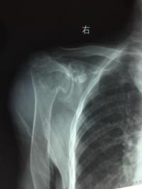
Case data in this group, 9 cases was male and 7 cases were female, with a total of 16 cases, aged from 58 to 78, with an average age of 64.4 years old. Clinical symptoms: shoulder joint pained repeatedly, clearly at night, with swelling, joint effusion and activity obstacle. The X-ray of shoulder joint showed that the head of the humerus was degenerated and hardened with osteophyte,the lower part of the glenoid cartilage was distroyed, and the joint space narrowed down (Figure 1). In the cases all the patients suffered discomfort able with right shoulder, and they were all right-handed. The shortest course was nine years, and the longest was 23 years, with an average of 16.7 years. Oral administration and local drug injection therapy was invalid for them. Because of economic reasons, the patients from the pastoral areas can't afford to have the shoulder joint replacement surgery operation.
Surgical Method
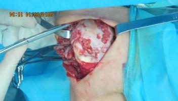
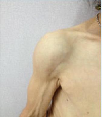
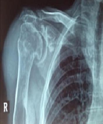
The patient maintained the beach-chair posture, the doctor made a decision along the ditch between the deltoideus muscle, protected the cephalic vein, cut the joint capsule, removed the thickened synovial membrane. Than exposed the humerus head fully (Figure2), use sharp osteotome to remove the osteophyte around the head of the humerus. Drilled on the defect of humerus head cartilage by kirschner wire with a diameter of 1mm as micro fracture treatment. Below the surface of humerus head articular using the needle with a diameter of 3 mm to drill 3-5 holes at a angle of 45 degrees to reduce the pressure of the humerus head. After repetitious irrigating, stitch joint capsule closely, placed the drainage tube, closed the incision, and let the wounded limb suspended. Half an hour before and 24 hours after the surgery conventionally giving cefazolin to prevent infection. After 24 to 48 hours if volume of drainage reduced conventionally, doctor removed the tube and guided the patients to do functional exercise,and take out stitches after 14 days. After 6 months, doctor should examine the X-ray (Figure 3) and assess shoulder function again (Figure 4).
Results
All the wounds healed primarily. Patients’ shoulder joint function was evaluated by constant score [1]. Constant score (maximum score of 100 points) Consists of four parts: Pain score (0~15 points), Daily activity score (0~20 points), Shoulder joint activity score (0~40 points)and Shoulder joint outreach power score (0~25 points). Before the surgery all the patients got the constant score as 32.35±14.78 points, and after the surgery of 12 months got the constant score as 68.95±16.78 points.
Statistical Methods
Contrasting the function score that 12 months before and after the surgery through t-test,P<0.05, the score 12 months after surgery compared with preoperative score is more statistically significant.
Discussion
Glenohumeral joint osteoarthritisis caused by the repeated friction between the head of humerus and glenoid cavity cartilage, characterized by articular cartilage degeneration, soften, peeling and gradually disappearance ,the lower layer of cartilage bone sclerosis, osteophyte formation, and trigger off arthrosynovitis, narrowing of the joint space,shoulder dysfunction. It mostly occurs in the elderly patients. The patient shows the shoulder arthralgia. Activity obstacle and extort difficultly, hyper osteophyte, obvious joint clearance narrowing, sclerosis of sub chondral bone, proximal humeral and glenoid cavity cystic changing around the head of humerus and at the margins of glenoid cavity marked osteophyte formation [2, 3]. Literature reported the occurrence of the disease is more related to the shoulder joint trauma [4], such as dislocation of shoulder, the humeral head or humeral neck fractures, rotator cuff tear and rheumatoid arthriti [5]. Sankaye [6] reported shoulder joint as a non weight- bearing joints primary osteoarthritis was uncommon. So serious progress of shoulder joint osteoarthritis without the history of trauma should raise the clinician’s alert, doctors should perfect relevant examination and rule out other diseases. Considering the author lifed in Xinjiang Yili valley at that time where all the 16 shoulder joint osteoarthritis cases in this paper came from, nomadic activities was given priority for local residents. They are more frequent to use shoulder joint in daily life than residents living inland areas. Higher-intensity bring about a high incidence of shoulder joint osteoarthritis. Although there were no reports of public literature to support the conclusion, the phenomenon was reflected in the patients who the author treated in inland areas and in Xinjiang Yili valley,and the incidence of shoulder joint osteoarthritis in Xinjiang Yili valley is indeed higher than it in Jiangsu Province. Literature reported high prevalence of shoulder joint osteoarthritis, second to knee joint and hip osteoarthritis in the United States. It is more often appeared in female,and the onset of disease tends to be younger [7]. Shoulder joint osteoarthritis is clinically classified by Walch classification [8] or the improved Walch taxonomy [9]. The classification is in accordance with the 3D scan form of patients’ glenoid cavity, which is widely used in clinical and makes some improvement. The treatment in the early stage often gives priority to oral non-steroidal anti-inflammatory analgesic drugs, combined with physical therapy and other methods. If the methods above can't improve symptoms, clinical doctors would use topical medications, mainly using hyaluronate [10] and ocorticoids [11, 12]. It was reported that therapy combined with intra-articular injection could control the onset of acute inflammation [13], Giuseppe’s [14] multi- center study showed intra-articular injection in shoulder joint of sodium hyaluronate could obviously reduce the pain of shoulder. Statistics showed that the VAS pain score of treatment group patients within 6 months decreased from 66.1mm to 46.5 mm significantly and rotary activities were improved obviously without evident complications. In the late lesion further progress the debridement of shoulder joint under arthroscopy can ease the pain in the shoulder in a minimally invasive environment, improve the function of shoulder joint,and delay the time for shoulder joint replacement [15]. Although there are clear evidence to prove that the treatment can improve the symptoms of glenohumeral joint, shoulder joint replacement is always as the last treatment choice for the serious shoulder joint osteoarthritis [16, 17]. According to the shoulder joint osteoarthritis treatment guidelines of American Osteopathic Physicians for the early physical therapy, medication, glucocorticoid drugs, minimally invasive, or open debridement of shoulder joint, the strength of recommendations is not sure; For sodium hyaluronate and shoulder joint replacement, the strength of recommendation is weak [7]. All the patients of this group were from remote mountainous area farmers and herdsmen, whose symptoms showed that the shoulder joint pained repeatedly. Loss of joint function and decline of the life quality encouraged them to see a doctor. Conservative treatment effect is poor. For economic reasons, they were unable to bear the high cost of shoulder joint replacement. The medical conditions in remote areas can not develop the arthroscopic technique. After it is invalid to use physical therapy, oral medications, sodium hyaluronate, and steroid local injections were given .We informed patients the treatment options in detail, and patients who chose the open debridement of shoulder joint to treat glenohumeral joint osteoarthritis were satisfied with functional recovery and pain remission. The sample size of cases is small, so the judgment whether this operation method curative is effect or not remains to be further summarized and follow-up visit.
References
-
Constant CR (1997) Assessment of shoulder function. In: Gazielly DF, Gleyze P, Thomas T (Eds.) The cuff 1st (Edn.) Paris: Elsevier 39-44.
-
Edwards TB, Kadakia NR, Boulahia A, Kempf JF, Boileau P et al. (2003) A comparison of hemiarthroplasty and total shoulder arthroplasty in the treatment of primary glenohumeral osteoarthritis: results of a multicenter study. J Shoulder Elbow Surg 12: 207-213.
-
Iannotti JP, Norris TR (2003) Influence of preoperative factors on outcome of shoulder arthroplasty for glenohumeral osteoathristis. J Bone Joint Surg Am 85: 251-258.
-
Carbone A, Rodeo S (2016) A review of current understanding of post-traumatic osteoarthritis resulting from sports injuries. J Orthop Res 35(3): 397-405.
-
Li Li (2013) Medical experience of shoulder joint osteoarthritis, World Latest Medicine Information 36(13): 23-24.
-
Sankaye P, Ostlere S (2015) Arthritis at the shoulder joint. Semin Musculolskelet Radiol. 19(3): 307-318.
-
Liqun Bai (2012) Interpretation of the shoulder joint osteoarthritis treatment guidelines of American Osteopathic Physicians ,Chinese medical journal Frontiers 4(11): 63-65.
-
Nowak DD1, Gardner TR, Bigliani LU, Levine WN, Ahmad CS (2010) Inter observer and intra osberver reliability of the Walch classification in primary glenohumeral arthritis. J Shoulder Elbow Surg 19(2): 180-183.
-
Bercik MJ, Kruse K, Yalizis M Gauci MO, Chaoui J et al. (2016) A modification to the Walch classification of the glenoid in primary glenohumera osteoarthritis using three-dimensional imaging. J Shoulder Elbow Surg 25(10): 1601-1606.
-
Christopher Gross, Aman Dhawan, Daniel Harwood Eric Gochanour, Anthony Romeo (2013) Glenohumeral joint injections. Sprots Health 5(2): 153-159.
-
Sascha Colen, Pieter Geervliet, Daniel Haverkamp Michel PJ Van Den Bekerom (2014) Intra-articular infiltration therapy for patients with glenohumeral osteoarthritis: A systematic review of the literature, Int J Shoulder Surg 8(4): 114-121.
-
Liang Zhao,Chunli Zhang (2012) Three cases of articular cavity injection with cut blood cream topical treatment of senile stubborn shoulder arthritis, Journal of Hubei University of Chinese Medicine 14(3): 63-64.
-
Li Li (2013) Medical experience of shoulder joint osteoarthritis,World Latest Medicine Information 36(13): 23-24.
-
Porcellini G, Merolla G, Giordan N, Paladini P, Burini A et al. (2015) Intra-articular glenohumeral injections of HYADD®4-G for the treatment of painful shoulder osteoarthritis: a prospective multicenter open-label trial. Jonits 3(3): 116-121.
-
Willian R. Mook, Maximilian Petri, Joshua A Greenspoon Peter J Millett (2015)The comprehensive arthroscopic management procedure for treatment of glenohumeral osteoarthritis. Arthrosc Tech 4(5): e435-e441.
-
Kanglai Tang,Qihong Li,Peter Habermer (2006) A new generation of artificial shoulder joint prosthetic replacement therapy, after brachial joint osteoarthritis,National Medical Journal of China 86(10): 2741-2748.
-
Michel P, Pieter C, Matthijs P, van den Borne PJ, Ronald Boer (2013) Total shoulder arthroplasty versus hemiarthroplasty for glenohumeral arthritis: A systematic review of the literature at long-term follow-up. Int J Shoulder Surg 7(3): 110-115.
- Return to Work Among Manual Workers After the Latarjet Procedure: A Cohort Study of 43 Patients
- Refractory Pelvic Collection Following Modified Stoppa Approach for Both-Column Acetabular Fracture Fixation: A Case Report
- Comparative Study of Dynamic Knee Phenotypes Under Loaded and Unloaded Conditions: Clinical Impact
- Locked Intramedullary Nailing of the Tibia Using a Humeral Nail: A Care Case Report
- Subtalar Dislocation: About a Case Report
- Surgical Site Infection in Orthopedics in a Country with LimitedResources: Indications, Treatment and Results