Primary Subcutaneous Hydatid Cyst Of The Elbow: An Exceptional Location
Hydatid disease in humans is an endemic zoonotic infestation caused by the larval form of Echinococcus species. The most commonly afflicted organs are the liver and lungs, but any tissue other than hair, nails and teeth may be involved. However the involvement of soft parts is exceptionnal, especially subcutaneous location.
Introduction
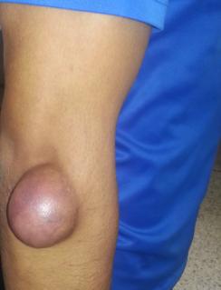
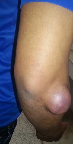
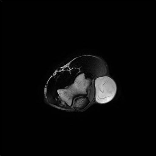
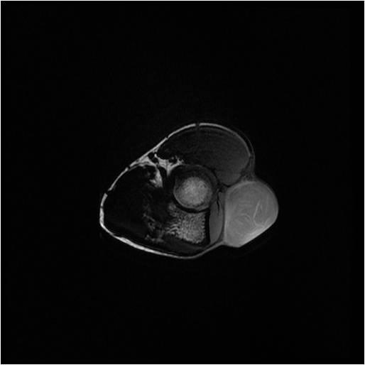
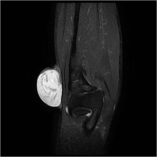
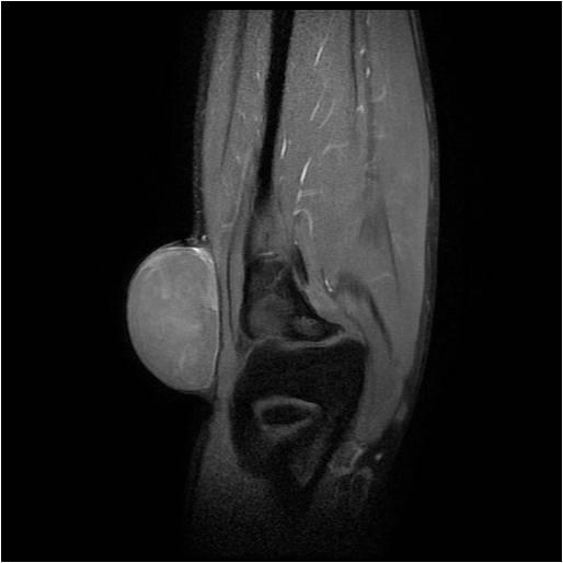
The treatment of hydatid cysts in unusual locations is challenging; because firstly the clinical presentation can be insidious and the diagnosis can be missed, and then althought the removal of the cyst combined with medical treatment is the optimal treatment choice, the risk of recurrence is important. We report a rare case of subcutaneous hydatid cyst in a 32-year-old patient having prolonged exposure to dogs. The patient presented with a painless mass on the postero-lateral aspect of the right elbow for 7 months gradually increasing in volume. On examination, there was a smooth skin-colored mass of size 4 cm × 5 cm without fistulas (Figure 1). Biological examination revealed hypereosinophilia. The patient underwent MRI that objectived a subcutaneous mass in the external aspect of the elbow hyperintense in T1 hypointense in T2 without contrast enhancement, this mass is surrounded by a hypointense capsule and contains serpiginous linear structures (Figure 2). The diagnosis of hydatid cyst type 3 was made, and the patient was operated; the lesion was excised in its entirety, macroscopic examination revealed a cystic mass. The histopathologic examination confirmed the diagnosis.
Image Article
Surgical treatment was followed by medical treatment with Albendazol 400 mg twice a day for 28 days. A thorough radiologic evaluation of other organs including standard radiography of lungs and abdominal ultrasonography didn’t detect any other focus. Clinical evolution was satisfactory with no recurrence at 10 months retreat.
a
b
c
- Return to Work Among Manual Workers After the Latarjet Procedure: A Cohort Study of 43 Patients
- Refractory Pelvic Collection Following Modified Stoppa Approach for Both-Column Acetabular Fracture Fixation: A Case Report
- Comparative Study of Dynamic Knee Phenotypes Under Loaded and Unloaded Conditions: Clinical Impact
- Locked Intramedullary Nailing of the Tibia Using a Humeral Nail: A Care Case Report
- Subtalar Dislocation: About a Case Report
- Surgical Site Infection in Orthopedics in a Country with LimitedResources: Indications, Treatment and Results