Bone Involvement in Classic and Agressive Kaposi Sarcoma
Kaposi sarcoma (KS) is an endothelial proliferation described for the first time by moritz kaposi in 1872. It is commonly associated with human herpes virus 8 (HHV8) and human immunodeficiency virus (HIV). Skin and mucous membranes are the most common sites. Osseous involvement in kaposi sarcoma is rare and occurs either by direct spread of mucocutaneous lesions or through dissemination. There are 4 different variants of KS including : African (endemic) KS, classic KS, acquired immune deficiency syndrome (AIDS)-related (epidemic) KS, and transplantation (or immunosuppression)-associated KS.
Introduction
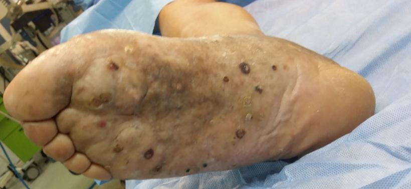
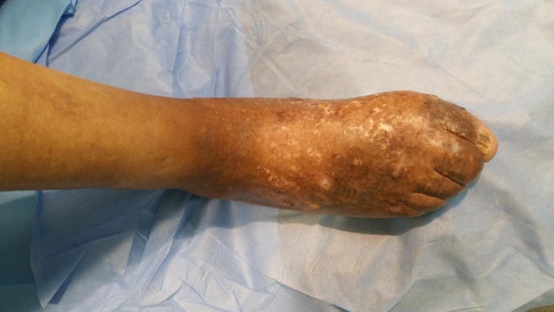
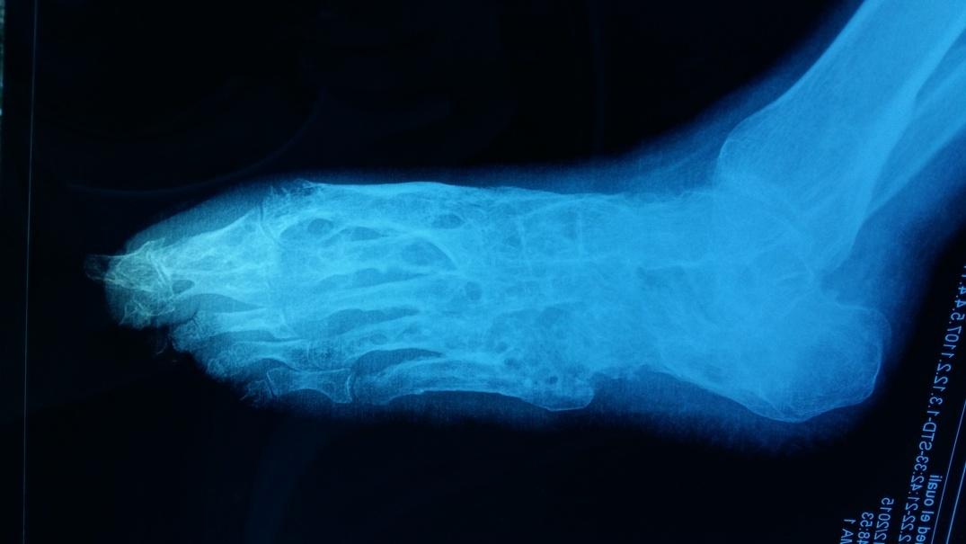
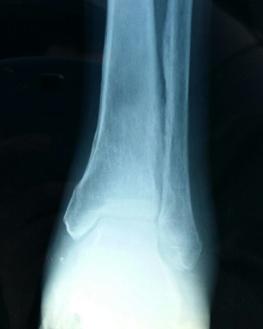
We report a case of 65-year-old male, presented with a 10-year history pain of the right foot. After 6 years of evolution, it was associated with fistulas. Physical examination revealed lymphoedema of the foot with multiple keratosic plaques and nodules, associated to multiple fistulas in dorsal and plantar aspects of the right foot (Figure 1,2). Standard radiography of the feet revealed a massive osteolysis of the right foot skeleton, there was also a Image Article periosteal reaction of the distal tibia and fibula (Figure 3, 4). A skin lesion biopsy was performed, the histopathologic examination confirmed the diagnosis of KS. HIV and Syphilis serology tests were negative. General examination didn’t reveal other locations. The decision of transtibial amputation was made, immunohistochemistry examination showed a dermal nodular proliferation with few mitoses.
- Return to Work Among Manual Workers After the Latarjet Procedure: A Cohort Study of 43 Patients
- Refractory Pelvic Collection Following Modified Stoppa Approach for Both-Column Acetabular Fracture Fixation: A Case Report
- Comparative Study of Dynamic Knee Phenotypes Under Loaded and Unloaded Conditions: Clinical Impact
- Locked Intramedullary Nailing of the Tibia Using a Humeral Nail: A Care Case Report
- Subtalar Dislocation: About a Case Report
- Surgical Site Infection in Orthopedics in a Country with LimitedResources: Indications, Treatment and Results