Glomic Tumors of the Finger: Diagnostic and Therapeutic Approach (About 24 Cases)
Glomus tumor according to MASSON is a benign neuro-myo-arterial proliferation. It accounts for approximately 1%-5% of all hand tumors. Pain is the main clinical sign. The diagnosis of certainty is based on a bundle of arguments: clinical and radiological, but only histology allows confirmation. A series of 24 patients was reviewed retrospectively, the average age was 35 years with an average follow-up of 4 years , and extremes of 21 and 60 years. Surgical excision was performed in all patients. This strategy allowed us to obtain satisfactory results.
Introduction
Glomus tumors of the hand correspond to hyperplasia of the glomus apparatus of the fingers, characterized by the presence of an arteriovenous anastomosis which controls the local temperature. We report through this work the experience of the Traumato-Orthopedics department of the Ibn Sina Hospital in Rabat in the management of glomus tumors of the nail region from 24 cases reviewed with a 4-year follow-up.
Material and Results
This is a retrospective study of 24 patients operated on for GT of the nail region between 2013 and 2019 with an average follow-up of 4 years. The female predominance was clear with 75% of cases. The average age of our patients was 35 years old with extremes ranging from 21 to 60 years old. The average consultation period was 3 years. The tumor was located most often in the subungual in 18 patients, in 4 patients in the latero-pulpal and pulpal in 2 patients. The thumb was affected 10 times, the index 6 times, the middle finger 4 times, the ring finger and the little finger respectively twice each. Both sides were affected equally. Pain was the main symptom, found in all our patients, most often triggered by shock. Thermosensitivity was found in 67% of patients. Love’s test was positive in 22 of the patients. As for the physical signs, in the subungual localizations, 16 out of 18 patients had a bluish or reddish spot. In the pulpal localization, 4 patients had a small bluish zone and one patient a hard swelling. At the end of the clinical examination, the preoperative diagnosis was affirmed in all our patients. Systematic standard radiography found a regular notch on the distal phalanx in 10 patients.
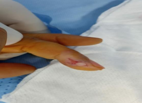
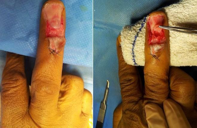
All of our patients received surgical treatment. For pulp or lateral pulp tumours, the approach was direct (Figure 1). For the subungual location, the approach was transungual (Figure 2) in 8 cases where the location was under the central ungual. The 10 other patients benefited from a periungual approach mainly motivated by their rather lateral location. The excision of the tumor was complete, the diagnosis had been confirmed by the histological study.
The immediate postoperative consequences were simple. Healing with functional recovery was obtained in 3 weeks on average after the peri-ungual approach and 5 weeks on average in the case of a trans-ungual approach, the regrowth of the normal nail after this approach required 6 to 8 months depending on the proximity of the injury to the matrix. We did not note any nail dystrophy, whether the approach was trans or peri-ungual, nor any recurrence at 4 years’ follow-up.
Discussion
The first description of a glomus tumor of the hand `was made by Wood and Dimmick in 1812 [1, 2] in the form of “painful subcutaneous nodules”, and it was not until 1924 that Masson [3] clarified its origin and gave the name of glomus tumor. It is a benign tumor formed at the expense of the neuromyoarterial tissue sitting at the level of the capillary- venous regulation anastomoses, particularly numerous at the level of the digital extremities. These tumors are rare, but not exceptional [4], their frequency varies from 1.6 to 5% of all tumors of the soft tissues of the hand [5, 6].
The female predominance reaches 82% of cases. The typical localization is dermal in order of frequency the nail bed, then the palmar surface of the fingers [7] especially at the level of the thumb, on the other hand the localization at the level of the 5th finger is not frequent [1].
Pain is the main reason for consultation in all series [8, 9, 10, 11], it can be spontaneous, exacerbated by cold, throbbing or even disabling, caused by contact or even the slightest touch that triggers violent pain [8, 9, 10, 11]. The tumor only becomes visible or palpable after a long evolution [9]. Local signs are noted three times out of four, in the form of a spot of a few millimeters that appears bluish or reddish, rounded or oval [12]. Three tests have great diagnostic value:
- The cold water immersion test [8] which triggers a paroxysm of pain when the patient plunges their hand into cold water
- The Love Test [13] which awakens pain on localized pressure by applying a pencil tip
- The Hildreth ischemia test [14], pathognomonic of the glomus tumour, based on the disappearance of pain in the tumor area by the creation of an ischemia of the order of 15 seconds with an inflated cuff at the root of the limb around 300 mm Hg.
Arteriography, which is not very sensitive, is of very limited use [10]. It may reveal a cluster of arterio- capillary hypervascularization with sharp borders which is characteristic, with sharp borders [15]. The digitalized video angiography can highlight a late vascular lake typical of the glomus tumor and allows to judge its extent. Some authors recommended it in cases where localization was imprecise or in the event of recurrence, but it could give non-characteristic images [16]. High-resolution MRI shows a hyposignal in T1 and a hypersignal in T2 [17].
The long delay in the diagnosis of glomus tumors is explained by the rarity of the pathology and the lack of information. You have to think about it and the clinical examination makes it possible in most cases to diagnose them. Excision of the tumor is always immediately effective. Recurrences are generally due to incomplete excision, hence the interest of operating on patients without emptying the limb and under magnifying glasses. It is also important to locate the lesions precisely by high-resolution MRI after the preoperative injection of gadolinium. Indeed, MRI can reveal lesions nearly 2 mm in diameter, thus constituting the best means of diagnosis of both glomus tumors that are difficult to locate, as well as recurrent forms [1].
Conclusion
The glomus tumor is a rare but not exceptional benign tumor that is often overlooked by practitioners. The diagnosis is mainly clinical and the treatment is exclusively surgical, the anatomopathological examination confirms the diagnosis. Recurrence is rare if the excision was complete [18, 19].
References
-
Amirat L, Youcefi MFA, Bougueni YSI (2017) Tumeur glomique du 4e, 5e doigt (A propos d’un cas). Hand Surgery and Rehabilitation 36(4): 475.
-
Wood WS, Dimmick JE (1977) Multiple infiltrating glomus tumors in children. Cancer 40(4): 1680- 1685.
-
Le MP (1924) Glomus neuromyo-artériel des régionstactiles et ses tumeurs. Lyon Chir 2: 257-280.
-
Raimbeau G, Mallet J, Fondimare A, Tirouflet D (1984) Tumeurs glomiques des doigts (à propos de 6 cas personnels). Ann Orthop Ouest 16: 85.
-
Glicenstein J, Ohana J, Leclercq C (1988) Tumeurs de lamain. Berlin: Springer Verlag pp: 143-149.
-
Vandevender DK, Daley RA (1995) Benign and malignant Vascular tumors of the upper extremity. Hand Clin 11(2): 161-181.
-
Bureau H, Jouglard PJ, Thion A, Tramier H, Pierre M (1978) Tumeurs glomiques. In: L’ongle. Monographie du GEM Paris Expansion scientifique franc aise pp: 102- 106.
-
Mansat M, Bonnevialle P, Gay R, Urroux R (1985) Tumeurs glomiques de la main. À propos de quatorze cas. Ann Chir Main 4: 43-50.
-
Carroll RE, Berman AT (1972) Glomus tumors of the hand: review of the literature and report on twenty- eight cases. Bone Joint Surg 54(4): 691-703.
-
Dupuis P, Pigeau I, Ebelin M, Barbato B, Lemerle JP (1994) Apport de l’IRM dans l’exploration des tumeurs glomiques. Ann Chir Main Memb Super 13(3): 358-362.
-
Van Geertruyden J, Lorea P, Goldschmidt D, De Fontaine S, Schuind F, et al. (1996) Glomus tumors of the hand. A retrospective study of 51 cases. J Hand Surg Br 42(3): 295-301.
-
Baran RL, Dawber RR (1995) Les traitements chirurgicaux: Tumeur glomique. In: Guide médicochirurgical des onychopathies. Paris: Arnette Blackwell pp: 78.
-
Love JG (1944) Glomus tumors: diagnosis and treatment. Proc Staff Meet Mayo Clin 19: 113-116.
-
Hildreth DH (1970) The ischemia for glomus tumor: a new Diagnostic testRev Surg 27: 147-148.
-
Boudjemaa B, Glock Y, Boccalon H, Ginestet MC, Puel P, et al. (1987) Tumeur glomique sous unguéales, à propos de 2 cas. Arch Mal CoeurVaiss 80: 227-230.
-
Mantero R, Auxilia E, de Albertis P, Ferro C (1984) Vidéoangiographie digitalisée de la main. Ann Chir Main 3: 160-164.
-
Drapé JL, IdyPeretti I, Goettmann S, Guérin-Surville H, Bittoun J (1996) Standard and high resolution magnetic resonance imaging of glomus tumors of toes and fingertips. J Am AcadDermatol 35(5): 550- 555.
-
Sorene ED, Goodwin DR (2001) Magnetic resonanceimaging of a tiny glomus tumour of the fingertip: a case report. Scand J Plast Reconstr Surg Hand Surg 35(4): 429-431.
-
McDermott EM, Weiss AP (2006) Glomus tumors. J HandSurg Am 31(8): 397-400.
- Return to Work Among Manual Workers After the Latarjet Procedure: A Cohort Study of 43 Patients
- Refractory Pelvic Collection Following Modified Stoppa Approach for Both-Column Acetabular Fracture Fixation: A Case Report
- Comparative Study of Dynamic Knee Phenotypes Under Loaded and Unloaded Conditions: Clinical Impact
- Locked Intramedullary Nailing of the Tibia Using a Humeral Nail: A Care Case Report
- Subtalar Dislocation: About a Case Report
- Surgical Site Infection in Orthopedics in a Country with LimitedResources: Indications, Treatment and Results