Regeneration of Cartilage Tissue by Comparing the Natural and Synthetic Biomaterials during ADSCs Differentiation
Objectives: Human adipose-derived stem cells have shown chondrogenic differentiation for cartilage engineering. In the present study, we evaluated different scaffolds for chondrogenic differentiation of Human adipose-derived stem cells. Methods: cells were cultured and chondrogenic differentiation was investigated in Polycaprolactone, Platelet Rich Plasma, and Fibrin glue scaffolds by MTT, Real time and histology. Results: Cell proliferation and collagen levels in Platelet Rich Plasma scaffolds were significantly higher than other scaffolds. The levels of Sox9 and col II were up regulated in Platelet Rich Plasma. Histological examination revealed that the pores of the scaffolds were filled cells. Conclusion: The results show that 3D Platelet Rich Plasma scaffolds provide a better environment for chondrogenic differentiation than others.
Introduction
There is a complex dynamic biological process, and in this process, homeostasis, inflammation, proliferation and remodeling help toward the improvement of organs. Cell types, extracellular components, growth factors and cytokines are involved [1]. The scaffolds have become necessary to facilitate in vivo and in vitro tissue healing and reconstruction, to provide the appropriate substrate for migration, cell attachment, growth and differentiation, and for the delivery carriers and replacement of new tissue [2]. One of the sources of mesenchymal stem cells (MSCs) is adipose-derived stem cells (ADSCs), which have some advantages. These can be obtained through invasive methods from abdominoplasty surgeries [3]. Their potential use has been demonstrated in in vitro [4], in vivo [5] and clinical experimentation [6]. ADSCs show pluripotent stem cell characteristics and have been identified by tissue culture plastic adhesion, self-renewal, proliferation in vitro, differentiation potential [7], cell surface expression of CD73, CD90 and CD105 [8]. Different categories of biomaterials are emerging for tissue engineering and regenerative medicine [9]. Advantages of natural biomaterials include high biocompatibility, ease of obtaining and availability, mimicry of the natural extracellular matrix of tissues, and non-toxicity [10]. Disadvantages are rapid degradation in vivo and poor mechanical properties [11]. Synthetic polymers are used for tissue engineering because of controllable fabricating and physical trait [12]. Positive effects of these biomaterials are good biodegradable and biocompatible [13]. Negative
effects are inflammatory response possible, poor cell adhesion, poor compression strength [14]. Platelet-rich plasma (PRP) has recently appeared as a natural hydrogel construct for injectable soft-tissue augmentation [15]. PRP can form a gel-like 3D construct to carry seeded cells. Some of the studies evidenced that PRP has appreciated for tissue engineering as an autologous injectable carrier [16]. PRP has the significant advantages: 1) autologous PRP is safe, it prevents disease- transfer and potential immunogenicity [17]; there are not any complications about PRP such as carcinogenesis or tumor growth; 2) moderate distribution of BMSCs in PRP is presented by scanning electron microscopy (SEM) examination [18]; 3) Cell migration, proliferation and differentiation are reinforced by native autologous growth factors in PRP. This hydrogel can protect nutrition for pre- seeded and recently migrated cells. Thus, in this study, we decided to design appropriate constructs for regeneration of cartilage tissue by comparison of natural and synthetic biomaterials. The purpose of this study is to accelerating more rapid advances in the use of biomaterials in pre-clinical researches and clinical therapies.
Materials & Methods
Isolation of hADSCs
This study was confirmed by the academic center for education, culture and research- Qom branch ethics committee (No1395-23). All patient studied and signature the informed consent. Human ADSCs were isolated from adipose tissue of 4 patients (After consulting with a statistical and epidemiologic consultant) during an abdominal simple surgery procedure (Nikan Hospital). The adipose tissue was collected and transferred to the laboratory in saline containing 1% antibiotic (penicillin-streptomycin) (Sigma, Germany). Samples were rinsed with sterile phosphate buffer saline (PBS) (Sigma, Germany), sliced into small pieces and digested with type I collagenase for 45-60 minutes at 37°C. After neutralization, isolated cells were cultured in Dulbecco’s Modified Eagle’s medium (DMEM (containing penicillin-streptomycin and 10% FBS (Sigma, Germany) and incubated at 37°C with 5% CO2 and 95% humidity.
Assay of Cellular Surface Marker
In monolayer culture, hADSCs were analyzed by the expression of cellular surface marker of CD44, CD90, CD105 and CD34 after third passage using flow cytometry technique. At first, hADSCs were trypsinized, rinsed and fixed with phosphate-buffered saline twice and 4% paraformaldehyde respectively. After washing again, cells suspended in PBS/ antibody.
Cell fluorescence was evaluated by flow cytometry using a FACS Calibur instrument (Becton Dickinson).
Fabrication of PCL-based Scaffolds
Poly (ɛ-caprolactone) (PCL) (Aldrich, USA; Mw= 80,000 g/mol) particles were dissolved in methylene chloride (CH2CL2) prior to mixing with NaCl. A plastic mold, 3mm in height and 6mm in diameter were then filled with the polymer/NaCl particles and frozen at -20˚С. The frozen polymer scaffold was placed in sterilized de-ionized water for 2 days to remove the particles. Finally, samples were sterilized by using UV. Cultivated hADSCs were trypsinized from the secondary passage (P2), and seeded onto the scaffolds.
Fabrication of Fibrinogen and Thrombin
Fresh-frozen plasma and fibrinogen were obtained from the Qom blood transfusion organization. Fresh-frozen plasma was added to calcium gluconate at 5:3 ratios, incubated for one hour at 37°C and centrifuged at 220 rpm for 10 min. After centrifugation, the supernatant was discarded and thrombin was separated. Fibrinogen and thrombin were then prepared for cell culture. For this purpose, ADSCs were converted to a soluble form at the concentration of 5×10⁶ cells in 1cc of thrombin, and fibrinogen was added to the solution. The solution was incubated at 37° C/5% CO2/99% humidity in expansion medium (EM; DMEM-HG supplemented with 50 mg/ml bovine serum albumin, 5 µg/ ml ascorbate-2-phosphate, 1% insulin-transferrin-selenium, 1 nM dexamethasone, 5 µg/ml linoleic acid, 1% penicillin- streptomycin and 10 ng/ml TGF-β1) for 14 days.
Preparation of Platelet-Rich Plasma (Prp)
PRP was prepared by placing peripheral blood into silicone tubes containing 3.8% sodium citrate at ratio 9.1 and centrifuging at 1000 rpm for 5 min. The number of platelets was determined by the sysmex counter system. The obtained product is usually divided into 3 layers, the under layer consists of erythrocytes; the middle layer contains leukocytes and inflammatory cytokines; and the top layer content plasma, platelets and growth factors. The PRP was then activated using 10% CaCl2 solution.
Chondrogenic-MTT Assay
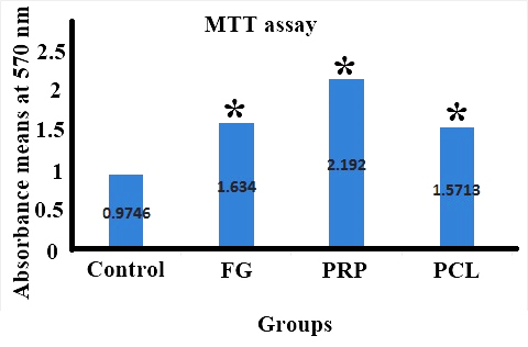
The numbers of viable hADSCs attaching to the scaffolds were evaluated using an MTT assay. hADSCs (at a density of 500,000 cells/scaffolds) were cultured on the scaffolds in chondrogenic medium and incubated at 37°C in 5% CO2. After 14 days, all of the samples were incubated with an MTT solution for 4h at 37°C and dimethylsulfoxide (DMSO, Sigma, USA) was added to evaluate the formation of formazan crystal. Finally, the optical density was assessed at 570 nm using an ELISA (plate reader) (biokit, ELx800 READER, Spain)
Chondrogenic Differentiation of hADSCs
After three passages, ADSCs were obtained and seeded on to scaffolds at cell density 1×10⁶ cells/ml. All of the scaffolds were incubated in chondrogenic medium containing of DMEM by great Glucose supplemented with 50 mg/ml bovine serum albumin, 5 µg/ml ascorbate-2-phosphate,1 nM dexamethasone, 1% insulin-transferrin-selenium, 5 µg/ ml linoleic acid, 1% penicillin-streptomycin (Sigma-Aldrich) and 10 ng/ml TGF-β1) for 2 weeks. The culture medium was changed every 2-3 days.
Real time-PCR for Evaluating the Expression of Chondrogenic Genes
Fourteen days after chondrogenic differentiation, RNA samples were isolated from each scaffold, separately. The scaffolds were digested in fluid nitrogen. Extraction of RNA and reverse transcription of RNA to cDNA was performed using AccuZol™ Total RNA Extraction Solution (Bioneer) and AccPower®RT Premix (Bioneer), respectively, according to manufacturer’s instructions. Real time-PCR performance using SYBRGreen PCR Master Mix (Amplicon) and Rotor Gene 6000 Real-Time PCR Machine (Corbett Life Science, Australia), and primers of each gene were designed as follows utilizing primer 3 program that it’s primer sequences is mentioned in Table 1.
The reaction was initiated by heating to 95°C for 15 min., followed by 40 cycles of elongation at 59°C for 30 sec and denaturation at 95°C for 15 sec. The mRNA expression level of Target gene was normalized based on GAPDH housekeeping gene as reference. The level of expression of every target gene was assessed using 2−ΔΔCt.
| Primer Name | Sequences (5’ -> 3’) | Melting Temperature (Tm) | The Genbank (Refseq) accession number |
|---|---|---|---|
| collagen, type I, alpha 1 (COL1A1) Forward | TGCTAAAGGTGCCAATGGT | 19 | XM_011524341.1 |
| collagen, type I, alpha 1 (COL1A1) Reverse | ACCAGGTTCACCGCTGTTAC | 20 | |
| Aggrecan (ACAN) Forward | GAATCAACTGCTGGAGACCA | 20 | XM_011521314.1 |
| aggrecan (ACAN) Reverse | CCACTGGTAGTCTTGGGCAT | 20 | |
| SRY-box 9 (SOX9) Forward | AGTACCCGCACTTGCACAAC | 20 | NG_012490.1 |
| SRY-box 9 (SOX9) Reverse | CGTTCTTCACCGACTTCCTC | 20 | |
| collagen, type II, alpha 1 (COL2A1) Forward | CTCCTGGAGCATCTGGAGAC | 20 | XM_011537935.1 |
| collagen, type II, alpha 1 (COL2A1) Reverse | GTCTCACCACGATCACCCTT | 20 | |
| collagen type X alpha 1 (COL10A1) Forward | AGCTTCAGAAAGCTGCCAAG | 20 | NG_021351.1 |
| collagen type X alpha 1 (COL10A1) Reverse | TAGTGGGCCTTTTATGCCTG | 20 | |
| GAPDH gene Forward | GAGTCAACGGATTTGGTCGT | 20 | KJ896860.1 |
| GAPDH gene Reverse | GACAAGCTTCCCGTTCTCAG | 20 |
Table 1: Primer sequences used for Real-time PCR.
Histological Evaluation
At 14 days, samples of ADSCs which were seeded on fibrin glue, PRP and PCL scaffolds were prepared for histological analyses. After fixation with 10% neutral-buffered formalin for at least 24 h, specimens (N = 4) were inculcated within paraffin blocks with a microtome and sectioned at 5 μm thickness. To assess tissue morphology, sections were deparaffinized, rehydrated and stained with Hematoxylin and Eosin (H&E).
Immunohistochemical Staining
The consecutive specimens (4 mm) were done from the tissue samples vertically throughout the exposed chondrocytes where the plane of the sampling was perpendicular to the primary incision line. The sections were incubated with anti-collagen type II (specific for chondrocyte) antibody (Collagen II (ab6308), Abcam, Tokyo, Japan); anti- Aggrecan antibody (Aggrecan; [sc-146], Santa Cruz, CA, USA) at room temperature for 1 h, then washed with PBS. Finally, All the photographs were taken at the same scale. The green color ratios in the groups were evaluated using a histogram tool (mean intensity value of green in pixels) of the software, Adobe Photoshop v7.0.
Statistical Analysis
To evaluate difference between scaffolds, Statistical analysis was performed by using Least Significant Difference (LSD) and SPSS (Statistical Package for the Social Sciences) software (version 17). The differences at p < 0.05 were considered significant.
Results
hADSCs Characterization
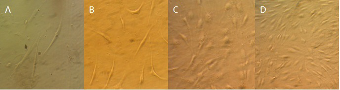
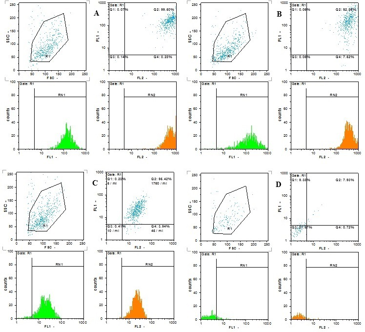
Human ADSCs are identified by spindle shape, fibroblast-like morphology in the primary culture (Figure 1). Homogeneous cell population was determined at passage 3. In our study, hADSCs expressed specific surface markers including CD44 (Hyaloronate receptor), CD90 (Thy-1) and a CD105 (Endogelin) are more than %90. Also, they confirmed with low levels of CD34 (Leucocyte common antigen) (Figure 2).
In Vitro Evaluation of hADSCs on PRP, FG and PCL
The hADSCs have the capability to attach on 3 scaffolds that evaluated by MTT assay (Figure 3).
PRP and FG structure represented a higher number of attached hADSCs compared to control group and PCL structure (Figure 2).
Quantitative RT- PCR
The chondrocyte genes expression has been shown in (Figure 4). Β-actin was considered as the house keeping gene.
Three positive markers for chondrogenesis consist of SOX-9, collagen type II and aggrecan certified by the significant difference in PRP and FG scaffolds compared with the control group. The level of collagen type I mRNA decreased remarkably in PRP scaffold. As a negative marker for chondrogenesis, the mRNA level of collagen type X was reduced significantly in all groups (P<0.05).
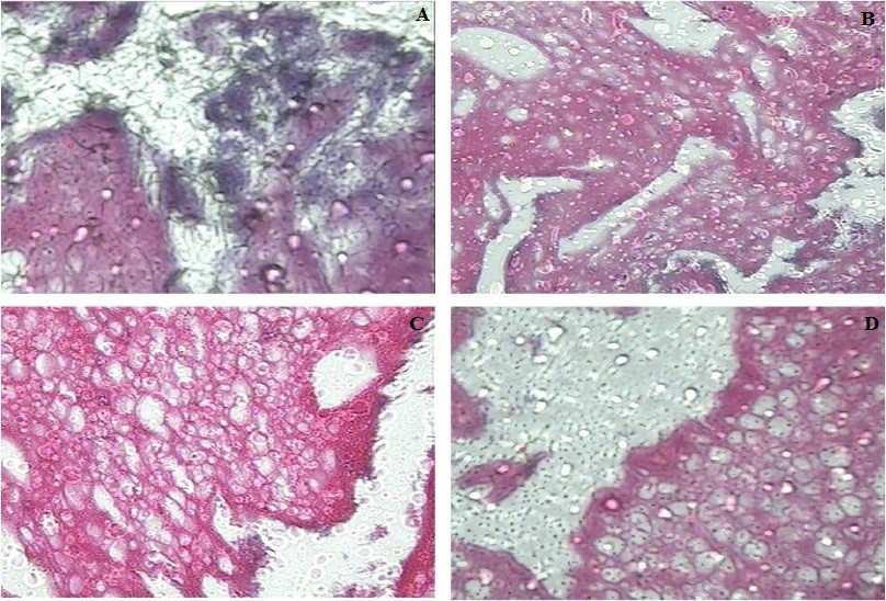
Immunohistochemistry and Histological Evaluation
Upon immunohistochemical analysis, ECM components accumulation within the construct indicated the presence of type II collagen. This marker was monotonically spread in PRP scaffold and FG and PCL shown less homogenous (Figure 5). Also, two weeks after chondrogenic differentiation, microscopic evaluation of the ADSC + PRP group showed well-formed chondrocyte surfaces, but other scaffolds included chondrocyte weak formation (Figure 6).
![Figure 4: Effect of different scaffold on chondrogenic gene expression. After 14 day of chondrogenic differentiation, gene expression was separately assayed for AD-MSCs seeded on the different scaffold. Data are expressed as mRNA expression for Aggrecan [A], Collagen II (Col2) [B], SOX9 [C], Collagen X (Col10) [D], and Collagen II (Col2) [E] genes (ng/μl) relative to β-Actin (ng/μl). Values are expressed as mean ± SD of triplicate assays. *p<0.05; **p <0.01. The results showed significant difference between the PRP and FG scaffolds compared control and PCL scaffolds.](/fulltextimages/10014/fig_4.png)
Figure 4: Effect of different scaffold on chondrogenic gene expression. After 14 day of chondrogenic differentiation, gene expression was separately assayed for AD-MSCs seeded on the different scaffold. Data are expressed as mRNA expression for Aggrecan [A], Collagen II (Col2) [B], SOX9 [C], Collagen X (Col10) [D], and Collagen II (Col2) [E] genes (ng/μl) relative to β-Actin (ng/μl). Values are expressed as mean ± SD of triplicate assays. *p<0.05; **p <0.01. The results showed significant difference between the PRP and FG scaffolds compared control and PCL scaffolds.
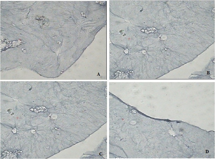
The similar results obtained for aggrecan in all of the scaffolds. Also, two weeks after chondrogenic differentiation, microscopic evaluation of the ADSC + PRP group showed well-formed chondrocyte surfaces, but other scaffolds included chondrocyte weak formation (Figure 6).
Discussion
At present time, there are many biomaterials to U.S. Food and Drug Administration (FDA) to apply in regeneration medicine. In this study, we investigated three constructs of natural and synthetic materials for the production of 3-D structures related to cartilage repair. We have been shown that the expression of Collagen I, II, X, Aggrecan and SOX9 significantly changed in the PRP scaffold. Thus, the development of tissue in the gel structure showed excellent results compared to the synthetic polymer. Many studies supported our results and demonstrated that the implantation of natural and synthetic materials can repair cartilage lesions by absorbing the bone marrow-derived MSCs [19]. Matsunaga, et al., described a novel protocol to prepare a Platelet-Rich Fibrin Scaffold (PRFS) resembling a sheet and illustrated that the healing effect of that on tendons and ligaments. But studies of Barbon S in 2019 showed that plenty of benefit observed in PRF over PRP as a result, postponed GFs and cells diffuse profiles, such as the denser fibrin network than it which permit for more comfortable handling and suturing, also slower degradation speed after utilization [20]. Moreover, this blood-derived membrane is enriched with leukocytes, which play a key role not only in immune and antibacterial responses but also in the wound healing process [21]. These scaffolds will be able to provide suit adhesion and differentiation of stem cells [22]. Ghiasi, et al. in same study compared five scaffolds containing: alginate, poly lactic-co-glycolic acid, fibrin glue, inactive platelet-rich plasma, and active platelet-rich plasma (APRP) and reported that the APRP is better than others [23]. These productions are effective in localized inflammation and cell death. Fibrin gel prepared from patients’ blood and can be applied as biological matrices and autologous scaffold, without the potential risk of a foreign body reaction. Fibrin gel can regulate the matrix synthesis via the secretion of platelet- derived growth factors and the transforming growth factor beta that this result is indicated through in vitro studies [24]. PRP is considered as a biological tool in regenerative medicine. It is shown that PRP has critical growth factors and mediators of tissue reconstruction pathways and caused, including proliferation and differentiation of stem cells in vitro and contained strong growth factors such as TGF-β and vascular endothelial growth factor. PRP (containing platelets and leukocytes) has antimicrobial effects which can decrease the hazard of graft infection. The formation of new cartilage has identified after implantation of ADSCs /FG in vivo experiment. This practice has confirmed that ADSCs / FG can repair the knee osteochondral defects in rabbit [25]. Another investigation has noted that the presence of PRP acts as an increasing element in expression of specific genes for ADSCs differentiation [24]. In 2006, Akeda et al. Found that PRP increase porcine chondrocyte proliferation and matrix Kocaomer, et al. and Vogel et al. identified that PRP can induce MSCs proliferation [26]. In 2003, one study reported that 10% PRP enhances growth of BMSCs [27]. By this approach, we accelerate more rapid advances in the use of biomaterials in pre-clinical researches and clinical therapies. In our study, an upregulation in chondrogenic-related genes (aggrecan, collagen type II and Sox9) was noted in differentiated ADSCs within the platelet-rich plasma and fibrin gels. The study finds that cell proliferation, and collagen levels were significantly higher in platelet-rich plasma (PRP) scaffolds than in other scaffolds. Also, transcript levels of SOX9 and type II collagen were upregulated in PRP group. Because ADSCs have higher expression of cartilage-specific genes (Collagen type II, Aggrecan and Sox9 genes) in PRP scaffold than other scaffolds, this result suggests that PRP scaffold is better than other scaffolds in terms of cartilage-specific gene expression.
Conclusion
The results suggest the three-dimensional PRP scaffolds provided an improved environment for chondrogenic differentiation compared to other scaffolds.
Acknowledgements
• Not applicable.
Conflict of interest
• Not applicable.
References
-
Cox TR, Erler JT (2011) Remodeling and homeostasis of the extracellular matrix: implications for fibrotic diseases and cancer. Dis Model Mech 4(2): 165-178.
-
Metcalfe AD, Ferguson MW (2007) Tissue engineering of replacement skin: the crossroads of biomaterials, wound healing, embryonic development, stem cells and regeneration. J R Soc Interface 4(14): 413-437.
-
Ghiasi M, Farzaneh S, Bigdelo M. Assessment of human cartilage regeneration in a patient with knee osteoarthritis using autologous adipose-tissue- derived stem cells and Platelet-rich plasma: a case study. Journal of Surgery and Trauma. 2020;8(2):73-8.
-
Baraniak PR, McDevitt TC (2010) Stem cell paracrine actions and tissue regeneration. Regen Med 5(1): 121- 143.
-
Chang KA, Lee JH, Suh YH (2014) Therapeutic potential of human adipose-derived stem cells in neurological disorders. J Pharmacol Sci 126(4): 293-301.
-
Grayson WL, Bunnell BA, Martin E, Frazier T, Hung BP, et al. (2015) Stromal cells and stem cells in clinical bone regeneration. Nat Rev Endocrinol 11(3): 140-150.
-
Bunnell BA, Flaat M, Gagliardi C, Patel B, Ripoll C (2008) Adipose-derived stem cells: isolation, expansion and differentiation. Methods 45(2): 115-120.
-
Ghiasi M, Mehdizadeh M, Khatib shad L (2022) Designing Nanofiber Multilayer Composite Scaffolds and Lyophilized Blood Growth Factors in the Process of Osteogenesis. J Mazandaran Univ Med Sci 32(210): 1-12.
-
Horst OV, Chavez MG, Jheon AH, Desai T, Klein OD (2012) Stem cell and biomaterials research in dental tissue engineering and regeneration. Dent Clin North Am 56(3): 495-520.
-
Mano JF, Silva GA, Azevedo HS, Malafaya PB, Sousa RA, et al. (2007) Natural origin biodegradable systems in tissue engineering and regenerative medicine: present status and some moving trends. J R Soc Interface 4(17): 999-1030.
-
Croisier F, Jérôme C (2013) Chitosan-based biomaterials for tissue engineering. European Polymer Journal 49(4): 780-792.
-
Dhandayuthapani B, Yoshida Y, Maekawa T, Sakthi D (2011) Polymeric Scaffolds in Tissue Engineering Application: A Review. International Journal of Polymer Science Article ID: 290602-290620.
-
Farraro KF, Kim KE, Woo SL, Flowers JR, McCullough MB (2014) Revolutionizing orthopaedic biomaterials: The potential of biodegradable and bioresorbable magnesium-based materials for functional tissue engineering. J Biomech 47(9): 1979-1986.
-
Butterfield TA, Best TM, Merrick MA (2006) The dual roles of neutrophils and macrophages in inflammation: a critical balance between tissue damage and repair. J Athl Train 41(4): 457-465.
-
Kretlow JD, Young S, Klouda L, Wong M, Mikos AG (2009) Injectable biomaterials for regenerating complex craniofacial tissues. Adv Mater 21(33): 3368-3393.
-
Ba R, Wei J, Li M, Cheng X, Zhao Y, et al. (2015) Cell- bricks based injectable niche guided persistent ectopic chondrogenesis of bone marrow-derived mesenchymal stem cells and enabled nasal augmentation. Stem Cell Res Ther 6: 16.
-
Li H, Li B. (2013) PRP as a new approach to prevent infection: preparation and in vitro antimicrobial properties of PRP. J Vis Exp (74): 50351.
-
Giannotti S, Trombi L, Bottai V, Ghilardi M, D’Alessandro D, et al. (2013) Use of autologous human mesenchymal stromal cell/fibrin clot constructs in upper limb non- unions: long-term assessment. PLoS One 8(8): e73893.
-
Maia FR, Carvalho MR, Oliveira JM, Reis RL (2018) Tissue Engineering Strategies for Osteochondral Repair. Adv Exp Med Biol 1059: 353–371.
-
Barbon S, Stocco E, Macchi V, Contran M, Grandi F, et al. (2019) Platelet-Rich Fibrin Scaffolds for Cartilage and Tendon Regenerative Medicine: From Bench to Bedside. Int J Mol Sci 20(7): 1701.
-
Fioravanti C, Frustaci I, Armellin E, Condò R, Arcuri C, et al. (2016) Autologous blood preparations rich in platelets, fibrin and growth factors. Oral Implantol 8(4): 96-113.
-
Ghiasi M, Farzaneh S, Bigdelo M, Vosoogh M (2021) The Effects of Allogeneic cADSCs on an Experimental Ear Auricular Defect to Evaluate Cartilage Regeneration in a Canine Model. Jour Clin Med Res 2(1) :1-11
-
Ghiasi M, Kalhor N, Tabatabaei Qomi R, Sheykhhasan M (2016) The effects of synthetic and natural scaffolds on viability and proliferation of adipose-derived stem cells, Frontiers in Life Science 9(1): 32-43.
-
Ghiasi M, Tabatabaei Qomi R, Kalhor N, Mehdizadeh M, Sheykhhasan M (2015) Survival Potential Investigation of the Adipose-Derived Mesenchymal Stem Cell in the Natural Scaffolds as a Suitable Growth Medium. JCT 6(1): 23-29.
-
Ghiasi M, Mehdizadeh M, Taghi Joghataei M (2022) Transplantation of Osteochondral Acellular graft/ Adipose derived stem cells/Fibrin glue for Reconstructing of Knee Defects in rabbit. PAS, 43.
-
Akeda K, An HS, Okuma M, Attawia M, Miyamoto K, et al. (2006) Platelet-rich plasma stimulates porcine articular chondrocyte proliferation and matrix biosynthesis. Osteoarthritis Cartilage 14(12): 1272-1280.
-
Liu F, Xu H, Huang H (2019) A novel kartogenin-platelet- rich plasma gel enhances chondrogenesis of bone marrow mesenchymal stem cells in vitro and promotes wounded meniscus healing in vivo. Stem Cell Res Ther 10(1): 201.
- Return to Work Among Manual Workers After the Latarjet Procedure: A Cohort Study of 43 Patients
- Refractory Pelvic Collection Following Modified Stoppa Approach for Both-Column Acetabular Fracture Fixation: A Case Report
- Comparative Study of Dynamic Knee Phenotypes Under Loaded and Unloaded Conditions: Clinical Impact
- Locked Intramedullary Nailing of the Tibia Using a Humeral Nail: A Care Case Report
- Subtalar Dislocation: About a Case Report
- Surgical Site Infection in Orthopedics in a Country with LimitedResources: Indications, Treatment and Results