Biomechanical Study of the Behavior of the Intervertebral Disc: Intact Lumbar Spine Versus Arthrodesis
Introduction: Arthrodesis is the most common solution for the treatment of degenerative pathology of the lumbar spine but it can expose to adjacent syndrome in the long term. However, this entity is currently debated, disc degeneration could simply be the consequence of degenerative evolution. The aim of our study was to make numerical simulations on finite element model of the lumbar spine, to analyze the behavior of the adjacent upper and lower intervertebral discs after an arthrodesis. Methods: Von Mises stress distribution on intervertebral discs was analyzed from a validated finite element model of lumbar spine (from L2 to L5), and compared to Von Mises stress distribution on intervertebral disks of the same subject, having an L3-L4 arthrodesis. We fixed the inferior surface of L5 vertebral body and applied increasing loads in axial compression and flexion on the upper surface of L2 vertebral body for the two models, with and without arthrodesis. Results: Von Mises stress in the adjacent discs increase, especially for the upper level, causing an accelerated degeneration of the disc responsible for adjacent syndrome. Our results showed also that L4-L5 disc support the maximum of pressure of the lumbar spine. Conclusion: Intervertebral discs adjacent to an arthrodesis are exposed to an increasing pressure and accelerated degeneration, proving the risk of adjacent syndrome.
Introduction
Arthrodesis is the most common solution for the treatment of spine’s degenerative pathology. However, it can expose to adjacent syndrome (AS) in the long term. Even though, this entity is currently debated, disc degeneration could simply be the consequence of degenerative evolution [1].
In order to understand the biomechanical behavior of discs adjacent to an arthrodesis, several experimental studies have been carried out, but these methods pose ethical problems, lack precision and are sometimes impossible to reproduce, and at a high cost. Finite element model (FEM) has therefore been proposed as a reliable alternative to calculate the stress undergone by intervertebral discs (IVD).
The aim of our study was to make numerical simulations on a very simple FEM of the lumbar spine, to study AS, and so to approve medical records.
Methods
Type of the study
This was a numerical study in silico. Von Mises stress distribution on IVD was analyzed from a validated FEM of lumbar spine (from L2 to L5), and compared to Von Mises stress distribution on IVD of the same subject, having an L3- L4 arthrodesis.
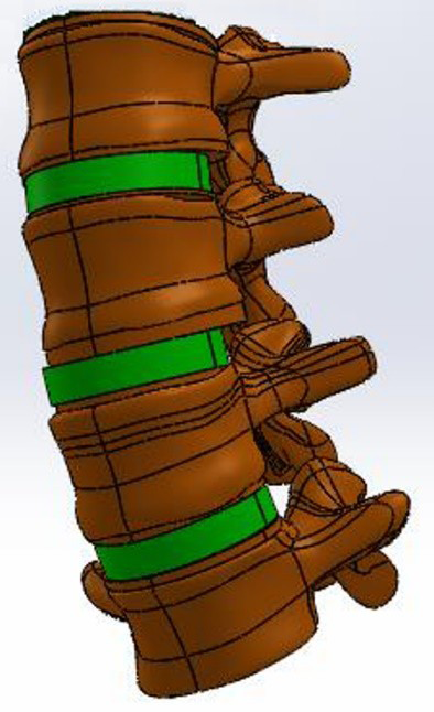
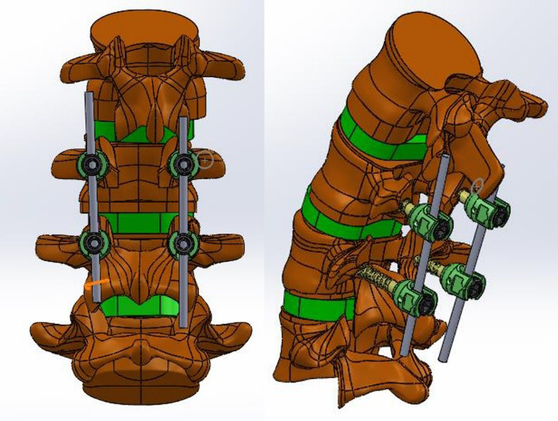
Geometrical model: Three-dimensional FEM is built up using scanner images (CT-scan Picker 5000) of a 40-year- old male. The geometrical model is generated from these files using SliceOmatic software. The finite element software Solidworks was used to conduct the numerical computing. Ligaments and muscles were not represented in the present model (Figure 1). Model with arthrodesis: We made modifications on the model to simulate an arthrodesis of L3-L4 stage: we replaced the L3-L4 disc with a bone graft, by changing the material properties of the disc to those of a vertebra, and fixed L3, L3- L4 space and L4 in one block by adding osteosynthesis with four pedicle screws and two rods (Figure 2). Material properties: The material properties of the model structures (Table 1) were taken from previous works [2]. Boundary conditions: Before proceeding to the application of loads and simulation, we defined boundary conditions which consisted in fixing all the elements of the lower base of the lumbar segment.
Loading
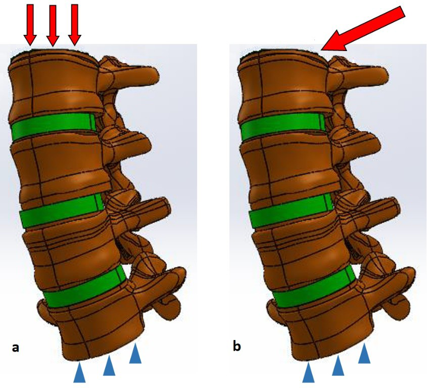
We applied loads on the upper surface of L2 (Figure 3) and observed Von Mises stress distribution on IVD for the two models. We studied the distribution of stress in axial compression after applying increasing loads of 400, 600, 800, 1000 and 1200 Newton (N). While knowing that the standing, relaxed position for a 70-kilogram man corresponds to a force of 400N, lifting an additional load of 20kg corresponds to a force of 600N and so on. We also studied the distribution of Von Mises stress in bending by using moments of force for the following values: 1.9 - 3.8 – 5.3 – 6.5 and 7.5 Newton- meter.
Results and Discussion
Validation of the Model
To validate our model, we compared the results with in vivo experimental data. We relied on the work of Wilke, et al. [3] who measured the pressure of the non-degenerated L4-L5 disc of a volunteer man, for several spatial positions and different load lifts. Von Mises stress on the healthy L4- L5 disc of our model were comparatively increased. This can be explained by the absence of ligaments and muscles, elements that allow movement and stabilize the spine, but are also essential for damping, transferring and distributing the loads. However, by comparing the evolution of stress as a function of loads between our model and experimental studies [4] or other FEM [2], we found that our model tends to follow the same linear behavior. We concluded that our model is reliable and valid for the study of IVD stress.
Von Mises Stress in Axial Compression
For the L2-L3 disc before and after arthrodesis, the stress increase linearly as the loads are increased for the two models. Stress increases significantly after arthrodesis (p=0.002). Likewise, for the L4-L5 disc stress increases linearly as the loads are increased for the two models. The difference was not significant between the models before and after arthrodesis (p=0.072). By comparing the sum of the stress undergone by the IVD on the healthy model, we find that the L4-L5 disc bears significantly more stress than the L2-L3 disc (p=0.008).
Von Mises Stress in Bending
The stress on L2-L3 and L4-L5 discs increase after arthrodesis, but the difference was not significant, with p equal to 0.151 and 0.44 respectively. However, the rate of stress increase at the L2-L3 disc was significantly higher compared to the rate of stress increase at the L4-L5 disc (p=0.032).
Discussion
AS does not have a consensus definition. It includes any radiologic changes in the adjacent stages of an arthrodesis, associated with clinical manifestations. This is still a hot topic in view of the increasing number of patients who have undergone surgery in the past, the monitoring of which will become a public health issue. Disc degeneration corresponds to the loss of the mechanical and biological properties of IVD. Although the disc aging process is physiological, it is considered pathological when it is premature [5]. It is currently established by mechanical, biochemical and biological approaches that mechanical stress has a role in the genesis of disc degeneration, thus increasing stress on the discs leads to premature degeneration. We found that the stress on IVD increase linearly with increasing compressive loads, causing degeneration. This may concern athletic disciplines and jobs involving prolonged vicious positions that are exposed to a risk of degeneration, leading to their classification as occupational diseases. We also found that L4-L5 disc supports significantly more constraints than L2- L3 disc in healthy subjects. It has already been established by Louis [6] that lumbar spine is subject to maximum stress in L4 and L5 vertebrae. Comparing the stress distribution before and after arthrodesis, we found that in axial compression, stress on the overlying disc increases significantly after arthrodesis, confirming that arthrodesis is a cause of premature degeneration of the overlying disc, and so the development of AS. This is consistent with several in vitro studies and numerical simulations that have shown an increase in the measured pressures in the adjacent discs after segmental lumbar fixation. For the underlying disc, we were unable to confirm the role of arthrodesis in the genesis of AS. This may be explained by the fact that AS predominates on the overlying rather than the underlying disc, as confirmed by the work of Cheh [7], finding that degeneration was present in 88% of cases in the overlying disc and only 7.5% of cases in the underlying disc.
Conclusions
IVD adjacent to an arthrodesis are exposed to an increasing pressure and accelerated degeneration, proving the risk of adjacent syndrome.
This simple model will enable us in future works to test certain hypotheses, characterize the risk factors of lumbar spine pathologies and propose solutions.
| Vertebrae | Disc | |
|---|---|---|
| Young Modulus (MPa) | 12000 | 8 |
| Poisson ratio (μ) | 0,3 | 0,45 |
| Shear Modulus (MPa) | 61,53 | 0,034 |
| Volumic mass (kg/m3) | 1020 | 1000 |
| Yield Stress (MPa) | 170 | 70 |
Table 1: Material Properties of the model structures.
References
-
Radcliff KE, Kepler CK, Jakoi A, Sidhu GS, Rihn J, et al. (2013) Adjacent segment disease in the lumbar spine following different treatment interventions. Spine J 13(10): 1339-1349.
-
Wang JL, Parnianpour M, Shirazi-Adl A, Engin AE, Li S, et al. (1997) Development and validation of a viscoelastic finite element model of an L2/L3 motion segment. Theor Appl Fract Mec 28(1): 81-93.
-
Wilke H, Neef P, Caimi M, Hoogland T, Claes L (1999) New in vivo measurements of pressures in the intervertebral disc in daily life. Spine 24(8): 755-762.
-
Schultz A, Andersson G, Ortengren R, Haderspeck K, Nachemson A (1982) Loads on the lumbar spine. Validation of a biomechanical analysis by measurements of intradiscal pressures and myoelectric signals. J Bone Joint Surg Am 64(5): 713-720.
-
Rannou F, Corvol M, Revel M, Poiraudeau S (2001) Degree scale and different scale: the mechanical contraindications. Rev Rhum Ed Fr 68(10): 908-912.
-
Louis R (1985) Spinal stability as defined by the three- column spine concept. Anat Clin 7(1): 33-42.
-
Cheh G, Bridwell KH, Lenke LG, Buchowski JM, Daubs MD, et al. (2007) Adjacent segment disease following lumbar/thoracolumbar fusion with pedicle screw instrumentation: a minimum 5-year follow-up. Spine 32(20): 2253-2257.
- Return to Work Among Manual Workers After the Latarjet Procedure: A Cohort Study of 43 Patients
- Refractory Pelvic Collection Following Modified Stoppa Approach for Both-Column Acetabular Fracture Fixation: A Case Report
- Comparative Study of Dynamic Knee Phenotypes Under Loaded and Unloaded Conditions: Clinical Impact
- Locked Intramedullary Nailing of the Tibia Using a Humeral Nail: A Care Case Report
- Subtalar Dislocation: About a Case Report
- Surgical Site Infection in Orthopedics in a Country with LimitedResources: Indications, Treatment and Results