Surgical Treatment of Distal Femur Fractures in Adults: Evaluation of Results
Introduction: Distal femoral fractures are defined as any bone gap in the femur whose focal point projects into the distal epiphyseal-metaphyseal line delimited by the “HEIM epiphyseal square”. The aim was to identify the anatomo-pathological forms of recent fractures of the distal extremity of the femur in adults, and to assess their treatment. Patients and Methods: This was a prospective, descriptive study of three (03) years, from January 1, 2013 to December 31, 2015, involving 45 patients at the Orthopedics-Traumatology Department of the Ignace Deen University Hospital. Adult patients received for recent distal femur fracture treated surgically, followed and evaluated, were retained for the study. Patients with fractures more than 21 days old and fractures on pathological bone, as well as patients treated orthopedically and those lost to follow-up, were excluded from the study. Results: We enrolled 45 patients, 16 men and 29 women, with a sex ratio of 0.5. We performed 24 cases (53.3%) of dynamic condylar screw fixation (DCS), 12 cases (26.7%) of 95° condylar plate blade, 3 cases (6.7%) of HOFFMANN I external fixator and 6 cases (13.3%) of screw fixation. Treatment was assessed according to CHIRON's anatomical and functional criteria, with a mean follow-up of 24 months. Conclusion: Distal femoral fractures are serious, often comminuted and involving a weight-bearing joint. Closed-focus surgical treatment needs to be popularized to reduce local care time and allow early rehabilitation, thereby improving the outcome of these patients.
Doukouré M¹, Kéita K¹, Camara A²*, Camara T¹, Soumah A¹, Cissé W¹, Bah ML¹ and Lamah L²
Keywords: Anatomopathology; Distal End of Femur; Fracture; Treatment
Introduction
Fractures of the distal end of the femur are defined as any solution of bony continuity of the femur whose focal point projects into the distal epiphyseal-metaphyseal line delimited by the “HEIM epiphyseal square”(a square whose side is equal to the greatest width of the epiphysis) [1].
They are considered to be either directly or indirectly articular, disrupting the integrity of the knee [2, 3]. They are serious fractures, often comminuted and involving a weight- bearing joint.
In the past, the treatment for these fractures was systematic amputation [4]. Gradually, radical treatment by lamputation gave way to orthopedic treatment, enabling limb preservation.
Today, treatment is virtually surgical. The principle: “anatomical reduction of the epiphysis with good limb axes to limit the risk of osteoarthritis, and a stable set-up allowing early rehabilitation to limit the risk of stiffness”[5].
The absence of previous studies on these fractures in the country and the increasing frequency of these injuries due to the advent of two-wheeled motorized vehicles as cabs in almost the entire country, motivated the choice of this study.
The aim of our work is to identify the anatomo- pathological forms of recent fractures of the distal extremity of the femur in adults, and to assess their treatment.
Patients and Methods
This was a prospective, descriptive study of three (03) years from January 1, 2013 to December 31, 2015, involving 45 patients at the Orthopedics-Traumatology Department of the Ignace Deen University Hospital, Conakry, Republic of Guinea.
All adult patients received for a recent fracture of the distal extremity of the femur, treated surgically, followed up in the department and evaluated, were included in the study.
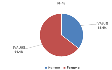
Patients with fractures more than 21 days old and fractures on pathological bone, as well as patients treated orthopedically, were excluded from the study. We enrolled 45 patients, 16 men and 29 women, with a sex ratio of 0.5 (M/F). The mean age was 61 years, with extremes of 22 and 96 years. The mean admission time was 48 hours, with extremes of 24 hours and 14 days.
We performed standard radiographs of the front, side and ¾internal and external knee, enabling us to identify anatomo-pathological forms. Fractures were classified according to the AO classification.
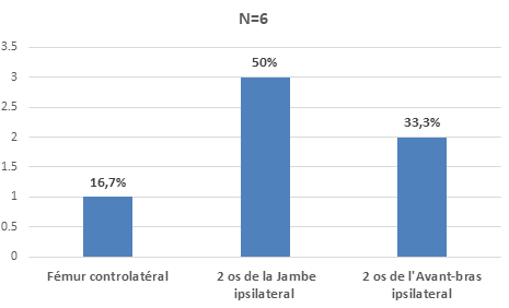
Associated lesions were: 1 case of closed diaphyseal fracture of the opposite femur; 2 cases of closed diaphyseal fracture of the 2 bones of the homolateral forearm; 3 cases of floating knees, of which 2 patients had a closed supra condylar fracture of the femur (33A1) associated with a closed diaphyseal fracture of the 2 bones of the leg. The 3rd patient had a closed supra condylar femur fracture (33A11) associated with a closed proximal metaphyseal comminuted fracture of the 2 leg bones.
There were 6 open fractures, including 2 type I, 3 type II and 1 type III according to the CAUCHOIX and DUPARC classification.
All patients were treated with transcalcaneal traction or elastoplast traction. Tetanus vaccination was performed for skin wounds and open fractures. Anti-coagulation with low-molecular-weight heparin and analgesics for delayed synthesis were administered. Surgical treatment was carried out in all our patients: the patient was installed in dorsal or lateral decubitus, depending on the surgeon’s habit. For floating knees and tibial fracture synthesis, we performed trimming and internal osteosynthesis of open CAUCHOIX type I fractures. The HOFFMANN-type external fixator was used for type II. The suction drain was removed on postoperative day 3, the first dressing was applied on postoperative day 5, and the sutures were removed on postoperative day 21.
Functional rehabilitation was started on postoperative day 1 with isometric contraction of the quadriceps, followed by the patient sitting at the edge of the bed, gradually flexing the knee under the effect of gravity. Support was allowed at D90 postoperatively in the majority of cases.
We recorded 2 cases of superficial sepsis and 1 case of deep sepsis postoperatively, with Staphylococcus aureus as the causative agent; 1 case of early disassembly of osteosynthesis material (DCS) at D45 postoperatively.
Treatment was assessed according to CHIRON’s anatomical and functional criteria [5], with a mean follow-up of 24 months.
Results
We enrolled 45 patients, including 16 men and 29 women, with a sex ratio of 0.5 (M/F) (Figure 1).
The mean age was 61 years, with extremes of 22 and 96 years. The mean admission time was 48 hours, with extremes of 24 hours and 14 days.
The associated lesions were: 1 case of closed diaphyseal fracture of the opposite femur; 2 cases of closed diaphyseal fracture of the 2 bones of the homolateral lower arm; 3 cases of floating knees, of which 2 patients had a closed supra condylar fracture of the femur (33A1) associated with a closed diaphyseal fracture of the 2 bones of the leg.
The 3rd patient had a closed supra condylar femur fracture (33A11) associated with a closed proximal metaphyseal comminuted fracture of the 2 leg bones (Figure 2).
There were 6 open fractures, including 2 type I, 3 type II and 1 type III according to the CAUCHOIX and DUPARC classification.
Anatomo-Pathological Forms
Supra condylar fractures (A) accounted for 32 cases (71.1%): A1 = 17 cases (37.9%), A2 = 6 cases (13.3%), A3=9 cases (20%). For uni-condylar fractures (B), we obtained 6 cases (13.3%): B2 = 5 cases (11.1%), B3 = 1 case (2.2%). Supra- and inter-condylar fractures (C) numbered 7 cases (15.6): C1 = 4 cases (8.9%), C2 = 2 cases (4.4%), C3 = 1 case (2.2%) (Table 1).
Open Focus: There were 6 open fractures (13.3%), 5 of which were localized to the femoral focus (2 CAUCHOIX type I and 3 type II). The last patient presented with a type III floating knee open at the femoral site.
Type of Treatment: All our patients were treated surgically in 45 cases (100%), including 24 cases (53.3%) with dynamic condylar screws (DCS: DYNAMIC CONDYLAR SCREW), 12 cases (26.7%) with 95° condylar plate blades, 3 cases (6.7%) with HOFFMANN I-type external fixators, and 6 cases (13.3%) with screw fixation (Table 2).
| Anatomopathological Forms | Types | Workforce | Percentage (%) |
|---|---|---|---|
| Supra-condylar fractures (A) | A1 | 17 | 37,9 |
| A2 | 6 | 13,3 | |
| A3 | 9 | 20 | |
| Unicondylar fractures (B) | B2 | 5 | 11,1 |
| B3 | 1 | 2,2 | |
| Supra- and inter- condylar fractures (C) | C1 | 4 | 8,9 |
| C2 | 2 | 4,4 | |
| C3 | 1 | 2,2 | |
| Total | 45 | 100 |
Table 1: Distribution of patients by anatomopathological form.
| Surgical Treatment | Workforce | Percentage (%) |
|---|---|---|
| DCS | 24 | 53,3 |
| Condylar plate blade | 12 | 26,7 |
| Screw fixation | 6 | 13,3 |
| Hoffmann I External Fixator | 3 | 6,7 |
| Total | 45 | 100 |
Table 2: Distribution of patients according to surgical treatment.
Treatment Evaluation
This takes into account the type of treatment in relation to each anatomopathological form. All our patients were evaluated, some of them physically during appointments (38), and the other patients (7) by telephone, as they were far from the Conakry region and its surroundings.
Anatomical Results: We obtained the following anatomical results: Very good: 33A=28 cases (62.2%), 33B=6 cases (13.3%), 33C=4 cases (8.9%), Good: 33A=2 cases (4.4%), Fair: 33A=1 case (2.2%), Poor: 33A=1case (2.2%), 33C=3 cases (6.7%) We observed 5 cases of callus, distributed as follows: varus 2 cases, valvus 3 cases (Table 3).
Functional Results: Functional results were assessed according to anatomopathological type and therapeutic method used.
These results were good overall, and we obtained: Very good: 33A=26cas (57.8%), 33B=5cas (11.1%); Good: 33A=4(8.9%), 33C=4(8.9%); Fair: 33A=1(2.2%); Poor: 33A=2(4.4%), 33C=3(6.7%) (Table 4).
| Vicious Callus | Degree | Nb | Degree | Nb | Degree | Nb | Degree | Nb | Total |
|---|---|---|---|---|---|---|---|---|---|
| Varus | 6° | 2 | 11° | - | 15° | - | 20° | - | 2 |
| Valgus | 8° | 3 | 16° | - | - | - | - | - | 3 |
| Recurvatum | 15° | - | - | - | - | - | - | - | - |
| Antecurvatum | 18° | - | - | - | - | - | - | - | - 5 |
| Total |
Table 3: Distribution of patients by type of callus.
| Functional Criteria | Types | Workforce | Percentage (%) |
|---|---|---|---|
| Very good | 33A | 26 | 57,8 |
| 33B | 5 | 11,1 | |
| Good | 33A | 4 | 8,9 |
| 33C | 4 | 8,9 | |
| Fair | 33A | 1 | 2,2 |
| Poor | 33A | 2 | 4,4 |
| 33C | 3 | 6,7 | |
| TOTAL | 45 | 100 |
Table 4: Patient distribution by functional criteria.
Overall Results: Whatever the anatomopathological type and treatment used, we obtained 97.8% good and very good results.
Complications: We recorded 3 cases of postoperative sepsis (6.7%), including 1 profound case requiring removal of the osteosynthesis material at D45.
One (1) case of disassembly of the post-synthesis material at D60 post-op was noted, involving the 95° plate blade.
Discussion
Anatomo-Pathologically
We found a predominance of supra-condylar forms (71.1%), with a clear proportion of A1 (37.9%). This predominance of supra-condylar forms was reported by Hoffmeyer P, et al. [3, 6].
On the other hand, CHIRON [5], excluding uni-condylar forms (B) from his study, found a predominance of supra- and inter-condylar forms.
In our series, uni-condylar forms are less frequent, accounting for 13.3%. Type B3 bi-condylar forms are extremely rare [7, 8].
The open fractures observed in 2 of our patients were secondary to an opening from medial to lateral generated by the proximal fragment, with sectioning of the vastus lateralis expansion (CAUCHOIX and DUPARC type I).
The other 3 presented an opening from the outside to the inside, with associated contusion and skin detachment (CAUCHOIX and DUPARC type II). The 6th patient presented with CAUCHOIX and DUPARC type III, with loss of bone substance and comminution of the tibial focus, creating a floating knee. This was a high-energy trauma. It is a traumatological emergency. It requires rapid management to avoid or minimize complications.
Vascular damage is rare, representing 0.5% to 3% depending on the series. We have not noted any vascular or nerve damage in our series.
Systematic opening of the joint at the time of lostosynthesis of type C joints enables exact assessment and repair of lesions.
In our study, no central pivot lesions were noted in any of the patients operated on. Type C patients underwent systematic arthrotomy.
Therapeutic approach
Current treatment of fractures of the distal end of the femur is resolutely surgical, allowing early functional rehabilitation to achieve patient autonomy. Traction remains an effective method of treatment in the event of delayed surgery, enabling pain to be controlled. It could be recommended in a few cases of major fracture with crushing, but it competes with the indications for external fixation.
As far as surgical treatment is concerned, whatever the equipment used and the technique chosen, surgical treatment is only conceivable if it allows perfect reduction of the epiphysis, correction of axial defects and stable fixation without additional postoperative external restraint. The therapeutic chain must be flawless, otherwise there is a risk of therapeutic failure, leading to serious sequelae.
Dynamic Condylar Screw (DCS) osteosynthesis was the most commonly used technique in our series, with 24 cases (53.3%). The 95° condylar plate blade was used in 12 patients (26.7%). It is the material of choice for fractures of the distal end of the femur [9, 10]. Although little or not used in advanced countries, it is still very much in vogue in our developing countries. It is inexpensive and readily available.
95° plate blades are used for supra condylar fractures, simple supra and inter condylar fractures and metaphyseal comminution.
We have performed 3 external fixations of open fractures of the distal end of the femur using a Hoffmann I-type fixator bridging the knee. Nowadays, closed or open fractures of the distal end of the femur are treated with an external fixator [11].
Six (6) of our patients underwent open screw fixation. These were type B2 fractures. Even non-displaced fractures should be treated surgically, with additional immobilization of the knee in a cast for 6 weeks to avoid secondary displacement [12].
We used an additional crural-pedal cast in 6 patients operated on: 4 with the 95° condylar plate blade and 2 with screw fixation. The reason for this was lost osteoporosis in 4 cases, and inadequate mounting in 2. Nowadays, retrograde centromedullary nailing of distal femoral fractures is recommended for types (A- C1-C2) [11].
Evaluation
In a series of 45 patients collected in our department from 2013 to 2015, we reviewed and evaluated 45 patients or 100%.
We obtained 88.8% of very good and good anatomical results. Functionally, we found 88.9% very good and good results.
These results led us to conclude that all fractures of the distal end of the femur should be treated surgically.
Lostosynthesis gave us good results (88.8% very good and good anatomical results and 88.9% very good and good functional results). These results are superimposed on those of the literature, with 81% of very good and good anatomical results and 71% of good functional results for CHIRON [5], 90% of very good and good anatomical results and 70% of very good and good functional results for NORDIN [13]. The Similar findings have been reported by other authors [14].
With regard to the opening of the fracture site, all results were similar to those of closed fractures, with the exception of one case of a CAUCHOIX type III open fracture with poor anatomical and functional results.
Correct surgical treatment followed by early and appropriate rehabilitation, whatever the type of osteosynthesis material used, is the ideal method for managing distal femoral fractures.
Complications
We noted 3 cases of sepsis. A revision of the surgical wound was performed in the operating room, with sampling for cyto-bacteriological examination + antibiotic susceptibility testing. The germs found were staphylococcus aureus, sensitive to lamoxicillin and clavulanic acid. Wound healing was achieved at D21, with a progressive fall in CRP and normalization of the white blood cell count.
The use of retrograde nails reduces the risk of sepsis [15, 16, 17].
We recorded one (1) case of disassembly of the osteosynthesis hardware at D60 post-op, concerning the 95° plate blade.
Conclusion
Distal femoral fractures are serious fractures, often comminuted and involving a weight-bearing joint. Closed- focus surgical treatment should be made more widely available, to reduce the time required for local care and allow early rehabilitation, thereby improving the outcome of these patients.
Conflicts of Interest
The authors declare no conflicts of interest.
References
-
Nazarian S (2005) Epidemiology, anatomical varieties and classification of fractures of the distal end of the femur: Knee fractures. Practical Approach in Orthopedics Traumatology. Springer Verlag France pp: 27-36.
-
Asencio G (1995) Fractures of the lower end of the femur. EMC, Musculoskeletal system, Editions techniques, Paris pp: 12.
-
Hoffmeyer P (2001) Fractures of the distal end of the femur in adults. EMC, Surgical Technique, Paris 11: 44- 80.
-
Chevallier JM (1998) Anatomy, musculoskeletal system. Medicine, Sciences, Flammarion, Paris pp: 291-352.
-
Chiron PH (1995) Recent fractures of the lower end of the femur in adults. SOFCOT teaching conference. Scientific expansions, Paris 52: 147-166.
-
Andreas X (2004) Papadopoulos, m.d. Operative treatment of unilatéral Bi Condylar Hoffa fractures. Journal of Orthopaedic Trauma 18(2): 119-122.
-
Carret JP (1991) Biomechanics of the knee joint. SOFCOT teaching conference. Scientific Expansions, Elsevier, Paris 40: 189-208.
-
Hahn U (2001) A condylar plate in distal fémoral fractures. Kongressbd Ges chir Leougr 118: 362-366.
-
Sanders R (1991) Double plating of comminuted, unstable fractures of the distal part of. J. Bone joint Surg 73A(3): 341-346.
-
Arazi M (2001) Ilizarov external fixation for severely comminuted supracondylar and intercondylar fractures of the distal femur. J Bone Joint Surg Br 83(5): 663-667.
-
Calmet J (2004) Open Bi condylar Hoffa Fracture Associated with extensor of mechanism injury. Journal Orthopaedic Trauma 18: 323-325.
-
Dunlop DG (1998) The supracondylar intramedullary nail in Eldertly patients with distal femoral fractures. Journal of Bone et Joint Surgery 80B (15): 51.
-
Nordin JY (1989) Early osteosynthesis in principle. Rev Chir Ortho Sup 75(1): 180-181.
-
Siliski JM (1989) Supracondylar, intercondylar fractures of the fémur. Treatment by internal. J Bone Joint Surg Am 7(1): 95-104.
-
Armstrong R (2003) Retrograd interlocked intramedullary nailing of supracondylar distal femur fractures. An average 76 years old patient population orthopedics 26(6): 627-629.
-
Seligson LD, Kentucky (2006) Supra-condylar fractures of the femur: Intramedullary nailing. Indications and contraindications. Master’s degree in orthopedics. Technical pp: 1-15.
-
Vichard P (2002) Retrograde nailing of the femur. SOFCOT teaching conferences. Scientific expansion, Paris 79: 125-140.
- Return to Work Among Manual Workers After the Latarjet Procedure: A Cohort Study of 43 Patients
- Refractory Pelvic Collection Following Modified Stoppa Approach for Both-Column Acetabular Fracture Fixation: A Case Report
- Comparative Study of Dynamic Knee Phenotypes Under Loaded and Unloaded Conditions: Clinical Impact
- Locked Intramedullary Nailing of the Tibia Using a Humeral Nail: A Care Case Report
- Subtalar Dislocation: About a Case Report
- Surgical Site Infection in Orthopedics in a Country with LimitedResources: Indications, Treatment and Results