A Physical Barrier Model for Preventing the Enamel Demineralization
The incidence and prevalence of dental caries in Indonesia has increased from year to year. The main cause is often associated with bad tooth brushing habits. Caries occurs because there is a process of demineralization of the surface of hard tooth tissues such as email due to the presence of acids produced by bacteria that metabolize the substrate. Coating the tooth surface with an acid-resistant material is an emerging idea for caries prevention in the future, Preliminary studies with extracted tooth models demonstrated that enamel surface coating was able to prevent demineralization through physical barrier. This method is possible as novel strategy of caries prevention on clinical level.
Introduction
Dental caries is a disease affecting hard tissue of the tooth that takes place due to demineralization process. This process is caused by acid, which is a by-product of substrate metabolism by microorganism. Dental caries is one of the most common and the oldest infectious disease known to man [1, 2]. Prevention efforts with different approaches have been conducted, such as reducing the number of microorganisms, either chemically (using antiseptic) or mechanically by tooth brushing, fluoridation of tap water and dental sealant application [2, 3]. Unfortunately, those approaches mentioned above have not showed the desirable result. According to a research conducted by health ministry of Indonesia in 2018 (riset kesehatan dasar), there were 57.6% out of 1.2 million people that suffered from dental caries [4].
Perspective
Studies about caries prevention using immunological approach have also been carried out, such as immunoglobulin making or antibody recombinant [5, 6]. Despite all the approaches attempted, so far there isn’t any particular caries prevention technique or method that can be used effectively on grass root level. The most recent study in dental caries prevention evaluated the usage of chlorhexidine antimicrobial coating on tooth surface to reduce bacterial colonies in oral cavity. Unfortunately, this method could result in oral flora imbalance which in turn, could cause another disease, such as candidiasis [7].
Theoretically, dental caries takes place due to interaction of tooth structure, diet (glucose), microorganism, and also with adequate time [2, 3]. Substrates metabolism by cariogenic bacteria will result in acid by-product, which then will initiate demineralization process on tooth surface.
The acidic environment causes dissolution of calcium and phosphate ion, resulting in porous tooth surface and milky appearance [8]. Dental caries occurs as the result of physical interaction between cariogenic microorganism and substrates, with acid as its product. Inhibiting of said interaction is difficult to achieve, while inhibiting physical interaction between acid and tooth surface is relatively more feasible. Based on the concept that dental caries happens due to demineralization process caused by physical contact between acid and tooth surface, layering the surface with certain coating material in order to prevent said physical contact could be taken into consideration as novel method to inhibit dental caries process.
Physical contact inhibition conducted in this research showed that manipulating the tooth surface by coating it could significantly inhibit demineralization process of tooth enamel, which naturally happens after contact with acid. In this pilot study, we experimented on extracted human teeth acquired from dental clinic.
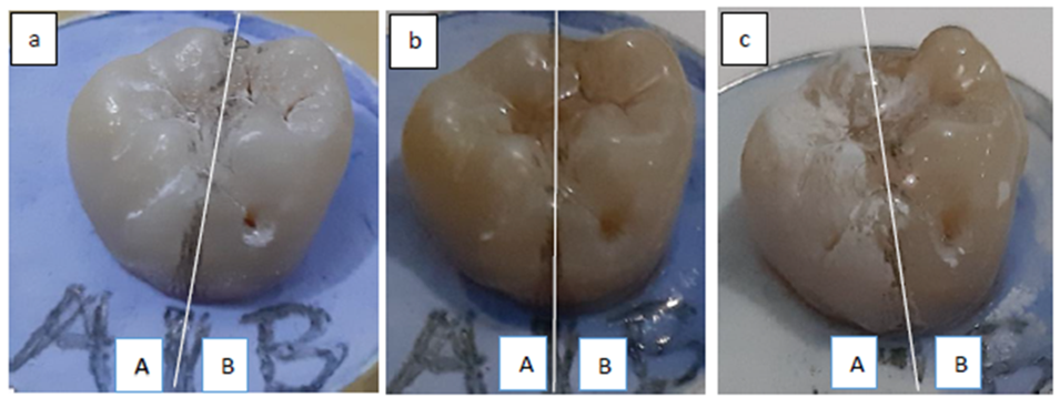
First, the teeth were cleansed carefully from debris and then mounted into dental stone. Imaginary midline was drawn to divide the teeth into two equal parts; the uncoated surface as control group and the coated surface as the experimental group (Figure 1a). The teeth then soaked into acid solution for 1- 3 hours to imitate demineralization process. The samples were dried using air syringe to be evaluated visually. Visual observation showed that there was milky appearance on the uncoated surface, indicating there was visible demineralization (Figure 1b), while the coated surface showed no changes (Figures 1c).
Soaking the teeth in acid solution (pH 3.0-4.8) for 1-3 hours caused demineralization process; this happened because in acidic environment, crystal apatite Ca10(PO4)6(OH)2 released Ca2+ ion and PO43-. Upon the ion release, the uncoated tooth enamel became porous and showed milky appearance. Initial experiments have been carried out on 3 extracted teeth, and have the same characteristics of results. This event didn’t happen on the coated surface. This result suggests that demineralization process can be inhibited physically.
The result from this study emerges the need to find a coating material that has to meet certain properties from several aspects. Physically, it has to be transparent, thin and yet has adequate hardness and smooth level. Chemically, it has to be acid resistant, strong bonds to email and insoluble in saliva. Biologically, it should not cause any teeth discoloration, non-toxic, and must have a good biocompatibility. Mechanically, it has able to withstand masticatory loads.
The progress of nano-technology development lately enables us to provide materials corresponding with aforementioned standards, one of the is nanoceramic composite [9, 10].
Conclusion
In summary, our finding that coating the tooth surface can inhibit demineralization process of enamel through physical barrier described in this “perspective” is open to translation into novel strategy of caries prevention on clinical level.
Conflict of Interest
The author has no conflict of interest relevant to this article.
Acknowledgement
The author would like to acknowledge the support from Indonesian Dentist Association (PDGI) and Indonesia Ministry of Health for the collections of caries data (Riskesdas 2018).
References
-
Richard JL, Paul GE (2015) Molecular Medical Microbiology 2nd (Edn.), Elsevier, pp: 945-955.
-
Selwitz RH, Ismail AI, Pitts NB (2007) Dental caries. Lancet 369(9555): 51-59.
-
Jolan B, Andrew RG (2013) Epidemiology and Prevention of dental Caries. Acta Med Acad 42(2): 105-107.
-
Indonesian Ministry of Health (2018) Riskesdas Basic Health Research.
-
Abiko Y (2000) Passive immunization against dental caries and periodontal disease: development of recombinant and human monoclonal antibodies. Crit Rev Oral Biol Med 11(2): 140-158.
-
Li Y, Jin J, Yang Y, Bian Z, Chan Z, et al. (2009) Enhanced in immunogenicity of an anti caries vaccine encoding a cell-surface protein antigen of Streptococcus mutans by intranasal DNA prime- protein boost immunization. J Gene Med 11(11): 1038-1047.
-
Mogens K (2018) The Oral Microbiome-Friend or Foe? European Journal of Oral Sciences 126(S1): 5-12.
-
Korishettar BR, Pathak S, Poornima P, Neena IE (2015) White spot lesions: A literature review. Journal of Pediatric Dentistry 3(1): 1-7.
-
Sakthivel S, Saritha D, Baskaran V (2014) Preparation, properties and applications of Nano Glass Ceramics 4(2): 21-25.
-
Bilandzic MD, Wollgarten S, Stollenwerk J, Poprawe R, Esteves Oliveira M, et al. (2017) Glass-ceramic coating material for the CO2 laser based sintering of thin films as caries and erosion protection. Dental Materials 33(9): 995-1003.
- Diagnosis and Management of Mental Nerve Paresthesia Secondary to Apical Periodontitis of Mandibular Second Premolar: A CBCT Based Case Report
- A Randomized, Double Blinded Clinical Trial to Compare the Effect of Oral Premedication (Diclofenac Potassium or Dexamethasone) on Post-Operative Pain Following Pulpectomy
- Modified Lip Repositioning Technique for the Management of Excessive Gingival Display
- Integral Role of Non-Dental Providers and Fluoride Dissemination
- Root Canal Treatment Rate in Deciduous Teeth Among 6-Year- Olds in the Era of Discontinuing Water Fluoridation - Historical Cohort Study
- The Impact of the Notch1 on the Migratory Capacity and the Expression of E-Cadherin and CyclinD1 in Ameloblastoma Cells