The Mechanism by Which Chemotherapy with use of Alkylating Agents Cause Follicular Activation: Is there any Further Mode for the Loss of the Primordial Follicles? A Short Communication
A lot of women In their reproductive life go through treatment with a lot of chemotherapeutic drugs of which the alkylating agents which are inclusive of cyclophosphamide, busulphan, along with dacarbazine, more specifically cyclophosphamide, whose utilization is considered for a lot of chemotherapy protocols like In breast cancer, In cases of chronic lymphoblastic leukaemia in children and we encounter survivors of these cancers with usual manifestation of Premature ovarian insufficiency(POI). Thus detailed evaluation was attempted by Shai et al., In case of human ovaries immediately In which fresh ovarian tissue that was obtained from ladies following receipt of alkylating agents, non-alkylating agent in addition to that collected from those who did not get treatment. They corroborated the existence of significant depletion of primordial follicles (PMF’s), in addition to significant escalation of the absolute quantities of growing follicles in contrast to ovaries collected from those who did not get treatment. Staining done for FOXO3A demonstrated reduction in the nuclear existence in the PMF oocytes in cases where ovaries removed were from patients receiving treatment with alkylating agent in the ultra-short period following alkylating agents. No escalation in the expression of the cleaved caspase 3 was observed in PMF’s within the ovaries that got alkylating agents or non-alkylating agents exposure. Marked stromal fibrosis in addition to neovascularization was seen in alkylating agents treatment group only following loss of PMF had occurred earlier (4-6mths). This is a great study despite them unable to demonstrate the phosphorylated FOXO3A. Although it was anticipated that they would further show the part of PI3K/Akt pathway that possesses major part in the follicular activation although they explained in view of immediate preservation of ovaries In formaldehyde in addition to embedding In paraffin. Thus this study goes a long way in explaining the cause of follicular loss following treatment with alkylating agents and aid in the strategies we need to use to avoid the same once feasible.
Introduction
We possess the knowledge with regards to the harmful action of the chemotherapeutic agents on female reproduction for practically last 50 yrs. Despite that our insight in the context of the way chemotherapy impacts the function of the gonads remains a significant problem. Of the alkylating drugs cyclophosphamide, busulphan, along with dacarbazine represent the ones that possess the maximum significant ovotoxicity [1, 2]. Their utilization gets done In a lot of chemotherapy protocols, that have demonstrated to result in stimulation of depletion of ovarian follicle reserve that is correlated with the amount of dose utilized [3] that causes premature ovarian insufficiency (POI) in addition to reduction in fertility in a lot of subjects who have survived from cancer [4]. One specific alkylating drug i.e. cyclophosphamide (Cy), that causes stimulation of DNA crosslinking in addition to finally avoiding DNA replication, is utilized commonly in the treatment of cancer. Escalation of proof with regards to implication of alkylating drug in the deleterious action on ovarian function in addition to exposure of granulosa cells in the generating follicles to these drugs results in apoptotic demise along with subsequent, abrupt reduction in anti mullerian hormone (AMH) amounts. For offering an explanation for the deletion of the primordial follicles (PMF’s), secondary to alkylating drugs, various modes have got posited as reviewed by Spears et al., in 2019 [5],that have got obtained basically from observational studies conducted in nonhuman animal models, namely direct apoptotic death, indirect stimulation of activation of the follicles which then gets subsequently followed by deletion of the follicles (alias ‘’burn-out’’ mode), besides indirect deletion secondary to injury to the stroma that suirrounds the oocyte in addition to/or interference with the blood supply [5].
As per the deletion of the primordial follicles (PMF’s), a lot of modes have been take into account, that is inclusive of exaggerated activation, atresia, apoptosis in addition to stromal injury, like inflammation as well as vascular injury. Meirows group in the last 10 years has robustly correlated that with alkylating drugs resulted in the upregulated phosphatidyl inositide 3-kinase(PI3K) /protein kinase B(AKT) signalling pathway, that cause regulation of follicles remaining In quiescent state, that ultimately caused s activation of the PMF’s that resulted in PMF’s deletion. They posited that AMH amounts reduction occurred within 12 h of Cy therapy, that was subsequently followed by an escalation to double the concentrations that were observed at baseline. Since this escalation got sustained for 14 days following Cy therapy, it was speculated that it was secondary to an escalation in the quantity of the generating follicles that was secondary to the over stimulation of the quiescent PMF’s. The biochemical pathways that were implicated In the PMF’s stimulation was inclusive of growth factors that work via different pathways like phosphatidyl inositide 3-kinase(PI3K) / Phosphatase as well as TENs in (PTEN) Akt [protein kinase B(PK alias Akt) in addition to Hippo pathways.
Hence in their recent publication to find out the exact mode of follicular depletion Shai, et al. [6], evaluated fresh ovarian tissue that was obtained from ladies receiving treatment with alkylating agent, non-alkylating agent in addition to that collected from those who did not get treatment. A significant depletion of PMF’s, nevertheless, a significant escalation In the absolute numbers with regards to the growing follicles In the group that belonged to the alkylating agent group, in contrast to the ones that belonged to the group comprising of those not getting treatment. This escalation In the quantity of the growing follicles possessed an inverse association with the timing of chemotherapy besides that as per the researchers was agreeable with a burst of activation of the follicles recruited just subsequent to the delivery of alkylating agents, an observation that was earlier seen in ovaries of mouse that had received Cy exposure [7]. Evaluation of the placement of Forkhead box O3(FOXO3A), that avoids the stimulation of the follicles, when its existence is within the nucleus, yet causes stimulation of the growth of the oocyte only on its phosphorylation under the implication of the PI3K pathways in addition to getting exported toward the cytoplasm, they observed that in addition to getting expressed In the oocyte nuclei of 77% as well as 80% of the PMF’s in the ovaries of those ladies untreated as well as those getting treatment with non-alkylating drugs, respectively [6] (Figure 1). Conversely its expression existed in under 47% of the nuclei of PMF’s in the ovaries of ladies who received treatment with alkylating agents. Nevertheless, the variation did not achieve statistical significance in addition to as agreed by Shai, et al. [6] they could not illustrate the existence of FOXO3A that is phosphorylated In the cytoplasm whose requirement exists for starting the growth of the oocyte. Anticipation of greater validation of this was done by Dolmans in addition to Donnez [8], that was dependent on the analysis of the PI3K/Akt pathway that possesses a key part in the follicle getting activated since its isolation is required within 24h of exposure to Cy by the group of Shai et al. In view of their bad luck as detailed by Shai, et al. [6], molecular analysis of the Akt pathway was not feasible secondary to the whole samples of tissue that had got obtained with regards to this study had been fixed in formaldehyde, besides getting embedded In, paraffin that ensured that molecular evaluation became tough. Nevertheless, knowledge is possessed by us that the stimulation of the PMF’s all through the reproductive life gets Controlled by the PI3K/ PTEN)/ Akt in addition to Hippo pathways [9] (figure2). Shai, et al. [6], need to get motivated for continuation of persuing their experimental work by probing further In depth with regards to the Akt pathway, besides its association with FOXO3A, Akt in addition to Hippo pathways along with further its signaling effectors like -Yes-associated protein (YAP) and transcriptional co- activator with post synaptic density protein, drosophila disc large tumor suppressor [PDZ] and zonula occludens- 1-binding motif (TAZ) that have got illustrated to aid In this process of follicle burn, out that has been seen following transplantation [9] (figure3).
![Figure 1: Courtesy ref no-6-Fibrosis and neovascularization in ovaries at different time points after treatment with alkylating agent and nonalkylating agent chemotherapy compared with untreated ovaries. (A) Representative images showing Sirius red staining for collagen deposition (a–e; positive staining in red) and CD34 staining for neovascularization (f–j; positive staining in brown) in untreated ovaries compared with ovaries 0–2 months or 4–6 months after treatment with alkylating agent chemotherapy or nonalkylating agent chemotherapy. (B) Quantitative Image analysis for collagen content and neovascularization was performed on fields of tissue up to an average of 300 μm from cortex edge with the use of Image J software. Left: Total collagen content per field was calculated (collagen = R; counterstained tissue = G) and average R/(G+R) value for each treatment group calculated (n = 3–5 per group, fields from individual patients are represented by different shaped icons; total fields evaluated: untreated, 27; 0–2 months after alkylating chemotherapy, 21; 4–6 months after alkylating chemotherapy, 30; 0–2 months after nonalkylating chemotherapy, 43; 4–6 months after nonalkylating chemotherapy, 41). Data are shown as mean [R/(G+R)]/field ± SEM. ∗∗∗P<.001. Right: The number of CD34-positive blood vessels (BVs) per field was evaluated and the average number for each treatment group was calculated (n = 3–5 per group; fields from individual patients are represented by different shaped icons; total fields evaluated: untreated, 22; 0–2 months after alkylating chemotherapy, 30; 4–6 months after alkylating chemotherapy, 22; 0–2 months after nonalkylating chemotherapy, 19; 4–6 months after nonalkylating chemotherapy, 22). Data are shown as mean number of CD34-expressing BVs/field ± SEM. ∗∗∗P<.001.](/fulltextimages/7761/fig_1.png)
Figure 1: Courtesy ref no-6-Fibrosis and neovascularization in ovaries at different time points after treatment with alkylating agent and nonalkylating agent chemotherapy compared with untreated ovaries. (A) Representative images showing Sirius red staining for collagen deposition (a–e; positive staining in red) and CD34 staining for neovascularization (f–j; positive staining in brown) in untreated ovaries compared with ovaries 0–2 months or 4–6 months after treatment with alkylating agent chemotherapy or nonalkylating agent chemotherapy. (B) Quantitative Image analysis for collagen content and neovascularization was performed on fields of tissue up to an average of 300 μm from cortex edge with the use of Image J software. Left: Total collagen content per field was calculated (collagen = R; counterstained tissue = G) and average R/(G+R) value for each treatment group calculated (n = 3–5 per group, fields from individual patients are represented by different shaped icons; total fields evaluated: untreated, 27; 0–2 months after alkylating chemotherapy, 21; 4–6 months after alkylating chemotherapy, 30; 0–2 months after nonalkylating chemotherapy, 43; 4–6 months after nonalkylating chemotherapy, 41). Data are shown as mean [R/(G+R)]/field ± SEM. ∗∗∗P<.001. Right: The number of CD34-positive blood vessels (BVs) per field was evaluated and the average number for each treatment group was calculated (n = 3–5 per group; fields from individual patients are represented by different shaped icons; total fields evaluated: untreated, 22; 0–2 months after alkylating chemotherapy, 30; 4–6 months after alkylating chemotherapy, 22; 0–2 months after nonalkylating chemotherapy, 19; 4–6 months after nonalkylating chemotherapy, 22). Data are shown as mean number of CD34-expressing BVs/field ± SEM. ∗∗∗P<.001.
Does chemotherapy stimulated activation of the follicle represent the sole mode that aids In the explanation of the depletion of PMF’? For evaluation of apoptosis, Shai et al. [6], analyzed cleaved caspase 3 in the sections of ovaries that had got collected following an ultra-short time duration Subsequent to the alkylating agent delivery (i.e 4-12days) (Figure 2). This particular group demonstrated that 79% of the follicles that were growing got the classification as apoptotic, yet no escalation of staining for apoptosis was picked up in PMF’s although significant PMF’s depletion was not seen within this time period.
Nevertheless, Luan, et al. [10], illustrated that the amounts of Ki67 positive cells could be compared to 8weeks old mice that had received phosphate buffered saline along with those that received Cy that pointed to that Cy isn’t implicated In the activation of PMF’s into the pool of follicles that are growing. They demonstrated, in an experimental model that Cy stimulated the PMF’ depletion via apoptosis that resulted in oocyte depletion, besides loss of ovarian reserve in addition to which inhibitors of the apoptotic pathway constitutents, confers protection to the follicles. Their posit was that Cy stimulation of the PMF’ loss was secondary to pataxia telagiectasia as well as Rad3- correlated/p-checkpoint kinase i/p checkpoint kinase 2-p63 apoptotic pathway. They did not find escalation of activation of PMF’s associated with Cy delivery along with pointed to apoptosis pathway inhibitors might be the agents that resulted in gonadal protection against the toxic actions of Cy. Actually, in an ovarian in vitro culture, two apoptotic inhibitors were shows to efficaciously protect the PMF’s from the chemotherapy stimulated injury. Rest of the modes like Oxidative stress (OS) as well as Cy stimulated inflammation have further been held responsible as illustrated by greater concentrations of pro-inflammatory cytokines , interleukin-6, IL-8, as well as Tumor necrosis factor alpha(TNFα) .
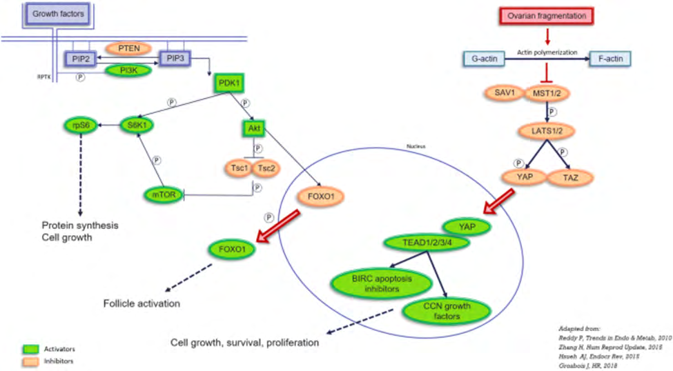
Figure 2: Courtesy ref no-9-Representation of the PI3K and Hippo pathways involved in follicle activation.PIP2: phosphatidylinositol 4, 5-bisphosphate; PIP3: phosphatidylinositol (3,4,5)-trisphosphate; PTEN: phosphatase and tensin homolog deleted on chromosome 10; PI3K: phosphoinositol-3-kinase; PDK1: pyruvate dehydrogenase lipoamide kinase isozyme 1; Akt: protein kinase B; FOXO: forkhead box O; TSC1 and TSC2: tuberous sclerosis proteins 1 and 2; mTOR: mechanistic target of rapamycin; S6K1: ribosomal protein S6 kinase beta-1; RPS6: ribosomal protein S6. MST1/2: mammalian Ste20- like 1/2; SAV1: protein salvador homolog 1; LATS1/2: large tumor suppressor kinase 1/2; YAP: yes-associated protein, TAZ: transcriptional coactivator with PDZ-binding motif; TEAD 1/2/3/4: TEA domain family members 1/2/3/4; BIRC: baculoviral inhibitors of apoptosis repeat containing.
Hence concluding that a lot of modes have been suggested to reason out the PMF’s depletion that is inclusive of exaggerated activation, atresia, apoptosis, inflammation in addition to vascular injury. Their total microenvironment that these elements are constituting creates an unfavorable surroundings for the follicles. In the context of injury to the vasculature in addition to its action on PMF’s death, Shai et al. [6], saw fibrosis in the ovarian cortex along with neovascularization do take place long subsequent to PMF’s depletion, that points to that stromal injury can’t be the etiology of immediate loss of follicles. Nevertheless, the query ‘’chemotherapy-induced ovarian damage: apoptosis and or in vitro activation? [11], that was put In the’’ Fertile Battle’’ area In the ’’Fertil Steril‘’ journal has not got its reply till now, despite positing that both might be implicated.
Despite no existence of any query this very good publication by Shai, et al. [6] gives strong proof that alkylating agents have a significant role in the activation of follicles. However, further evaluation is needed for the exploration of the PI3K/ PTEN/ Akt pathway in addition to Hippo pathways, besides the apoptotic pathway In greater details for offering newer propositions for the protection of the ovarian reserve [8]. Recently we had reviewed the crosstalk among the various MAPK/ERK along with Hippo/ MST signaling pathways in addition to their crosstalk with the PI3K/Akt/mTOR signaling pathways for avoidance of generation of chemo resistance, besides using agents targeting Hippo/MST signaling further to avoid the detterent actions of PI3K/Akt/MTOR signaling on the MAPK/ERK signaling, besides explaining a lot of complications like DM, MetS, Other body conditions with role of ERK as final common effectors of bodily functions that are significant for a lot of cellular events that is inclusive of reproduction, with the role of cyclophosphamide impacting on YAP/TAZ signaling aid in proving the burn out action of Cy [12].
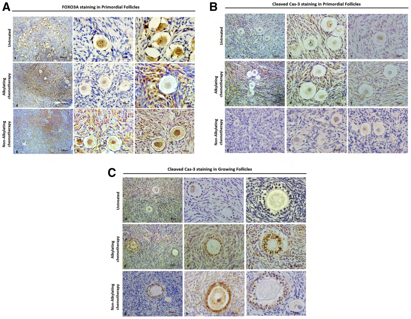
Figure 3: Courtesy ref no-6-Foxo3A and cleaved caspase-3 (Cas-3) immunostaining of ovarian sections from untreated women and women 4–12 days after treatment with alkylating agent chemotherapy. (A) Representative images showing localization by immunofluorescence of FOXO3A in ovarian tissue from each treatment group. Positive staining (brown) indicates FOXO3A expression in PMF oocyte nucleus 4–12 days after alkylating agent (d–f) and nonalkylating agent (g–i) chemotherapy treatment compared with untreated control samples (a–c). Scale bars: a, b, d, e, g, h = 20 μm; c, f, i = 100 μm). (B) Representative images of primordial follicles showing no positive staining for cleaved caspase-3 (brown) in oocytes or granulosa cells in sections from untreated women (a–c), women treated with alkylating agent chemotherapy (d–f), and women treated with nonalkylating agent chemotherapy (g–i). Scale bars: a, d, g = 50 μm; b, c, e, f, h, i = 25 μm. (C) Representative images showing positive staining for cleaved caspase-3 (brown) in granulosa cells of primary and secondary follicles in sections from untreated women (a–c) or women treated with alkylating agent chemotherapy (d–f), and women treated with nonalkylating agent chemotherapy (g–i). Scale bars: a, d, g = 50 μm; b, c, e, f, h, i = 25 μm
Refereces
1. Chemaitilly W, Li Z, Krasin MJ, Brooke RJ, WilsonCL, et al. (2017) Premature ovarian insufficiency In childhood cancer survivors: a report from the St Jude Lifetime Cohort. J ClinEndocrinol Metab 102(7): 2242-2250.
2. Lower EE, Blau R, Gazder P, Tummala R (1999) The risk of Premature menopause induced by chemotherapy for breast cancer. J Women’s Health Gend Based Med 8(7): 949-954.
3. Oktem O, Oktem K (2007) Quantitative assessment of the impact of chemotherapy on ovarian folliclereserve and stromal function. Cancer 110(10): 2222-2229.
4. Anderson RA, Brewster DH, Wood R, Nowell S, Fischbacker C, et al. (2018) The impact of cancer on Subsequent chance of pregnancy:a population based analysis. Hum Reprod 33(7): 1281-1290.
5. Spears N, Lopez F, Stefanddottir A, Rossi V, Anderson RA, et al. (2019) ovarian damage from chemotherapies and current approaches to its protection. Hum Reprod Update 25(6): 673-693.
6. Shai D, Aviel-Ronen S, Spector I, Raanani H, Shapira M, et al. (2021) Ovaries of patients recently treated with alkyl- ating agent chemotherapy indicate the presence of acute follicle activation, elucidating its role among other pro- posed mechanisms of follicle loss. Fertil Steril 115(5): 1239-1249.
7. Kalich Phlosoph L, Roness H, Carmey A, Fishel Bar- tal M, Ligumski H, et al.(2013)Cyclophosphamidetrig- gers follicle activation and burnout, AS101 prevents fol- licle loss and preserves fertility. Sci Trasl Med 5(185): 185ra62.
8. Dolmans MM, Donnez J (2021) Chemotherapy with alkylating agents: is follicle activation the only mechanism responsible for the loss of primordial follicles? Fertil Steril 115(5): 1166-1167.
9. Masciagelo R, Hossay C, Chiti MC, Maavella DD, Amorim CA, et al. (2020) Role of the PI3K and Hippo pathways in follicle activation after graftingof human ovarian tissue. J Assoc Reprod Genet 37(1): 101-108.
10. Luan Y, Edmonds ME, Woodruff TK, Kim SY (2019) Inhibitors of apoptosis protect the ovarian reservefrom Cyclophosphamide. J Endocrinol 240(2): 243-256.
11. Dolmans MM, Taylor HS, Rodriguez-Wallerg KA, Blumenfeld Z, Lamertini M, et al. (2020) Utility of gonadotropin releasing hormone agonists for fertility preservation in women receiving chemotherapy: pros and cons. Fertil Steril 114(4): 725-738.
12. Kulvinder KK, Allahbadia GN, Singh M (2021) Escalation of efficacy & Prevention of chemoresistance in various Cancer Therapies by the utilization of targeting the crosstalk amongst MAPK/ERK along with Hippo/MST signaling-A Comprehensive Review.
- Postpartum Maternal Mental Health - A Narrative Review
- Beta HCG in Cervico-Vaginal Secretion as a Predictor of Preterm Delivery
- Successful Management of Mid Trimester Foetal Death with Major Placenta Previa by Expectant Management Followed by Induction of Labour
- To Evaluate the Expression of Egr2 Gene in Term Low Birth Weight Newborns
- Impact of Maternal Obesity on Maternal and Foetal Outcomes: A Prospective Cohort Study from Northern India
- ‘’Benefit of Pulsatile GnRH Therapy in Treatment of Functional Hypothalamic Amenorrhea (FHA) and Congenital Hypogonadotropic Hypogonadism(CHH) in Infertile Patients Over Canonical Gonadotropins with IVF –A Short Communication’’