Isolation, Identification and Characterization of Fungus causing Coconut’s Bud Rot Disease: Inhibition using Silkworm Fecal Matter Mediated Synthesized Nanoparticles
Coconut is one the major economic crop in India; a considerable amount of crop will be lost every year due to bud rot disease. Phytophthora palmivora is an omnipresent pathogen which causes many different diseases on a wide range of plants including bud rot of coconut. In the present investigation, we have successfully isolated bud rot disease causing fungus from the infected coconut plant samples. The isolated fungus was primarily identified by observing in microscopy further the same sample was sent for molecular identification. The presence of Phytophthora palmivora was confirmed in 18s rRNA sequencing. The growth of isolated fungus was effectively inhibited using biosynthesized Silver and Copper nanoparticles. The inhibition effects of nanoparticles against Phytophthora palmivora were observed excellent in dose-dependent manner. The silver nanoparticles synthesized using Silkworm fecal matter was shown superior inhibition activity towards Phytophthora palmivora compare to standard fungicide Fluconazole. Hence, these silver Nano particles could be successfully used in inhibiting the pathogenic fungus causing bud rot disease to coconut.
Introduction
Nanomaterials are cornerstones of Nano-science and nanotechnology. We specifically considered nanoparticles as clusters of atoms in the size of 1-100nm. ‘Nano’ is a Greek word synonymous to dwarf meaning extremely small. Nanostructure science and technology is expansive and interdisciplinary area of research and development activity that has been growing explosively worldwide in the past few years. The idea of nanotechnology was coined by physicist Professor Richard Feynman (known as “Father of Nanotechnology”) in his historic talk “there’s plenty of room at the bottom” Feynman RP, et al. [1], though the term nanotechnology was introduced by Tokyo Science University Professor Norio Taniguchi Taniguchi N, et al. [2]. Nano-biotechnology has emerged as integration among biotechnology and nanotechnology for developing biological synthesis and environmental-benign technology for synthesis of nanomaterials. Nano-biotechnology is one of the most promising areas in modern Nano-science and technology. Biological synthesis is a green and environmental friendly approach of synthesizing nanoparticles which are biodegradable and non-toxic to the environment [3]. Biosynthesis includes using of plants, algae, fungi, actinomycetes, yeast, bacteria, etc. along with precursors to synthesize nanoparticles alternative to convention chemicals for capping and bio reduction purposes. The biologically synthesized nanoparticle finds their own way in biomedical applications due to their unique and enhanced properties [4]. The wide variety of core materials available, coupled with tunable surface properties, make nanoparticles an outstanding platform for a broad range of biomedical and biological applications.
The coconut tree (Cocos nucifera) is a member of the palm tree family (Arecaceae) and the solitary living species of the genus Cocos. The term “coconut” (or the archaic “coconut”) can refer to the whole coconut palm, the seed or the fruit, which botanically is a drupe, not a nut. The term is derived from the 16th-century Portuguese and Spanish word coco meaning «head» or «skull» after the three indentations on the coconut shell that resembles facial features. Coconut belongs to Kingdom: Plantae, Order: Arecales, Family: Arecaceae, Subfamily: Arecoideae, Genus: Cocos, Species: nucifera. In India it is cultivated in an area about 1.93million hectare with an estimated production of 12,147million nuts, has tremendous influence on the socio-economic property of million people. Coconut palm is dominant and essential component of the homesteads and garden lands along the coastal parts of southern India and it plays a vital role in the sociocultural and economic life of huge number of small and marginal farmers Dagar JC, et al. [5]. Coconut tree has much economic importance as food, drink, health, shelter, medicine, fuel, aesthetics and wealth. Because of its versatile nature, it is known as “Kalpa-Vriksha”, “Tree of abundance”, “Tree of Heaven”, “Tree of Life”, “Nature’s super market”, “King of palm”, “Tree of virtues “ and by many names. It is also known as lazyman’s crop Gupta S, et al. [6]; Loomba S, et al. [7]; Lima EBC, et al. [8].
Coconut production is limited by several factors, among them diseases play a major havoc role. Coconut crop diseases are a major biotic stress, play a great havoc causing considerable reduction in yield level. Due to microbial infections, there were several major diseases reported from our country, causing severe damage to the plant body such as Basal stem rot (Thanjore wilt), Bud rot, Leaf blight, Leaf rot disease, Root wilt, Stem bleeding Nambiar KKN, et al. [9]; Louis H, et al. [10]. Among those Bud rot disease caused by Phytophthora palmivora was known to be severe of coconut palm. Palms of all ages were susceptible to bud rot disease, but the young palms were observed to be more susceptible, particularly during monsoon season when the temperature is low and humidity is very high. Briton-Jones HR, et al. [11] described the disease symptoms first. The first visible symptom was the withering of the spindle marked by pale colour. The spear leaf or spindle turns brown and bends over. A basal tissue of leaf rots quickly and can be easily detached from the crown. Spindle withers and droops down and one by one, the inner leaves also fall away, leaving only fully matured leaves in the crown. A foul smell is emitted by the rotting tissue [9]. The palms succumb to the disease with the death of the spindle Briton-Jones HR, et al. [11]; Menon KPV, et al. [12]; Lingaraj DS, et al. [13]. Later, infection spreads to the older leaves, causing sunken leaf spots covering the entire leaf blade spreading both up and down. Spot margins are irregular and water soaked and when the leaves are unfolded, the characteristic irregular spots are conspicuous on the blade. In severely affected trees, the entire crown may rot and in few months the trees wilt. The heart leaf becomes wilts, chlorotic and collapses. The disease may spread to older, adjacent leaves and spathes, producing a dead center with a fringe of living leaves. Light brown to yellow, oily, sunken lesions may be seen on leaf bases, stipules or pinnae. Internally, the tissues beneath the bud rot discolored pink to purple with a dark brown border. Infected nuts showed brown to black necrotic areas with yellow border rising on the surface. Internally, they have a mottled appearance. Young nuts are highly susceptible to disease and fail to mature and then fall off from the tree. Older, infected nuts ripen normally [14, 15]. Ultimately, the entire crown falls down and the palm dies. In the 1920s “bud rots diseases” were identified in Jamaica, Puerto Rico, Africa, Peninsular Malaysia and the Philippines [12]. Later, it was also reported in India, Sri Lanka, Central America, West Indies, Fiji, and Vanuatu etc. Several practices like spraying chemicals, keeping the garden clean, application of talc powder formulation on rotten areas, etc., were made to avoid the spreading of bud rot disease [16].
Phytophthora is a genus of plant- damaging oomycetes (water molds), whose member species are capable of causing huge economic losses on crops worldwide, as well as environmental damage in natural ecosystems. Phytophthora palmivora is an oomycete which causes bud-rot of palms, fruit-rot orkole- roga of coconut and areca nut. These are among the most severe diseases caused by fungi and moulds in South India. The causative organism was first identified as Phytophthora palmivora by Butler in 1917. P. palmivora belongs to Phylum: Heterokontophyta, Class: Oomycota, Order: Peronosporales, Family: Peronosporaceae, Genus: Phytophtora, Species: palmivora. The pathogen was assumed to have originated in Southeast Asia but is now pantropical. It causes significant losses to farmers of tropical fruit and vegetable crops. P. palmivora infects a thousand or more plant species including horticultural, ornamental and agricultural crops. It is also a common soil inhabitant. Important horticultural hosts include cocoa (black pod, canker, cherelle wilt), papaya (fruit rot), durian (fruit rot, canker) pineapple (heart rot, citrus (canker), black pepper (foot rot) and coconut (bud rot). P. palmivora grow vigorously under humid wet conditions and as a result can cause significant losses in many economically important tropical fruit crops [16]. The main distinguishing feature of the pathogen includes conspicuous papillate sporangia, which can be distinguished from other Phytophthora species because they are caducous and have short pedicels. Primary inoculum originates from soil and infected plant material. The pathogen is circulated through rain splash, insects and human activity into the canopy of trees, where symptoms appear. Secondary inoculum spreads rapidly through wind and rainsplash, contact and vector activity in humid weather [17, 18]. Phytophthora palmivora can be managed using cultural and chemical methods. The use of resistant varieties, sanitation (including complete harvesting and the removal of infected planting material and weeds), and improved nursery hygiene, pruning to improve air flow and reduce humidity and boosting soil health by increasing organic matter can be used as part of an integrated management strategy. Chemicals used in the control of P. palmivora include metalaxyl, phosphonates or copper hydroxide to paint cankers, and phosphonates as a soil drench, trunk injection or foliar spray.
In present investigation, we have undertaken inhibition studies against isolated bud rot disease causing fungi using novel metal nanoparticles synthesized using Silkworm fecal matter, which would lead to develop a new drug and treatment for bud rot disease of coconut. This would in turn may helpful for uplifting the coconut growing farmers economically.
Materials and Methods
All the materials used in the present study were used as received. Chemicals used in the study were of analytical grade and highest possible purity. They were purchased from Hi-Media Chemicals (Mumbai, Maharashtra, India) and Sigma Chemical Company (St. Louis, Mo. USA).
Sample Collection
During rainy season, bud rot disease infected lesion samples were randomly collected from different regions of Chikkamagaluru districts, Karnataka, India. If the tissue is fresh and recently infected, isolation of fungus from plant tissue or coconut bud is quite simple. Successful isolation of pathogenic fungus from the disease tissue involves careful selection of freshly infected tissue, it is best to obtain it from actively growing lesions. Collected samples were placed in between the butter paper or newspaper and placed in separate small sealed envelopes and then they were taken to laboratory and stored at 40C for further fungal isolation.
Isolation and Identification of Pathogenic Fungus
The infected plant parts were surface sterilized by dipping in 70% ethanol for 30-60seconds or in 0.5-1% sodium hypochlorite for 3-5minutes, followed by rinsing with distilled water and blotting, then on sterile filter paper.
Primarily two media were used for the isolation of pathogenic fungus and the composition is listed below:
Composition of Carrot Agarmedium Composition of Potato Dextrose agar medium as follows
| Components | Quantity | Components | Quantity | |
|---|---|---|---|---|
| Carrot | 50gm | Potato | 20gm | |
| Dextrose | 2gm | Dextrose | 2gm | |
| Ampicillin | 20mg | Ampicillin | 20mg | |
| Agar | 2gm | Agar | 2gm | |
| d. H O 2 | 100ml | d. H O 2 | 100ml |
The infected part of coconut bud was cut into small bits using sterile stainless steel blade, these bits were surface sterilized using sterile distilled water followed by washing in 0.1% mercuric chloride (HgCl2) and finally washed with sterile distilled water (3times; 1min interval). The surface sterilized bits were inoculated onto the above mentioned sterilized culture plates, the pH of both the medium was adjusted to 7 previously, the plates were covered with radiation resistive laboratory barrier film (Parafilm M) (PPP Company, Menasha, WI) to prevent drying of agar and to avoid contamination of medium and kept for incubation at 24±1ºC in biological incubator for 6-7 days. Then the grown colonies were observed under microscope for the identification of pathogenic fungi and subcultured to obtain pure culture of desired fungus. The pure cultures of the fungi were maintained in Potato Dextrose Agar medium (PDA) slants at 4ºC by sub culturing at 2-3-week intervals.
Molecular Identification of Isolated Fungus
The fungal genomic DNA was extracted from one week old culture maintained in PDA medium. Polymerase Chain Reaction (PCR) was performed with the final volume of 100µl containing approximately 100ng of DNA template, 10mM dNTPs, 400ng of each primers, 10XChrom Taq DNA Polymerase assay buffer and 3UTaq DNA Polymerase enzyme.
Two Universal Fungal Specific Primers: 5’-GRAAGNAHADGTVGKAAYAWSG-3’ (forward) and 5’-TCCTNCGYTKATKGVTADGH-3’ (reverse) were used to amplify in a Biometric T personal thermocycler (Biometric GmbH, Goettingen, Germany). The initial denaturation at 95ºC for 300sec, followed by 35 cycles of denaturation at 94ºC for 30sec, primer annealing at 52ºC for 30sec, elongation at 72ºC for 45sec with final extension at 72ºC
for 420sec. The PCR products were run on 1% agarose gel and visualized under UV light. The amplified 1.5kb product was purified and directly sequenced on ABI 3500XL Genetic Analyzer using a mixture; 4µl of Big Dye Terminator Ready Reaction Mix, 1µl of Template, 2µl of Primers and 3µl Milli Q water as recommended by the manufacturer.
Inhibition of Phytophthora palmivora using Biosynthesized Nanoparticles
The antifungal activities of the biosynthesized nanoparticles were tested using radial growth technique [19]. Petri dishes (9cm diameter) containing 25ml of PDA medium were used for anti-fungal activity assay, performed in solid media by disc diffusion method. Different concentrations (1mg, 2mg and 3mg/ml) of CuNPs [20] and AgNPs [20] were added to the medium immediately before it was poured into the petri dish. Control was also maintained without any nanoparticles and fungicide (Fluconazole). A diameter of 6mm disc of Phytophthora palmivora was taken from 8-day culture of fungal strain and placed upside down in the centre of the Petri dish. The treatments were kept for incubation at 25ºC for 6-8days, time by which the growth of control would have reached the edge of petri dish. The inhibition of fungal growth was calculated as percentage of inhibition of radial growth related to control. Percentage of mycelial growth inhibition was calculated using the following formula, Mycelial growth inhibition(%)=(DC-DT/DC)×100 Where, DC- average diameter of fungal colony of control. DT- average diameter of fungal colony of treated.
Results
Isolation and Identification of Pathogenic Fungus
Mixed colonies were obtained when fungi were first isolated on Carrot agar and potato dextrose agar medium. Pure cultures of bud rot causing fungi were identified based on its routine cultural and morphological characteristics. The pathogenic fungus was sub-cultured on freshly prepared medium. The pure cultures of the fungi were maintained in Potato Dextrose Agar medium (PDA) slants at 4ºC by sub culturing at 2-3week intervals. Further pure culture of bud rot causing fungi was sent for molecular identification.
Molecular Identification of Fungus
Fungal isolate was identified and confirmed on the basis of its molecular characterization. Genomic DNA was successfully isolated from fungal species and the purity of the same was determined on 1% Agarose gel. After optimization of the PCR condition, the ITS1/ITS4 region DNA was successfully amplified from the genomic DNA using specific primers as described earlier. The PCR product of species had a length of ~700bp. The partial sequence of obtained 18S rRNA was aligned with the available 18S rRNA sequences on NCBI website and compared for homology. The isolated strain exhibited high level of 18s rRNA similarity of 98% with Phytophthora palmivora isolate (Genebank accession no. MH401199.1). hence, the molecular characterization confirmed the isolated fungus was Phytophthora palmivora (Figures 1 & 2) (Table 1).
The partial sequence of obtained 18SrRNA was aligned with the available 18SrRNA sequences on NCBI website and compared for homology.
| Description | Similarity | Gene Bank Accession Number | |
|---|---|---|---|
| 1 | Phytophthora palmivora strain PPC3614L 18S ribosomal RNA gene, partial sequence; internal transcribed spacer 1, 5.8S ribosomal RNA gene, and internal transcribed spacer 2, complete sequence; and 28S ribosomal RNA gene, partial sequence | 98% | MH401199.1 |
| 2 | Phytophthora palmivora isolate FG-12 small subunit ribosomal RNA gene, partial sequence; internal transcribed spacer 1 and 5.8S ribosomal RNA gene, complete sequence; and internal transcribed spacer 2, partial sequence | 98% | MF370567.1 |
| 3 | Phytophthora palmivora isolate FG-11 small subunit ribosomal RNA gene, partial sequence; internal transcribed spacer 1 and 5.8S ribosomal RNA gene, complete sequence; and internal transcribed spacer 2, partial sequence | 98% | MF370566.1 |
| 4 | Phytophthora palmivora strain PPG13 18S ribosomal RNA gene, partial sequence; internal transcribed spacer 1, 5.8S ribosomal RNA gene, and internal transcribed spacer 2, complete sequence; and 28S ribosomal RNA gene, partial sequence | 98% | KY475632.1 |
| 5 | Phytophthora palmivora isolate TW183 18S ribosomal RNA gene, partial sequence; internal transcribed spacer 1 and 5.8S ribosomal RNA gene, complete sequence; and internal transcribed spacer 2, partial sequence | 98% | KU682577.1 |
| 6 | Phytophthora palmivora genomic DNA containing ITS1, 5.8S rRNA gene and ITS2, strain C011 | 98% | LM650992.1 |
| 7 | Phytophthora palmivora isolate NRCPh-138 18S ribosomal RNA gene, partial sequence; internal transcribed spacer 1, 5.8S ribosomal RNA gene, and internal transcribed spacer 2, complete sequence; and 28S ribosomal RNA gene, partial sequence | 98% | KF010307.1 |
| 8 | Phytophthora palmivora genomic DNA containing 18S rRNA gene, ITS1, 5.8S rRNA gene, ITS2, 28S rRNA gene, strain CPPHN02 | 98% | HE580280.1 |
| 9 | Phytophthora palmivora isolate Dal1b 18S small subunit ribosomal RNA gene, partial sequence; internal transcribed spacer 1, 5.8S ribosomal RNA gene, and internal transcribed spacer 2, complete sequence; and 28S large subunit ribosomal RNA gene, partial sequence | 97% | MH219904.1 |
| 10 | Phytophthora palmivora voucher LSVM1405 small subunit ribosomal RNA gene, partial sequence; internal transcribed spacer 1 and 5.8S ribosomal RNA gene, complete sequence; and internal transcribed spacer 2, partial sequence | 97% | MG956799.1 |
Table 2: Sequences showing similarity with the isolated Fungus.
Inhibition of Phytophthora palmivora using Biosynthesized Nanoparticles
Inhibition study of Phytophthora palmivora_using synthesized AgNPs and CuNPs at different concentrations were showed strong dose-dependent antifungal activity (Figures 3 & 4). It was observed that, as the concentration of biosynthesized nanoparticles increases, there will be decrease in the mycelial growth of pathogenic fungi for both the nanoparticles. Comparatively, AgNPs were exhibited more inhibition effect on _Phytophthora palmivora than CuNPs. AgNPs has shown some remarkable better activity than the standard fungicide, which we can observe in the figure and table. The percentage of mycelial growth inhibition, which was calculated using the formula, is depicted in Table 2.
| Components | Concentration | MGI (%) |
|---|---|---|
| AgNPs | 1mg/ml | 30 |
| 2mg/ml | 58 | |
| 3mg/ml | 86 | |
| CuNPs | 1mg/ml | 18 |
| 2mg/ml | 48 | |
| 3mg/ml | 70 | |
| Fungicide | 1mg/ml | 32 |
| 2mg/ml | 53 | |
| 3mg/ml | 80 |
Table 3: Percentage of Mycelial Growth Inhibition (MGI). The percentage of MGI in _Phytophthora palmivora_ was observed to be mor
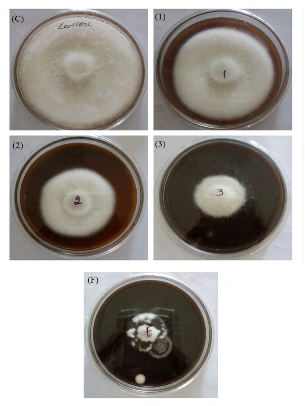
Antifungal effect of AgNPs was measured using Radial growth technique, Phytophthora Palmivora showing strong dose-dependent response, with the increase in the concentration of AgNPs there is an increase in the percentage of inhibition.
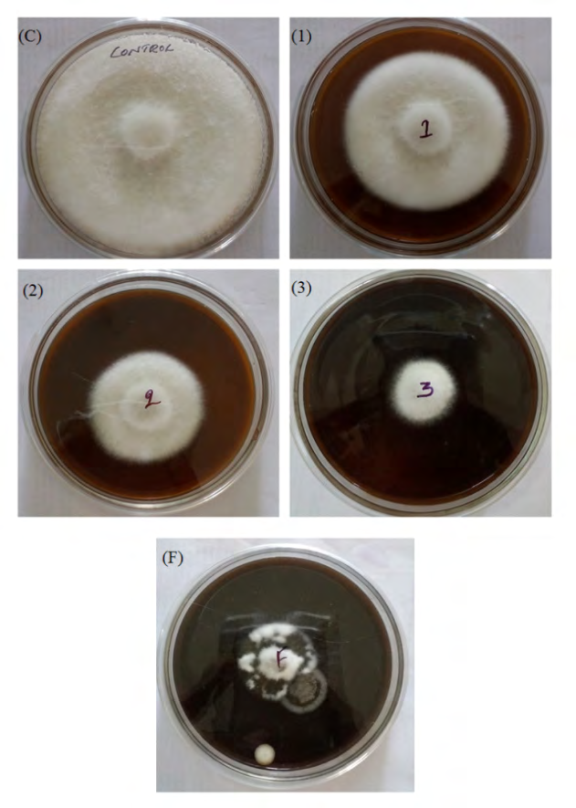
Antifungal effect of CuNPs was measured using Radial growth technique, Phytophthora Palmivora showing strong dose-dependent response, with the increase in the concentration of CuNPs there is an increase in the percentage of inhibition.
Discussion
In the present investigation, the fungal species was isolated from bud rot affected coconut plant using PDA medium. The fungal species was identified as Phytophthora palmivora by its molecular characterization. The sequence of the isolated has shown upto 98% identity with other Phytophthora species. It is well-known that molecular classification is a fast procedure which requires least management of pathogens and also supports in distinguishing morphologically, similar fungal species. Similar applications of PCR technology were used for detection and identification of fungi, by using an). Phytophthora palmivora is the main cause for Bud rot disease in coconut, which causes huge loss to economy of farmers. Rashmi AR, et al. [21] and Sharadraj KM, et al. [22] and many other researchers reported the isolation of Phytophthora palmivora which caused bud rot and root rot in coconut. In 2008, Srinivasulu B, et al. [16] made a complete report on Phytophthora palmivora isolation from bud rot caused tissue of coconut and well written about the disease management [16]. Including Bud rot of coconut, P.palmivora is one of the reasons for many deadly diseases of other plants. In the same way, Hung PM, et al. [23] identified P. palmivora from root rot symptoms of Citrus maxima in Thailand. Serious root rot disease of citrus and papaya caused by P.palmivora has been recorded in India Graham JH, et al. [24] and America Zitko SE, et al. [25] also isolated and characterized Phytophthora species which caused Citrus Gummosis in Kenya. Suskiri S, et al. [26] reported Phytophthora form Durian Orchard in Chumphon Province, Thailand. Root rot and stem rot are the major disease of durian orchard, which is caused by Phytophthora species. Root rot of Pomelo caused by this pathogen was observed in Thailand [23]. There are many other major diseases are associated with Phytophthora species which causing major loss to agricultural and ornamental crops. Hence, the fungus should be inhibited to avoid loss of economy.
Biosynthesized nanoparticles showed an effective range of antifungal activity against Phytopthora palmivora. Antifungal activity of synthesized nanoparticles was determined using Radial Growth Technique [19]. As P.Palmivora shows strong dose dependent response over the addition of AgNPs and CuNPs. There was an increase in the percentage of inhibition with the increase in the concentration of biosynthesized nanoparticles. In present study, both AgNPs and CuNPs were proven to be good antifungal agents, where AgNPs established remarkable inhibition activity against the pathogenic fungi P.palmivora. When compare to CuNPs and standard Fungicide (Fluconazole). As previous reports suggests, biosynthesized nanoparticles were already proved that they were remarkable antifungal agents against many pathogenic fungus like Candida albicans and Aspergillus niger [27], Rhizoctonia solani, Fusarium oxysporum, Sclerotiniasclerotiorum and Sclerotium rolfsii Kaman P, et al. [28]; Khan NT, et al. [29] reported antifungal activity against Candida glabrata, Candida albicans and Candida tropicalis using fungal mediated nanoparticles in disc diffusion method [29]. However, there is a report available on antifungal activity of nanoparticles against pathogenic fungus causing red root rot disease in tea plants [30]. This study is the first trial of biosynthesized nanoparticles as antifungal agents against the bud rot causing pathogenic organism Phytophthora palmivora. Our inhibition study stands in front with the previous reports on inhibition of P. palmivora reported by Gills-Alex P, et al. [31].
Conclusion
Bud rot causing fungus was isolated from infected tissue of coconut using suitable method. The molecular characterization of the isolated fungus species as Phytophthora palmivora. Where in the sequence has 98% homology with the similar Phytophthora species. The isolated species was found to be the main reason for causing bud rot disease to coconut trees [32, 33]. Therefore, there was a demand for investigating to inhibit the Phytophthora palmivora for protecting the disease. In present research, the inhibition effect of AgNPs and CuNPs synthesized using silk worm fecal matter was explored against Phytophthora palmivora using radial growth technique. The pathogenic fungus showed dose dependent inhibition response on addition of nanoparticles. Here we have observed, both synthesized AgNPs and CuNPs were very excellent antifungal agents. The Percentage of Mycelial Growth Inhibition of fungus was 86%, 70% and 80% for AgNPs, CuNPs and Standard Fungicide respectively. For the concentration of 3mg/ml and AgNPs were found as potent antifungal agent against Phytophthora palmivora, when compare to CuNPs and standard Fungicide. Plant cell wall-degrading enzymes play significant roles throughout the fungal life including acquisition of nutrients and decomposition of plant cell walls. Many plant pathogens are known to secrete a variety of PCWDEs to perceive weak regions of plant epidermal cells and penetrate the plant primary cell wall. Cell wall degrading enzymes are believed to help pathogens directly penetrate host root tissue. Therefore, may be essential for pathogenicity. Cell wall degrading enzymes produced by pathogenic fungus were found to play an important role in penetration of organism inside the plant tissue; hence it is critical to inhibit those enzymes using synthesized nanoparticles. Our Silver and copper nanoparticles have successfully inhibited the cell wall degrading enzymes produced by pathogenic fungi in a dose dependent manner. Therefore these nanoparticles could be used as potent inhibitor of Phytophthora palmivora in place of standard fungicide fluconazole.
References
-
Feynman RP (1960) There’s a Plenty of Room at the Bottom. Caltech Engineering and Science 23(5): 22-36.
-
Taniguchi N (1974) On the Basic Concept of Nanotechnology. Proceedings of the International Conference on Production Engineering, Tokyo. Scientific Research 5(2): 18-23.
-
Kuppusamy P, Yusoff MM, Govindan N (2016) Biosynthesis of metallic nanoparticles using plant derivatives and their new avenues in pharmacological applications-An updated report. Saudi Pharm. J 24(4): 473-484.
-
Hasan S (2015) A Review on Nanoparticles : Their Synthesis and Types. Research Journal of Recent Sciences 4: 9-11.
-
Dagar JC, Pandey CB, Chaturvedi CS (2014) Agroforestry: A Way Forward for Sustaining Fragile Coastal and Island Agro-Ecosystems. Agroforestry Systems in India: Livelihood Security and Ecosystem Services, Advances in Agroforestry 10: 185-232.
-
Gupta S (2013) “Coconut Palm (tree).” Encyclopedia Britannica Online 1: 1-7.
-
Loomba S, Jothi V (2013_) Cocos Nucifera_: Its Properties and Contributions to Dentistry. International Journal of Scientific Study 1(3): 138-140.
-
Lima EBC, Sousa CNS, Meneses LN, Ximenes NC, Santos MA, et al. (2015) A phytochemical and pharmacological review. Braz J Med Biol Res 1.
-
Nambiar KKN (1994) Diseases and disorders of coconut. Advances in Horticulture 1: 857-882.
-
Louis H (2002) Coconut the Wonder Palm. Hi-Tech Corporation Ramanputhoor Nagercoil 1: 206-218.
-
Briton-Jones HR (1940) The diseases of the coconut palm (book), London, Baillière, Tindall & Cox 01: 1-196.
-
Menon KPV (1958) The Coconut palm; a monograph. Ernakulam, India: Indian Central Coconut Committee 16: 384.
-
Lingaraj DS (1972) Disease of Coconut. Lal-Baugh 17: 25-31.
-
(2000) Agriculture: Coconut Woes. Economic & political weekly 35(51): 44-57.
-
Srinivasulu B, Aruna K, Rao DVR (2007) Biocontrol of _Ganoderma_ Wilt Disease of coconut palm. South Indian Horticulture 49: 240-243.
-
Srinivasulu B, Gautham B, Sujatha A, Kalpana M, Vijayalakshmi A, et al. (2008) Bud Rot Disease of Coconut. AICRP on Palms, HRS, Ambajipeta Technical Bulletin, pp: 1-24.
-
Drenth A, Sendall B (2004) Economic impact of Phytophthora diseases in Southeast Asia. Diversity and management of Phytophthora in Southeast Asia. ACIAR Monograph No. 114. Canberra: Australian Centre for International Agriculture 10-28.
-
Ramesh R, Maruthadurai R, Singh NP (2013) Management of bud rot disease in the coconut plantations of Goa. ICAR Research Complex for Goa.
-
Bajpai VK, Rahman A, Kang SC (2007) Chemical composition and antifungal properties of the essential oil and crude extracts of Metasequoia glyptostroboides Miki ex Hu. Ind Crops Prod 26(1): 28-35.
-
Avinash B, Neelagund SE (2017) An Investigation on Antibacterial and Free Radical Scavenging Efficacy of Biosynthesized Silver Nanoparticles Using Silkworm Fecal Matter _(Bombyx mori-L)_. Journal of Bionanoscience 11(6): 592-597.
-
Rashmi AR, Rohini L (2010) Characterization of Phytopthora palmivora isolates inciting but rot and nut rot in coconut. Indian Council of Agricultural Research 38(3): 188-193.
-
Sharadraj KM, Mohanan CR (2016) A new and simple baiting technique for easy isolation of _Phytophthora_ _palmivora_ Butl. from bud rot affected tissue of coconut. Journal of Applied Horticulture. 18(1): 44-47.
-
Hung PM, Wattanachai P, Kasem S Poaim S (2015) Biological Control of Phytophthora palmivora Causing Root Rot of Pomelo Using Chaetomium spp. Mycobiolog 43: 63-70.
-
Graham JH, Timmer LW (1992) Phytophthora diseases of Citrus. _In:_ Singh US, et al. (Eds.), Plant diseases of international importance: diseases of vegetables and oil seed crops. Englewood Cliffs: Prentice-Hall Inc 1: 250- 269.
-
Zitko SE, Timmer LW, Sandler HA (1991) Isolation of Phytophthora palmivora pathogenic to citrus in Florida. Plant Dis 75(5): 532-535.
-
Suksiri S, Laipasu P, Soytong K, Poeaim S (2018) Isolation and Identification of Phytophthora sp. and Pythium sp. from Durian Orchard in Chumphon Province, Thailand. International Journal of Agricultural Technology 14(3): 389-402.
-
Kumarasamyraja D, Jeganatan NS (2013) Antimicrobial activity of Biosynthesized Silver nanoparticles prepared from the leaf extract of Lantana camara. Int Res J Pharm 4: 203-207.
-
Kaman P, Pranab D (2018) Synthesis, characterization and antifungal activity of biosynthesized silver nanoparticle. Indian Phytopathology 10: 1-8.
-
Khan NT, Mushtaq M (2016) Determination of Antifungal Activity of Silver Nanoparticles Produced from _Aspergillus niger_. Biology and Medicine 9(1): 1-4.
-
Ponmurugan P, Manjukarunambika K, Elango V, Gnanamangai BM (2016) Antifungal activity of biosynthesised copper nanoparticles evaluated against red root-rot disease in tea plants. Journal of Experimental Nano science 11(13): 1019-1031.
-
Gilles-Alex P, Mpika J, Kone D, Ducamp M, Kebe I, et al. (2018) Inhibition of Phytophthora species, agents of cocoa black pod disease, by secondary metabolites of Trichoderma species. Environ Sci Pollut Res 25(30): 29901-29909.
-
Basavarajappa A, Shivayogeeswar E (2016) An Investigation on Antibacterial Efficacy of Biosynthesized Novel Copper nanoparticles using Silkworm fecal matter. Imperial Journal of Interdisciplinary Research 2: 1501- 1507.
-
Sankar S, Sukumari Nath V, Shekar Misra R, Lajapathy Jeeva M (2013) Inhibitory activity of plant growth regulators on Phytophthora palmivora causing cassava tuber rot. Archives of Phytopathology and Plant Protection 46(4): 402-409.
- Diversity of Candida sp and Antifungal Susceptibility Patterns in Digestive Candidiasis among People Living with HIV in CHU of Libreville, Gabon
- Vulvovaginal candidiasis: Retrospective study (2019- 2021) at the Centre Hospitalier National de Pikine, Suburban Dakar, Senegal
- Identification of Environmental Fungal Species in Clinical Services of University Hospital of Angre, Abidjan (Cote d’Ivoire)
- New Location of some Gasteroid Basidiomycetes in Western Kazakhstan
- Evaluation of Various Extracellular Enzymes of Ectomycorrhizal Mushrooms
- Morphology and Phylogeny of Lactarius Wallichianae sp. nov and Xerula magnispora sp. nov. from India