Uniteral Interstisial Keratitis Caused by Systemic Lupus Erythematosus
Purpose: To report a case of rare and unusual interstitial keratitis (kerato endothelitis) in a 46-year-old man with systemic lupus erythematosus (SLE). Methods: Images were obtained using biomicroscope photography, and direct photography.Results: The patient had a history of SLE and corneal opacification, decrease of visual acuity during three days in his right eye. Biomicroscopic examination was revealed a corneal opacification in superotemporal area. There was also an increase of corneal thickness at the area. In addition there were a few discoid erythematos lesions in his noise, ear and face. Conclusion: Interstitial keratitis (kerato endothelitis) is uncommon and immune complex accumulation may a role in the pathogenesis of the corneal lesions of SLE. It must be recognized as a definite clinical entity. Its prompt recognition and treatment with corticosteroids may quickly bring about improvement, whereas if it is unrecognized, corneal scarring and loss of vision may follow.
Introduction
Systemic lupus erythematosus (SLE) is a chronic systemic autoimmune disease, the exact etiology of which is unknown. It is characterized by the production of pathological auto antibodies which adhere to cellular surfaces or form immune complexes which deposit in tissue, leading to end-organ damage via inflammatory mechanisms including complement activation. Ophthalmic sites of involvement include the cornea, conjunctiva, sclera-episclera, uvea, retina, vasculature, optic nerve, and orbit [1]. Kerato conjunctivitis sicca is the most common finding in the eye, present in one-third [2].
Other anterior segment structures may be involved in patients with SLE, including the cornea, conjunctiva and episclera [3, 4, 5]. Diffuse deposits were also found in association with the epithelial basement membrane in the cornea were performed on ocular tissue obtained at autopsy patients with SLE [6]. These results suggest a role for immune complex localization in the pathogenesis of the ocular lesions of SLE. To the best our knowledge, there are only two case associated with kerato endotheliitis in the literature [3, 7]. We report the stromal keratitis (kerato endotheliitis), a rare ocular manifestation of SLE.
Case
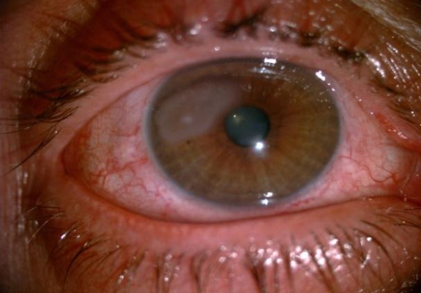
A 46-year old man complaining decrease of visual acuity in his right eye during three days and he had also realized an opacification in his right eye at the same time. The patient had a history of SLE for about ten years. On initial examination, he had a best-corrected visual acuity of 20/50 in the right eye and 20/20 in the left eye. Bio microscopic examination showed 2x3 mm wide corneal interstitial opacification in superotemporal area which is not staining with flourescein (Figure 1).
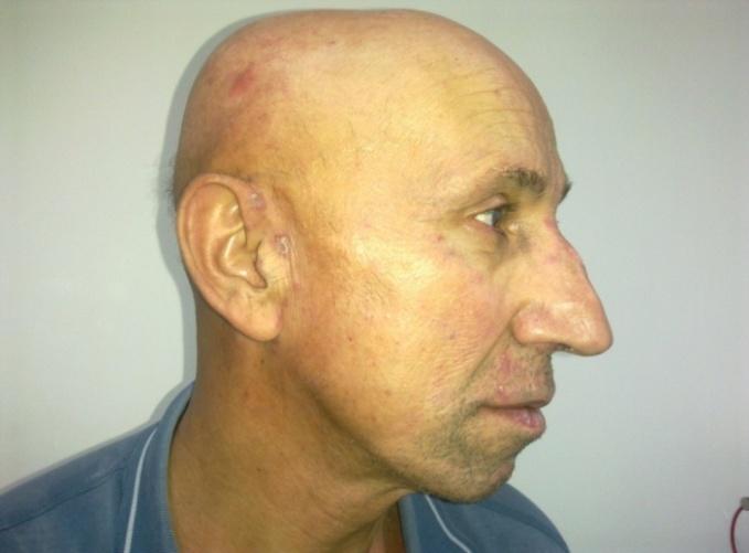
Figure 1: Corneal interstitial opacification in superotemporal area. The outer parts of cornea were clear and there was no cell in the anterior chamber. Intraocular pressure was unremarkable in both eyes and pupils were isochoric with brisk light reaction and no afferent pupillary defect. Her color vision was normal using Ishihara test. Funduscopic examination was also normal in both eyes. Systemic examination was revealed a few discoid erythematos lesions in his noise, ear and face (Figure 2).
Dexamethasone 0.1% eye drop was started in the right eye per hour and dexamethasone 0.1% ophthalmicpomad also started as 2x1. In addition oral metilprednisolone was started 1 mg/kg perday. Vision increased to 20/35 therefore corneal opacity was remained during the first week of treatment. At theend of the 2nd week corneal opacity became smaller and cornea cleared at around of lesion. Unfortunately corneal opacity was still unresolved at the end of the first month.
Discussion
SLE may result in manifestations in any portion of the eye. According to Jensen et al. [2] the most common ocular manifestation of SLE is kerato conjunctivitis sicca, present in one-third of patients. Jensen et al. [2] have also reported diagnoses of SLE, the median value obtained in unanesthetized Schirmer tests was 7.5mm at 5 minutes. In our case, unanesthetized Schirmer tests and BUT values were normal. Periorbital edema has been reported with SLE as a rare finding. Experimental studies in a mouse model of SLE induced by immunization with human monoclonal anti-DNA antibodies resulted in bilateral sub acute and chronic inflammation of the eyelids with immune complex IgG deposition and hyper trophic meibomian glands, providing evidence for the inflammatory nature of this finding [1]. There was no periorbital edema or chronic inflammation of the eyelids in ourcase. Corneal, conjunctival involvement, scleritis- episcleritis, üveitis are the other rare anterior segment manifestations in patients with SLE [1, 3, 4, 5]. Rainzman MB and Baum J have reported two patients with long- standing discoid lupus erythematosus developed acute, unilateral, corneal stromal infiltration and edema. No evidence of infection was found, and both responded rapidly to topical corticosteroid therapy [8]. Likewise there was an acute, unilateral, corneal stromalin filtration and edema in our case but it was not responded to topical corticosteroid therapy. Varga JH and Wolf TC have also reported bilateral transient kerato endotheliitis associated with systemic lupus erythematosus that was responsive to topical and systemic corticosteroid treatment [3]. In addition after successful laser in-situ keratomileusis (LASIK) in patients with SLE; stromal haze, melting, ulceration, and poor wound healing have been reported [9]. In our case, there was no history of LASIK or the other refractive surgical procedure. We suggest may a role for immune complex localization in the pathogenesis in this corneal lesion of SLE. Interstitial keratitis is rare in case of SLE and clinicians should consider it as a possible complication.
Open Access Journal of Ophthalmology
References
-
Read RW (2004) Clinical mini-review: systemic lupus erythematosus and the eye. Ocul Immunol Inflamm 12(2): 87-99.
-
Jensen JL, Bergem HO, Gilboe IM, Husby G, Axéll T (1999) Oral and oculars icca symptoms and findings are prevalent in systemic lupus erythematosus. J Oral Pathol Med 28(7): 317-322.
-
Varga JH, Wolf TC (1993) Bilateral transient keratoendotheliitis associated with systemic lupus erythematosus. Ann Ophthalmol 25(6): 222-223.
-
Heiligenhaus A, Dutt JE, Foster CS (1996) Histology and immunopathology of systemic lupus erythematosus affecting the conjunctiva. Eye 10(4): 425-432.
-
Nguyen QD, Foster CS (1998) Systemic lupus erythematosus and the eye. Int Ophthalmol Clin 38(1): 33-60.
-
Karpik AG, Schwartz MM, Dickey LE, Streeten BW, Roberts JL (1985) Ocular immune reactants in patients dying with systemic lupus erythematosus. Clin Immunol Immunopathol 35(3): 295-312.
-
Adan CB, Trevisani VF, Vasconcellos M, de Freitas D, de Souza LB, et al. (2004) Bilateral deep keratitis caused by systemic lupus erythematosus. Cornea 23(2): 207-209.
-
Rainzman MB, Baum J (1989) discoid Lupus keratitis. Arch Ophthalmol 107(4): 545-547.
-
Cua IY, Pepose JS (2002) Late corneals carring after photo refractive keratectomy concurrent with development of systemic lupus erythematosus. J Refract Surg 18(6): 750-752.
- Screening of Hospital Staff During World Glaucoma Week in a Tertiary Eye Care Centre
- Angioid Streaks with Macular Neovascularization: Clinical Insights from Two Cases
- Giant Kissing Naevus: An Oculoplastic Challenge
- Why Freedom of Vision Should Not Cost the Freedom of Feeling - LASIK in the Climate of Change
- Asymmetric Optic Nerve with Small Disc and Large Cup: A Rare and Challenging Case of Unilateral Optic Nerve Hypoplasia
- Large Angle Exotropia in a Child: A Case Report