Methods and Tools for Selecting Spectrum in Fundus Illumination
Aim: To develop a methodology of creating high-resolution images for vitroretinal surgery without increasing the intensity of light. Methods: A method of replacing a powerful light source whose spectrum is close to the solar one by a set of narrow-band sources with much smaller total energy, their bands are selected according to optical characteristic of informative areas of fundus. A selective spectrum emitter is designed. A method of spectrum optimization is proposed for video registration. The efficiency of the approach was verified by numerical simulation. Results: The experiments on real data the frames of video recording of the operating field using: a) using the Xenon Bright Star, b) a spectrally selective emitter, c) LED emitter have demonstrated the advantage of the proposed methodology. It improves the quality of images, and the intensity of light is even smaller. The analysis of real operations gives a material for selecting a trade-of between the quality and the light load on the eye. Conclusion: The intensity of light is not enhanced and hence, the method doesn’t damage the eye. An additional advantage of the proposed method is an increase in the robustness of video registration with respect to reducing the illumination.
Introduction
One of the problems of vitreoretinal surgery is the insufficient quality of illumination of the operating field [1]. This problem is especially acute when video recording and TV translation of the process of vitreoretinal surgery, both for educational purposes and iv telemedicine, since the requirements for the quality of TV images are high. It appears also in endoscopic optical coherence tomography (OCT), where, in addition to the radiation of the illuminating device, the powerful infrared OCT additionally affects the retina [2].
Modern systems are powerful light sources whose spectrum is close to the solar one, reserves of improving the illumination of the operating field only by increasing the intensity of light transmitted to the fundus are almost exhausted, since the density of light power that falls on the surface of the retina is practically maximal permissible one, in some cases it may exceed ~ 1 mW/mm2 [3]. Increasing the power causes damage of retina/hence, straightforward solution to the problem is unacceptable, and we need some nontrivial approaches.
The article presents the proposed methods and tools of illuminating the fundus operating field. They both improve the visibility of the operating field and reduce the light load on the fundus tissue. At the same time, considerable attention is paid to improving the quality of video recording in low light conditions.
Materials and Methods
The Idea of the Method
The principal idea is to replace a continuous spectrum light source by a small set of narrow-band light sources selected in such a way that the emission wavelengths correspond to the maximum visualization efficiency of the most informative areas of the fundus. In practice, informative objects are the vitreous, the colored and uncolored posterior hyaloid membrane, retinal and choroid tissues, venous and arterial blood. A performance criterion is the visibility to the density of light power ratio on the illuminated surface [4].
The block diagram of a selective spectrum illuminating device is shown in Figure 1. The device consists of several laser light sources, each emitting its wavelength from the set (λ1, λ2, λn), a multiplexer that combines the radiation of all lasers and transmits it into a single multimode optical fiber, a coherence noise removing element (speckle average), a driver for controlling laser light sources, and a B/W camera together with a video grabbers that provide high light sensitivity and high resolution in the registration of a color image [5].
![Figure 1: The device consists of several laser light sources, each emitting its wavelength from the set (λ1, λ2, λn), a multiplexer that combines the radiation of all lasers and transmits it into a single multimode optical fiber, a coherence noise removing element (speckle average), a driver for controlling laser light sources, and a B/W camera together with a video grabbers that provide high light sensitivity and high resolution in the registration of a color image [5].](/fulltextimages/3228/fig_1.jpeg)
Spectrum Optimization
The performance of the proposed method is defined by the emitting spectral components. It is easy to see that the problem of optimizing the spectral composition of the emitter in its general form reduces to the classical problem of detection and estimates described in terms of the theory [6]. Unfortunately, the number of informationally significant objects (N) in our case is much larger than the number of spectral components in the radiation of the emitter (m), and it is impossible to obtain an arbitrary wavelength (λ) of light with the help of lasers. Therefore, the methods and approaches of the theory of detection and estimates, which are based on the Neyman-Pearson optimal detection criteria, are unsuitable for solving our problem. However, it can be shown that the constrained problem reduces to splitting the graph describing the relations among the objects, $$ N (i, j) = \sum_ {k = 1} ^ {m (i)} N _ {1} (i, k) N _ {2} (k, j) = \sum_ {k = 1} ^ {m (t)} \left| E _ {i} ^ {k} \right| $$ | ( ) |(⋃ ( ) )|, where N – The matrix of connections among the elements, N1 –the partition matrix that shows which elements k are the results of the partition of i, m(i) –the number of such elements, N2 –the matrix of connections of the elements k to unseparated elements j, E – The sets of partitions.
For numerical solution, we used a greedy algorithm. The spectra of informative objects are from, and available laser sources, from the national and racial features of pigmentation were not taken into account [7, 8, 9, 10, 11, 12]. Numerical imitation showed that, the visualization efficiency of the topology of the retinal blood vessels and its differentiation using the degree of oxygenation is close to the maximum if the emitter is composed of only two lasers whose wavelengths are λ1 = 577 nm and λ2 = 640 nm. Addition of a third light source with λ3 = 520 nm gives an extra possibility to visualize the vitreous, the posterior hyaloid membrane, the retinal detachment, and the pigment epithelial defects. The energy needed and the light power density required for visually comparable image quality is more than halved.
Specific Features of Video Registration
The reduced total energy of the light reaching the fundus results in an unacceptably low quality of the operating field images the color video camera registers. The reason is the insufficient photosensitivity of modern color video cameras.
Since the sensitivity of a B/W video camera is about an order of magnitude greater than that of a color one, we used a high-speed B/W camera for video recording of color images. Figure 2 presents an algorithm suitable for the registration color images with B/W cameras together with a spectrally selective emitter composed of several light sources (in Figure 2, three ones).
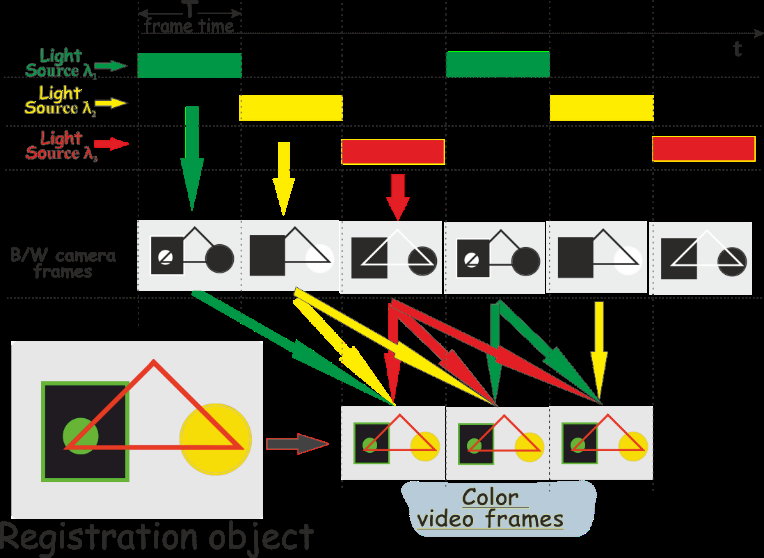
Light sources are switched on sequentially; each one is on during the time of registration of a frame (T) by the B/W camera. Since each source emits monochromatic light, each frame of the B/W camera registers only one color component of the registered object. A sequence of N black-and-white frames, N is the number of light sources, is a necessary and sufficient set of relevant color components in the color image of the registered object.
The graber buffers a sequence of N black-and-white frames and reconstructs a frame of the color image of the registered object using this sequence.
Results
The results of numerical simulation are tested in a medical experiment. In a spectrally selective emitter,
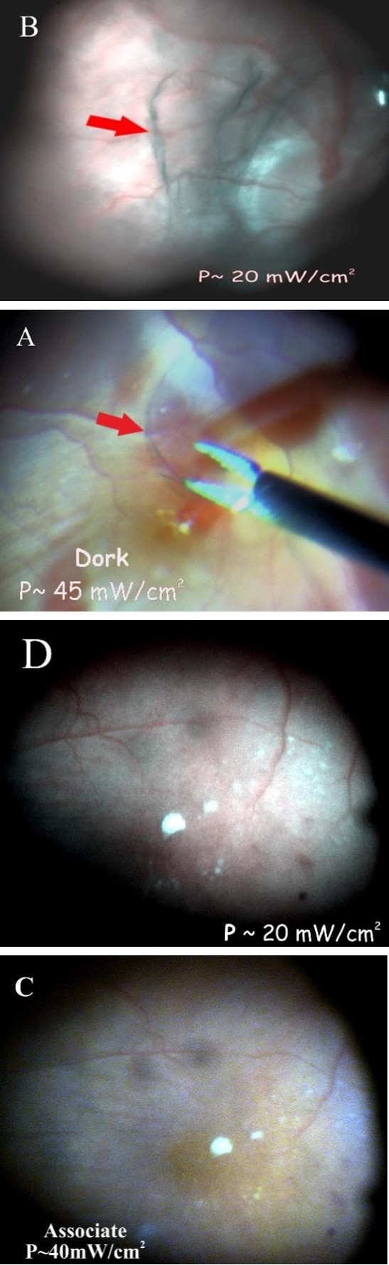
lasers with wavelengths of 520 nm, 577 and 650 nm was used as light sources. Figure 3 presents the results of video recording of the operating field on the fundus using: A) the D.O.R.C Xenon Bright Star, B and D) a spectrally selective illuminator, C) LED Illuminator Associate (DORC).
Frames A and Care obtained using the color CCD video camera B1641, the density of light power in the area of the arrow was 40 - 45 mW/cm2 and the frame frequency is 30 fpc. The arrow indicates the image of the colored posterior hyaloid membrane. Frames B and D are obtained using a B/W CMOS camera DMK 33GX236, the light power density in the central region of the frame is approximately 20 mW/cm2, the frame frequency is 50-60 fpc. To convert the frames of a black-and-white image into a color one, the algorithm described in was used the ratio of the powers of the individual light sources in the spectral selective emitter and the weight functions of the algorithm for processing the black-and-white video stream were selected using the criterion of maximum visual correspondence to the image obtained with the LED Associate (DORC) illuminator [5].
Figures 3C and 3B demonstrate the quality of visualization of the fundus and the choroid topology. It can be seen that, the image details of the retinal topology and defects of the choroid are better if the spectral selective illumination is used. This is mainly due to the large integral depth of light penetration, the elimination of transverse chromatic aberrations at the stage of conversion of a black and white image into a color one, and the robustness of video registration under a decrease in illumination.
Discussion and Conclusion
The proposed approach allows the surgeons to obtain fundus images whose resolution is sufficient for ophthalmologic operations. At the same time, the intensity of light is not enhanced end hence; the method doesn’t damage the eye. An additional advantage of the proposed method is an increase in the robustness of video registration with respect to reducing the illumination. An implementation and examples demonstrating the advantage of the approach are also presented.
References
-
Sakaguchi H, Oshima Y (2012) Considering the illumination choices in vitreoretinal surgery: continual improvements allow for better, safer outcomes. Retinal Phys 9: 26-31. [INLINE_TABLE:4:0] Optical Coherence Tomography in Macular Diseases. India: Springer, New Delhi.
-
Charles S (2008) Illumination and phototoxicity issues in vitreoretinal surgery. Retina 28(1): 1-4.
-
Yamgutdinov R (2017) Brightness and phototoxicity – two sides of endoillumination. Bulletin of Bashkir state medical University (7).
-
Patent RU# 2215463.
-
Van TH (1968) Detection, Estimation, and Modulation Theory, Part I. New York: John Wiley and Sons.
-
Tabulated Molar Extinction Coefficient for Hemoglobin in Water.
-
Schmitt J (1986) Optical Measurement of Blood Oxygenation by Implantable Telemetry. Technical Report G558-15, Stanford.
-
Moaveni M (1970) A Multiple Scattering Field Theory Applied to Whole Blood. Ph.D. dissertation, Dept. of Electrical Engineering, University of Washington.
-
Hammer V, Schweitzer D (2002) Quantitative reflection spectroscopy at the human ocular fundus. Phy Med Biol 47(2): 179-191.
-
Delori FC, Pflibsen KP (1989) Spectral reflectance of the human ocular fundus. Appl Opt 28(6):1061-1077.
-
Changchun New Industries Optoelectronics Technology.
- Screening of Hospital Staff During World Glaucoma Week in a Tertiary Eye Care Centre
- Angioid Streaks with Macular Neovascularization: Clinical Insights from Two Cases
- Giant Kissing Naevus: An Oculoplastic Challenge
- Why Freedom of Vision Should Not Cost the Freedom of Feeling - LASIK in the Climate of Change
- Asymmetric Optic Nerve with Small Disc and Large Cup: A Rare and Challenging Case of Unilateral Optic Nerve Hypoplasia
- Large Angle Exotropia in a Child: A Case Report