Posterior Capsule Rupture: Incidence Rate, Associated Characteristics and Visual Outcomes
Purpose: To see the Incidence rate, Risk factors, Associations, and Visual outcomes of posterior capsule rupture (PCR). Methods: A cross sectional study was done of all patients with PCR from June to November 2021 in a Tertiary eye care Hospital. In this study, distribution of the respondents by age, the morphology of cataract, risk factors for PCR, Surgeon’s experience, percentage of Phaco and SICS, in which steps of surgery PCR occurred, implantation of IOL following PCR were analyzed. The final visual outcome of cases with PCR was measured. Results: Among 7650 patients, who underwent cataract surgery during the study period, 142 had eventful cataract surgery with various per operative complications, and PCR was the most common (n = 94). Most of the study population was in the age group 61-70 years (37.2%). About 45% were male, and 55% were female. Among 94 cases, PCR occurred in 65 patients (69.1%) during Phaco and 29 (30.8%) cases during SICS. There was no statistically significant difference between Phaco and SICS for PCR (> 0.05). In most patients (26.1%), PCR occurred during cataract surgeries irrigation and aspiration step. The most common risk factors for developing PCR were small pupil (10.6%) and corneal opacity (9.5%). PCR was higher by trainee surgeons rather than by senior surgeons. There was a statistically significant improvement in postoperative visual acuity even when PCR occurred during surgery (P< 0.001). Conclusions: PCR occurs in all cataract surgeries and is the main intraoperative complication. This study identified the incidence rate and risk factors of occurring PCR during cataract surgery in a tertiary eye hospital of Bangladesh, which may assist in the application of preventive measures to decrease rates of PCR. Eyes having posterior capsule rupture at the time of cataract surgery have a significant risk of reduced visual acuity.
and Syeed Mehbub Ul Kadir3
Chamber Intraocular Lenses; ACIOL: Anterior Chamber Intraocular Lenses; SPSS: Statistical Package for Social Sciences; PCT: Posterior Capsule Tear; PPC: Posterior Polar Cataract.
Introduction
Cataract is the commonest cause of reversible blindness, and cataract surgery is the most routine intraocular surgery performed. Surgical problems do occur, despite advancements in the area of cataract surgery. Posterior Capsule Rupture (PCR) is the most common potentially sight‑threatening intraoperative complication during cataract surgery [1]. PCR may necessitate more surgical procedures, more postoperative follow-up visits, and a higher rate of postoperative complications, all of which could compromise the final visual outcome. Furthermore, PCR can lead to various other issues, including retinal detachment, macular oedema, and uveitis, glaucoma, and IOL dislocation. Today, however, the control provided by closed chamber current surgical procedures may allow for a final aesthetic outcome comparable to a straightforward instance [2]. It is preferable to be better prepared to avoid or effectively manage this difficulty. Whether it has a vitreous loss (VL) or not, a poorly managed PCR can compromise the great results associated with standard cataract surgery. Several essential surgical concepts apply universally to all patients with PCR and every cataract surgeon should learn these fundamental principles to avoid and manage the long-term consequences [1, 2]. With this thought, this study analyzed the risk factors that caused. It was associated with PCR and the visual outcome of patients who had PCR during cataract surgery in a Tertiary Eye care Hospital.
Materials and Methods
A cross sectional study was conducted in a Tertiary Eye care Hospital to study the incidence rate, risk factors, associations & visual outcomes of patients who had PCR as a complication during cataract surgery. Data was collected for age, risk factors for PCR, surgeon’s experience, a difference of incidence of PCR between Phaco and SICS, in which steps PCR occurred, Morphology of cataract, Implantation technique of IOL following PCR, and postoperative best- corrected visual acuity. Cataract surgeries are done by senior surgeons, Mid-level surgeons, Trainees. The study covered the period of June 2021 to November 2021. All ocular comorbidities such as pterygium involving the cornea, corneal opacities, noncooperative patient, Past Ocular Injury, High myopia, small pupil, Floppy Iris, Zonular dehiscence, past vitrectomy were noted. Lens-related categories Cortical, PSC, PPC, Mature cataract, HMC, Brown, Black, and NSI/II/ III were recorded. In all cases, the vitreous loss was managed by anterior vitrectomy. Posterior chamber intraocular lenses (PCIOL) are placed if there is an adequate posterior capsule to support it. Otherwise, anterior chamber intraocular lenses (ACIOL) are placed. The visual outcome was taken as the best- corrected visual acuity based on refraction done by hospital- based optometrists. The visual acuity was measured by using a Snellen chart. The visual acuity was recorded on the first postoperative day. The best-corrected outcome was divided into good vision (6/6-6/12), impaired vision (6/18-6/60) and poor vision (worse than 6/60). Small sample size, single centered study, and limited follow up of the patients were the limitations of the study.
Both quantitative and qualitative data were statistically analyzed with Statistical Package for Social Sciences (SPSS) program version 24(USA). P<0.05 was considered for a level of statistical significance.
Results
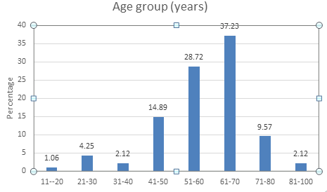
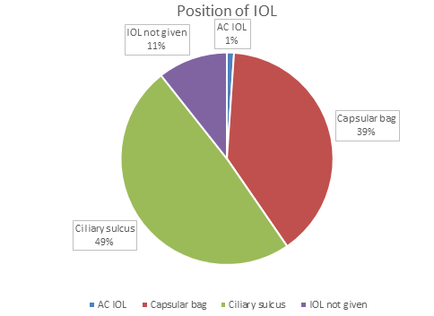
Among the 94 cases with PCR, Most of the study population was in age group 61-70 years (37.2%), 51- 60 years (28.72%), 71-80 years (9.5%) and 81-100 years (2.1%) (Figure 1). The age range was 16-98 years, and the mean age ±SD was 59.3±13.1years. About 45% of the study respondents were male, and 55% were female. PCR occurred in 65 (69.1%) patients during Phaco and 29 (30.8%) patients during SICS. The rate PCR was 1.18% in total 5480 patients who underwent phacoemulsification and 1.33% in the 2170 patients who underwent small incision cataract surgery (Table 1). There was no statistically significant difference between Phaco and SICS for PCR (>0.05). Different types of cataracts were responsible for developing PCR (Table 2). PCR occurred during the irrigation aspiration step of cataract surgery in most cases (26.1%) (Table 3). The associated risk factors for developing PCR where the small pupil was found in 10 cases (10.6%), and corneal opacity was found in 9 cases (9.5%) (Table 4). The occurrence rate of PCR was higher by trainee surgeons than by senior surgeons (Table 5). The implantation of IOL were placed in the ciliary sulcus (49%), capsular bag (39%), and in (1%) Anterior chamber IOL (Figure 2). But IOL was not given in 11% of cases due to lack of capsular bag support with the secondary SFIOL or Iris fixation lens. There was a statistically significant difference in visual acuity before and after cataract surgery (Table 6), even developing PCR during surgery (P< 0.001).
| Mode of Surgery | Posterior Capsular Rupture (n=94) | Odd Ratio | Chi-Square | P-Value | |
|---|---|---|---|---|---|
| Yes | No | ||||
| PE (n=5480) | 65(1.18%) | 5415(98.81%) | 0.887 (<1) | 0.289 | 0.59 |
| SICS(n=2170) | 29(1.33%) | 2141(98.66%) |
Table 1: Distribution of the PCR in Phacoemulsification (PE) and SICS (n=94).
| Types of Cataracts | Frequency | Percentage |
|---|---|---|
| NSII, PSC | 19 | 20.2 |
| NS III, PSC | 15 | 15.9 |
| Cortical | 5 | 5.3 |
| PSC | 17 | 18 |
| PPC | 3 | 3.1 |
| Mature cataract | 9 | 9.5 |
| HMC | 10 | 10.6 |
| Brown | 3 | 3.1 |
| Black | 2 | 2.1 |
| NS I/II/III | 11 | 11.7 |
| Total | 94 | 100 |
Table 2: Distribution of the study cases by the morphology of cataract (n=94).
| Steps of Surgery for PCR | Frequency | Percentage (%) |
|---|---|---|
| Rhexis extension | 5 | 5.3 |
| Rotation of lens | 3 | 3.1 |
| Phacoemulsification | 7 | 7.4 |
| IOL insertion | 12 | 12.7 |
| IOL dialing | 3 | 3.1 |
| Chopping | 3 | 3.1 |
| Quadrant removal | 11 | 11.7 |
| Epi nucleus removal | 7 | 7.4 |
| Cortex removal | 7 | 7.4 |
| Nucleus delivery | 11 | 11.7 |
| Irrigation aspiration | 25 | 26.5 |
| Total | 94 | 100 |
Table 3: Distribution of the study cases according to steps of surgery for PCR (n=94).
| Risk Factors | Frequency | Percent |
|---|---|---|
| Non-co-operative patient | 5 | 5.3 |
| Past Ocular Injury | 1 | 1 |
| High myopia | 1 | 1 |
| Corneal opacity | 9 | 9.5 |
| Nasal pterygium | 5 | 5.3 |
| Small pupil | 10 | 10.6 |
| Floppy Iris | 4 | 4.2 |
| Zonular dehiscence | 3 | 3.1 |
| Past vitrectomy | 1 | 1 |
| Unknown | 55 | 58.5 |
| Total | 94 | 100 |
Table 4: Associated Risk factors for PCR (n=94).
| Frequency | No. of PCR | Percent |
|---|---|---|
| Senior surgeon | 54 (out of 4960) | 1.08 |
| Mid-level surgeon | 33 (out of 2690) | 1.22 |
| Trainee surgeon | 7 (out of 314) | 2.22 |
| Total | 94 | 100 |
Table 5: Distribution of the study cases according to PCR done by a surgeon (n=94).
| VA | Pre-Operative | 1st POD | P Value |
|---|---|---|---|
| 6/6 to 6/12 | 27 (28.7%) | 52 (55%) | < 0.001 |
| 6/18 to 6/60 | 49 (52.1%) | 35 (37.2%) | |
| Worse than 6/60 | 18 (19.1%) | 7 (7.4%) |
Table 6: ** Visual acuity following PCR (n=94).
Data were analyzed using the Chi-square test Table 6: Visual acuity following PCR (n=94).
Discussion
Posterior capsule rupture (PCR) or posterior capsule tear (PCT) t is the most common extracapsular cataract surgery operating complication that impacts postoperative visual acuity. This retrospective observational study was conducted in a tertiary level hospital in Bangladesh to investigate occurrence rate, association, and visual outcome among patients of PCR during cataract surgery from June to November 2021. Here ninety-four PCR cases were selected from 142 cases of postoperative complication among 7650 patients who underwent cataract surgery during the study period.
The incidence rate of PCR was 1.2% and 66.1% of postoperative complications. In previous studies, the occurrence rate of PCR in cataract surgery has been calculated as 1.9-5.2% [3, 4]. Another Chen M, et.al. [5] Study found a 0.68% incidence rate of PCR.
Sixty-five (1.1%) patients had PCR who underwent Phaco while 29 (1.3%) patients in the group of SICS. There was no significant difference between Phaco and SICS for PCR (P>0.05). But Bhutto et al. found 3% PCR in the case of Phaco and 1.5% in the case of SICS [6]. In another study, Kyei S, et al. [7] mentioned a 3.8 % incidence of PCR among patients who underwent Phaco while the incidence rate was 1.8% among groups of SICS.
Different types of cataracts were found responsible for developing PCR in this study. But certain types of cataracts are at a higher risk for developing PCR. They are Posterior polar cataract (PPC), White cataract, Brunescent/black cataract in this hospital cataract surgery for PPC, black and hyper mature cataracts are mainly done by senior and experienced surgeons so that it may be the cause of lower incidence of PCR in these cases [6, 7].
In this study, PCR happened in different steps of Phaco and SICS, where 26.5% PCR occurred in the irrigation and aspiration phase. Thanigasalam T, et al. [8] found that PCR occurred more during segment removal & cortical removal. Bai H, et al. [9] study showed the occurrence of PCR was more common in Phaco or irrigation and aspiration phase.
Some risk factors for PCR were found in this study. Hyper mature cataract (8.5%), brown cataract (3.1%),black cataract (3.1%), corneal opacity (5.1%), small pupil (3.1%), zonular dehiscence (3.1%) were more commonly seen where PCR occurred Chen M, et al. [5] found restlessness of patient, small pupil, zonular dehiscence, pseudoexfoliation, floppy iris, shallow chamber as causative factors for PCR [5].
The occurrence rate of PCR is mostly by trainee rather than a midlevel and senior surgeon in this study. As it is a tertiary level hospital, the bulk of cataract surgery and referred case are done by the senior surgeon. So they managed the case properly following PCR with their experience. Similarly, another study by Bai H, et al. [9], Chen M, et al. [5] mentioned surgeons with the highest number of patients had the lowest rate of PCR than surgeons with the lowest number of cases. Ionides A, et al. [10] also mentioned PCR occurrence rate more by trainee surgeons than an experienced surgeon.
Among 94 PCR cases, only one IOL was implanted in the anterior chamber. In all other patients, the IOL was implanted in either the capsular bag (in 37 eyes) or the ciliary sulcus (in 46 eyes). Due to inadequate posterior capsule and ciliary sulcus support, in 10 cases, the IOLs were planned but were implanted later. Other studies Thevi T, et al. [11] also mentioned IOL placement in the anterior chamber, capsular bag, ciliary sulcus, and fixated scleral IOL due to inadequate posterior capsular support and planning of implantation of IOL at a later [11].
The visual study was significantly improved (P< 0.05) in this study following PCR due to being managed by a senior surgeon and small PCR where anterior vitrectomy required little. Ionides A, et al. [10] encountered those eyes with PCR were 3. 8 times more likely to get the vision worse than 6/12 [10]. Another study Thevi T, et al. [11] mentioned, there was no statistically significant reduction of visual outcome following PCR. Good visual outcomes were significantly higher among specialists than trainees following PCR [5, 11, 12].
PCR is the commonest intraoperative complication that may cause poor vision. No technique is superior to the other as it occurs significantly in all cataract surgeries. In combination, it is better to do filtering surgeries and various surgeries after trauma as separate procedures from cataract surgery [5, 13].
Conclusion
By recognizing predisposed conditions and modifying the surgical approach appropriately, the incidence of PCR can be considerably reduced. The key to a favorable postoperative outcome is early detection of a posterior capsular tear and rapid therapy of the capsular tear and vitreous prolapse. We recommend and encourage other researchers to conduct more studies about clinical investigations of posterior capsule rupture, its proper management and finally, good visual outcome.
Author Contributions
MR, MS, and MRR: Designed the research study, procured the samples, performed the experiments, and interpreted the results. MR, MS, and YJK: Designed and performed the statistical analyses. MR, SMRH, and CS: Provided critical input. MR, MRR, and YJK: Wrote the first draft of the manuscript with information from all co-authors. MR, MS, CS, SMK: Critical appraisal of the manuscript. All authors reviewed and approved the final version of the manuscript before submission.
Informed Consent
All subjects gave informed written consent before their surgery and gave the consent to use their clinical documents for the study.
Statement of Ethics
This study was performed with the highest ethical standards per the World Medical Association Declaration of Helsinki.
Funding Sources
The authors declared that this study received no financial support.
Data Availability Statement
Data are available upon request which can be directed to the corresponding author.
Conflict of Interest Statement
The authors declare no potential conflicts of interest.
References
-
Zainal M, Ismail SM, Ropilah AR, Elias H, Arumugam G, et al. (2002) Prevalence of blindness and low vision in Malaysian population: results from the National Eye Survey1996. Br J Ophthalmol 86(9): 951-956.
-
Evi an Amazon Company. The population of Malacca City in 2014 was around 474000.
-
Johnston RL, Taylor H, Smith R, Sparrow JM (2010) The national cataract dataset electronic multi-centre audit of 55567 operations: variation in PCR rates between surgeons. Eye 24(5): 888-893.
-
Narendran N, Jaycock P, Johnston RL, Taylor H, Adams M, et al. (2009) The cataract national dataset electronic multicentre audit of 55,567 operations: risk stratification for posterior capsule rupture and vitreous loss. Eye 23(1): 31-37.
-
Chen M, Lamattina KC, Patrianakos T, Dwarakanathan S (2014) Complication rate of posterior capsule rupture with vitreous loss during phacoemulsification at a Hawaiian cataract surgical centre: a clinical audit. Clin Ophthalmol 8: 375-378.
-
Bhutto IA, Memon MN, Ali I, Soomro AQ, Indhar I (2021) Comparison of Per-operative and Early Postoperative Complications between Manual Small Incision Cataract Surgery and Phacoemulsification in Patients with Senile Cataract. Pak J Ophthalmol 37(4): 384-387.
-
Kyei S, Zaabaar E, Assiamah F, Kwarteng MA, Asiedu K (2021) Comparison of the Outcomes of Manual Small Incision Cataract Surgery (MSICS) and Phacoemulsification (PHACO) in Ghana. Ann Afr Surg 18(3): 143-149.
-
Thanigasalam T, Sahoo S, Ali MM (2015) Posterior capsule rupture with /without vitreous loss during phacoemulsification in a hospital in Malaysia. Asia Pac J Ophthalmol (Phila) 4(3): 166-170.
-
Bai H, Yao L, Wang H (2020) Clinical Investigation into Posterior Capsule Rupture in Phacoemulsification Operations Performed by Surgery Trainees. J of Ophthalmol.
-
Ionides A, Minassian A, Tuft S (2001) Visual outcome following posterior capsule rupture during cataract surgery. Br J Ophthalmol 85(2): 222-224.
-
Thevi T, Maizura MZ (2016) Posterior capsule rupture- causes, associations and outcomes: eight-year analysis in a Malaysian General Hospital. Guoji Yanke Zazhi (Int Eye Sci ) 16 (4): 600-606.
-
Ti SE, Yang YN, Lang SS, Chee SP (2014) A 5-year audit of cataract surgery outcomes after posterior capsule rupture and risk factors affecting visual acuity. Am J Ophthalmol 157(1): 180-185.
-
Daien V, Le Pape A, Heve D, Carriere I, Villain M (2015) Incidence and characteristics of cataract surgery in France from 2009 to 2012: a national population study. Ophthalmology 122(8): 1633-1638.
- Screening of Hospital Staff During World Glaucoma Week in a Tertiary Eye Care Centre
- Angioid Streaks with Macular Neovascularization: Clinical Insights from Two Cases
- Giant Kissing Naevus: An Oculoplastic Challenge
- Why Freedom of Vision Should Not Cost the Freedom of Feeling - LASIK in the Climate of Change
- Asymmetric Optic Nerve with Small Disc and Large Cup: A Rare and Challenging Case of Unilateral Optic Nerve Hypoplasia
- Large Angle Exotropia in a Child: A Case Report