Cephalic Disorders with Neuro-Ophthalmic Manifestations
Purpose: To report three different cases of cephalic disorders with neuro-ophthalmic manifestations. Method: Descriptive case report. Case Summary Case 1: 14 years female came with Primary optic atrophy (POA) of both eyes associated with poor intelligence. MRI brain showed disproportionate dilatation of both occipital horn of lateral ventricle suggestive of Colpocephaly. Case 2: 16 years male presented with POA of eyes, horizontal jerky nystagmus and left sided hemiparesis with contracture of left hand since birth. MRI brain suggestive of Open lip Schizencephaly. Case 3: 12 years boy came with alternate exodeviation, left sided hemiparesis since birth. MRI brain suggestive of right sided Procencephalic cyst. Conclusion: Cephalic disorders caused by disturbance in development of fetal nervous system. For most cephalic disorder’s treatment is only symptomatic and supportive. Early diagnosis may ensure patient’s quality of living.
Introduction
Cephalic disorders are congenital condition that stem from damage to, or abnormal development of the budding nervous system. More common in premature baby specially those born with hypoxia or lung immaturity. It is caused by hereditary or genetic conditions or by environmental exposure during pregnancy such as medication, maternal infection or exposure to radiation [1].
In this study, we will report 3 different cases of cephalic disorders along with neuro-ophthalmic manifestations.
Case No 1
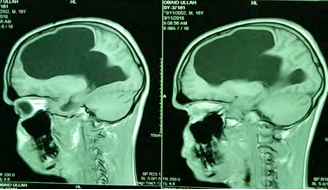
A 14 year female came with poor vision of both eyes since childhood. She was dumb with poor intelligence. Vision in right eye was 6/36, left eye 6/24, not improved with pin hole. Color vision was impaired in both eyes, pupillary reaction in both eyes were sluggish, both the optic discs were pale (Figure 1).
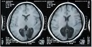
MRI of brain showed disproportionate dilatation of both occipital horn of lateral ventricle with hypoplastic corpus callosum, feature suggestive of Colpocephaly.
Case No 2
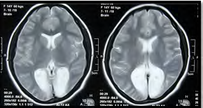
A 16 year male came with poor vision of both eyes along
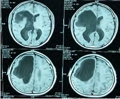
MRI of brain showed a large CSF intensity lesion involving right fronto-parietal lobe communicating to right lateral ventricle with moderate dilatation of both lateral ventricles, feature suggestive of Open lip Schizencephaly.
with weakness of left side of the body since childhood. Visual acuity in both eyes were 6/36, not improved with pinhole, color vision was significantly impaired, pupillary reaction in both eyes were sluggish, optic discs were pale and there was horizontal jerky nystagmus. The patient had left sided hemiparesis with deformity of left hand (Figure 2).
Case No 3
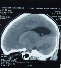
A 12 year boy presented with alternate outward deviation of both eyes, weakness of left side of the body since birth. There was alternate exodeviation, visual acuity in right eye was 6/6 and left eye 6/9, not improved with pin hole and left sided hemiparesis. Both the fundi were normal (Figure 3).
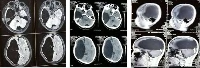
MRI of brain showed CSF filled large cavity in right cerebrum communicating with right lateral ventricle with thinning of overlying cerebral parenchyma, feature suggestive of Procencephalic cyst.
Discussion
Colpocephaly believed to occur as a result of neuronal migration disorders during early brain development, intrauterine disturbances, perinatal injuries and other central nervous system disorders and may present with various degrees of motor disabilities, visual defects, spasticity and moderate to severe intellectual disability [2].
Schizencephaly is a rare congenital malformation with prevalence of 1.48 in 100,000 live births [3]. The defect characterized by abnormal clefts or slits in cerebral hemisphere of brain which may be unilateral or bilateral, lined with grey matter. Common clinical features include microcephaly, hydrocephalus and intellectual disability, partial or complete paralysis and or hypotonia [4].
Procencephalic cyst is an extremely rare cephalic disorder, characterized by congenital or acquired cavity within cerebral hemisphere, containing cerebrospinal fluid [5]. It occurs due to disturbance of vascular supply leading to cerebral degeneration, also due to abnormal development (malformative), direct damage, inflammation or haemorrhage [6]. The patient may suffer from minor neurological deficit to severe disability even death.
Conclusion
The cause of cephalic disorder is multifactorial and neuro imaging is the investigation of choice. The condition is
usually nonfatal. There is no definitive treatment and visual prognosis is guarded. If physiotherapy, occupational therapy, speech therapy given in early state, may reduce morbidity and improve quality of living.
Conflict of Interest
There is no conflict of interest involved.
References
-
Cephalic Disorders Fact Sheet. The National Institute of Neurological Disorders and Stroke (NINDS).
-
Puvabanditsin S, Garrow E, Ostrerov Y, Trucanu D, Ilic M, et al. (2006) Colpocephaly: a case report. Am J Perinatol 23(5): 295-297.
-
Barkovich AJ, Norman D (1988) MR imaging of schizencephaly. AJR Am J Roentgenol 150(6): 1391- 1396.
-
(2014) NINDS Schizencephaly Information Page. National Institute of Neurological Disorders and Stroke.
-
Parker J (2004) The official parent’s sourcebook on porencephaly: A revised and updated directory for the internet age. ICON Health Publications.
-
Debus O, Kosch A, Strater R, Rossi R, Nowak-Gottl U (2004) The Factor V G1691A Mutation is a Risk for Porencephaly: A Case-control Study. Annals of Neurology 56(2): 287-290.
- Screening of Hospital Staff During World Glaucoma Week in a Tertiary Eye Care Centre
- Angioid Streaks with Macular Neovascularization: Clinical Insights from Two Cases
- Giant Kissing Naevus: An Oculoplastic Challenge
- Why Freedom of Vision Should Not Cost the Freedom of Feeling - LASIK in the Climate of Change
- Asymmetric Optic Nerve with Small Disc and Large Cup: A Rare and Challenging Case of Unilateral Optic Nerve Hypoplasia
- Large Angle Exotropia in a Child: A Case Report