Moon in the Eye
A 78-year-old male visited our OPD with a history of painless, progressive diminition of vision in his left eye for the past four months. He was diagnosed as having posterior capsular opacificaton (PCO) of the left eye at some other center, and was referred to us for YAG laser capsulotomy. He gave a history of undergoing bilateral cataract surgery with intraocular lens (IOL) implantation three years back, but there were no old records. There was no history of any systemic diseases. His best corrected visual aquity was 6/6 in the right eye and 6/24 in the left eye. Torch and slit lamp examination revealed bilateral pseudophakia with left eye having opacified IOL, resembling a moon in the eye (Figure 1) with fundal glow being appreciated between the IOL margins and the pupil. The patient was advised IOL exchange, but asked for some time before making a decision for undergoing the procedure.
Image Article
A 78-year-old male visited our OPD with a history of painless, progressive diminition of vision in his left eye for the past four months. He was diagnosed as having posterior capsular opacificaton (PCO) of the left eye at some other center, and was referred to us for YAG laser capsulotomy. He gave a history of undergoing bilateral cataract surgery with intraocular lens (IOL) implantation three years back, but there were no old records. There was no history of any Image Article systemic diseases. His best corrected visual aquity was 6/6 in the right eye and 6/24 in the left eye. Torch and slit lamp examination revealed bilateral pseudophakia with left eye having opacified IOL, resembling a moon in the eye (Figure 1) with fundal glow being appreciated between the IOL margins and the pupil. The patient was advised IOL exchange, but asked for some time before making a decision for undergoing the procedure.
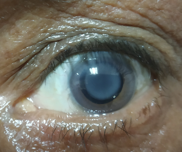
IOL opacification is also called as ‘Tertiary cataract’ as it resembles a white cataract and is often misdiagnosed as posterior capsular opacification. It is a rare, late postoperative complication of cataract surgery and is seen commonly with hydrophilic IOLs. The cause of this opacification is probably due to the disturbance of the blood‐aqueous‐barrier [1]. IOL opacification has been seen in patients with diabetes mellitus and hypertension. The diagnosis is easily clinched by a thorough slit‐lamp examination. The definitive management is explanation of opacified IOL followed by implantation of a new IOL [2].
Source of Support-None
The paper being submitted has not been published, simultaneously submitted or already accepted for publication elsewhere.
Conflict of Interest
The authors declare that they have no competing interest.
Financial Disclosures
The authors have no proprietary or commercial interest in any material discussed in this article.
References
-
Gupta G, Goyal P, Bal A, Jain AK, Malhotra C (2020) Pearly white intraocular lens opacification: “Tertiary cataract”. Indian J Ophthalmol 68(1): 188-189.
-
Jain P, Pattnaik A (2021) Intraocular lens opacification: A rare enigma. J Clin Ophthalmol Res 9: 51-54.
- Screening of Hospital Staff During World Glaucoma Week in a Tertiary Eye Care Centre
- Angioid Streaks with Macular Neovascularization: Clinical Insights from Two Cases
- Giant Kissing Naevus: An Oculoplastic Challenge
- Why Freedom of Vision Should Not Cost the Freedom of Feeling - LASIK in the Climate of Change
- Asymmetric Optic Nerve with Small Disc and Large Cup: A Rare and Challenging Case of Unilateral Optic Nerve Hypoplasia
- Large Angle Exotropia in a Child: A Case Report