Fungal Orbital Cellulitis by Candida Albicans Following Snake Bite -A Rare Case
Fungal orbital cellulitis is extremely rare in immune-competent individuals. An ocular manifestation of snake bite is rare and orbital cellulitis following snake bite is one of the rarest entities. A 10 year old boy presented with periorbital edema with lid necrosis following snake bite on upper lid of the right eye. Patient was treated with intravenous empirical antibiotic along with anti-snake venom. The following day, the patient developed proptosis of the right eye. CT scan suggested orbital cellulitis. Microbiological examination confirmed Candida species. Patient was started on intravenous and topical antifungal agent with resultant decrease of proptosis. But, despite ocular improvement, the patient succumbed due to respiratory failure. The present case highlights fungal orbital cellulitis in immune-competent patient as one of the rarest manifestation of snake bite. Clinical suspicion and diagnosis by imaging and microbiological examination and immediate treatment are necessary while treating such patients.
Introduction
Venomous snakebite is a medical emergency and orbital cellulitis is one of the ocular emergencies. Orbital cellulitis is an infection of the soft tissue surrounding the orbit. Orbital cellulitis of fungal origin is one of the most serious ocular infections with significant potential morbidity, including loss of vision, cavernous sinus thrombosis, intracranial spread of infection and occasionally death. Fungal orbital cellulitis is uncommon manifestation and is rare in an immune- competent person. To the best of our knowledge, literature does not show any case of fungal orbital cellulitis in immune- competent following snakebite.
Case Report
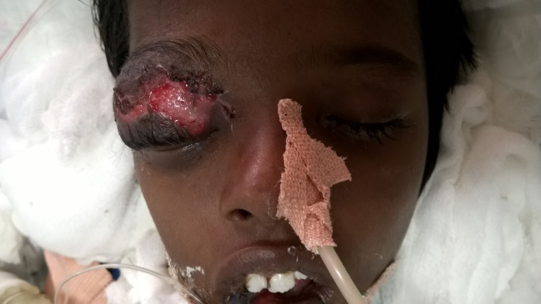
A 10-year-old male was bitten by a cobra over right upper lid and he presented 8 hours later in the emergency department with low grade fever, malaise, nausea and vomiting and altered sensorium. He had received first aid following the bite in the form of local wound incision. There was no sign of internal or external bleeding. Clinically, there was periorbital edema associated with local area of necrosis of right upper eyelid. Visual acuity could not be assessed. Anterior segment examination of right eye showed conjunctival congestion, a quiet anterior chamber of normal in depth. Pupils were briskly reacting to light. Extra ocular movements could not be assessed as the patient’s sensorium was altered. Intraocular pressures were 16 mm Hg for right eye and 14 mm Hg for left eye by Perkin’s tonometry. Fundus examination was normal. Patient was started on anti-snake venom and empirical intravenous antibiotic with topical antibiotic eye drop and eye ointment in view of preseptal cellulitis following snake bite. There was no history of chronic sinusitis and diabetes mellitus. Systemic evaluation did not reveal any evidence of an immuno-compromised state. On second day patient deteriorated systemically and was unable to maintain oxygen saturation and had to be intubated. Ocular examination revealed development of proptosis (Approximate Hertel’s exophthalmometry: 22mm of right eye and 16mm for left eye at 95mm) with worsening of periorbital edema and necrosis Figure 1 with normal reacting pupil.
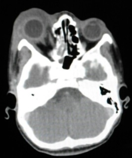
The anterior chamber did not reveal any inflammation. Fundus was within normal limits. Investigations like renal function test, bleeding time and clotting time were normal and blood culture was sterile. CT scan showed a collection in the preseptal area, intra and extraconal space of the superior portion of right orbit with inferolateral displacement of the eyeball Figure 2.
Incision and drainage was done of the right orbit. Wet mound preparation of pus from the wound showed pseudo hyphae and budding cells and culture in Sabouraud’s dextrode agar showed yeast-like colonies which was confirmed by Gram’s staining. Germ tube test identified the organism as Candida albicans. A diagnosis of fungal preseptal cellulitis with orbital cellulitis of right eye following snake bite was established. With these results, the treatment included intravenous amphotericin-B (one miligram per kilogram per day infusion in 5% dextrose) and topical clotrimazol ointment. On fourth day of this treatment, proptosis and periorbital edema started decreasing and so the same treatment continued. Unfortunately, despite ocular improvement, on fifth day patient deteriorated and succumbed due to respiratory failure.
Discussion
Poisonous snake bites result in multisystem toxicity. Ocular manifestations following snake bite are usually rare. Various ocular manifestations of snake bite are documented in literature including bilateral angle closure glaucoma [1], acute anterior uveitis, optic disc edema [2], direct injury to the eye causing open globe injuries, conjunctival and corneal lacerations [3], endophthalmitis [4], and vitreous haemorrhages [5], and exudative retinal detachment [1]. Fungal orbital infections are rare especially in immune- competent individuals. Fungal orbital cellulitis following snake bite has been reported uncommonly and a thorough review of the English literature found no reports of fungal orbital cellulitis following snake bite. Interestingly, there is paucity of literature of snakebite over eyelid. Proptosis and orbital cellulitis of the eye is commonly seen with rhino-orbital mucormycosis in the immunocompromised [6] and less commonly in individuals without predisposing medical conditions [7]. Trauma to the orbital region can also introduce fungi leading to an orbital cellulitis and proptosis [8]. The cause of fungal orbital cellulitis in this patient is unclear. It may have been due to the direct inoculation of fungal element in preseptal and postseptal area following snakebite or indirectly through the normal flora of the conjunctiva. Other proposed mechanism of fungal orbital cellulitis in this patient is the inflammatory response of host against the protein and nonprotein component of the snake venom with superimposed fungal infection. Concomitant systemic instability and antibiotic use also predisposes to fungal growth.
There is no conclusive evidence for any of these theories, just indirect evidence. Microbiological confirmation, response to anti-fungal therapy and absence of immunocompromised state support the diagnosis of fungal orbital cellulitis in immune-competent following snakebite. Eye infections that may be caused by Candida species range from extraocular (keratitis, orbital cellulitis) to intraocular (endophthalmitis, panophthalmitis). Most common Candida species isolated from ocular infection is Candida albicans. Orbital cellulitis by Candida has been rarely reported and only one case of orbital cellulitis due to Candida albicans has been found in literature by Motukupally, et al. [9]. This case reports the rare occurrence of orbital cellulitis in immune-competent by Candida species following a snake bite. Fungal etiology should be kept in mind while treating orbital cellulitis following trauma including snake bites and also fungal element could combine snake bites. Immediate suspicion, early microbiological diagnosis and prompt treatment are essential in the management of these cases.
References
-
Kumar KVP, Kumar SP, Kasturi N, Ahuja S (2015) Ocular Manifestations of Venomous Snake Bite over a One-year Period in a Tertiary Care Hospital. Korean J Ophthalmol 29(4): 256-262.
-
Kumar PK, Ahuja S, Kumar PS (2014) Bilateral Acute Anterior Uveitis and Optic Disc Edema Following a Snake Bite. Korean J Ophthalmol 28(2): 186-188.
-
Chen CC, Yang CM, Hu FR, Lee YC (2005) Penetrating ocular injury caused by venomous snakebite. Am J Ophthalmol 140(3): 544-546.
-
Iqbal M, Khan BS, Ahmad I (2009) Endogenous endophthalmitis associated with snake bite. Pak J Ophthalmol 25(2): 114-116.
-
Rao BM (1977) A case of bilateral vitreous haemorrhage following snake bite. Indian J Ophthalmol 25(2):1-2.
-
Sponsler TA, Sassani JW, Johnson LN, Towfighi J (1992) Ocular invasion in mucormycosis. Surv Ophthalmol 36(5): 345-350.
-
Elinav H, Zimhony O, Cohen MJ, Marcovich AL, Benenson S (2009) Rhinocerebral mucormycosis in patients without predisposing medical conditions: A review of the literature. Clin Microbiol Infect 15(7): 693-697.
-
Jacobson M, Galetta SL, Atlas SW, Curtis MT, Wule AW (1992) Bipolaris-induced orbital cellulitis. J Clin Neuroophthalmol 12(4): 250-256.
-
Motukupally SR, Nanapur VR, Chathoth KN, Murthy SI, Pappuru RR, et al. (2015) Ocular infections caused by Candida species: Type of species, in vitro susceptibility and treatment outcome. Indian J Med Microbiol 33(4): 538-546.
- Screening of Hospital Staff During World Glaucoma Week in a Tertiary Eye Care Centre
- Angioid Streaks with Macular Neovascularization: Clinical Insights from Two Cases
- Giant Kissing Naevus: An Oculoplastic Challenge
- Why Freedom of Vision Should Not Cost the Freedom of Feeling - LASIK in the Climate of Change
- Asymmetric Optic Nerve with Small Disc and Large Cup: A Rare and Challenging Case of Unilateral Optic Nerve Hypoplasia
- Large Angle Exotropia in a Child: A Case Report