Elevated Serum Hydrogen Sulfide in Age Related Macular Degeneration
Introduction: Age related macular degeneration is one of the major causes of global blindness and vision impairment. The study was done to determine the levels of hydrogen sulfide (H2S) in AMD subjects in the study cohort. Methods: Twenty-six blood samples were collected, 12 control and 14 AMD subjects, the serum obtained from these samples were used for the measurement of H2S by Methylene blue (MB) assay and IL6 using ELISA. Results: We observed that both the levels of H2S and IL6 to be elevated in AMD subjects when compared to control and H2S and IL6 showed a positive correlation. Conclusion: We propose H2S to be a marker in both dry and wet AMD.
Introduction
Age related Macular degeneration (AMD) is one of the leading causes of blindness globally. The prevalence rate of AMD is estimated to increase to 288 million by the year 2040 [1]. It is divided majorly into two type’s dry and wet AMD forms. The most common type, dry AMD is characterized by deposits called drusen in the sub retinal space. Advanced AMD can be classified as geographic atrophy (GA; i.e., dry, or non- exudative, AMD), characterized by a sharply delineated area of Retinal pigment epithelium (RPE) and the other choroidal neovascularization (CNV); i.e., wet, or exudative, AMD), which may involve sub retinal neovascular membranes; sub retinal fluid, exudates, hemorrhage, pigment epithelial detachment and sub retinal/intraretinal scarring [2]. Currently, there is no effective treatment for GA; nutritional antioxidant supplements are widely used as a strategy to prevent the disease progression [3]. On the other hand, in patients with wet AMD, there is severe vision impairment, intravitreal injection of anti-vascular endothelial growth factor (VEGF) agents such as ranibizumab and aflibercept, have been widely and effectively used worldwide in the clinical treatment of neovascular AMD via targeting CNV [4]. While several risk factors have been suggested including age, smoking and oxidative stress, the exact pathogenesis of the disease is still incompletely understood. Multitude of inflammation- related plasma proteins were detected in the drusen of the AMD patients and establish the involvement of systemic immunological processes in the pathogenesis of AMD. In our earlier study we have reported increased IL6 levels in the serum of AMD patients [5]. Various anti-inflammatory agents are under clinical trial for treating AMD [6]. H2S is produced endogenously in the body predominantly by three enzymes: Cystathionine β synthase (CBS), Cystathionine γ lyase (CSE) and 3-mercaptopyruvate sulfurtransferase (MST) [7]. Since the study of Abe and Kimura in 1996 showed the various enzymatic processes involved in the H2S generation, several studies has been reported on the role of H2S in neuroregulation, inflammation, endocrinologic regulation and vasodilation [8, 9]. H2S has been shown to inhibit inflammasome activation in uterine tissues, and inhibits the TLR4/NF-κB signalling pathway [10]. Endogenously produced H2S has been observed in various eye tissues as well with highest production in cornea and retina [11]. H2S is being studied as a therapeutic target in ocular diseases [9]. H2S-producing chemical donor GYY4137, has been reported to stabilize the intraocular pressure, and up regulate the intraocular glutathione (GSH) levels in animal models of glaucoma [12]. A recent study has proposed H2S as a potent molecular target to be explored in AMD [13]. However, there are no studies so far to our knowledge which has measured the serum levels of hydrogen sulfide in AMD where chronic inflammation is reported. We therefore conducted a pilot study to measure the serum levels of H2S in AMD subjects in comparison to control and evaluated the correlation if any with the inflammatory status.
Methodology
Sample Collection
Twenty-six blood samples were collected as part of the study. Out of the 26, 14 were AMD patients (7 Wet AMD and 7 Dry AMD; 6F and 8M; Mean age: 71±7 years) and 12 control (6M and 6F, Mean age: 54±10). The study was conducted in adherence to the principles of the Helsinki declaration and approved by the Institutional Ethics committee. Written consent was obtained from the participants. Detailed ophthalmic evaluation was done. Subjects with history of Smoking, Diabetes Mellitus (DM), renal dysfunction, hepatic disease were excluded and ocular disease other than AMD such as high myopia, retinal dystrophies, central serous retinopathy, vein occlusion, diabetic retinopathy, and uveitis or similar outer retinal diseases which have been present prior to the age of 50 years were excluded. AMD diagnosis was based on the Age-Related Eye Disease Study (AREDS) guidelines [14].
Hydrogen Sulphide (H2S) Estimation
The methylene blue method is the most reported method used in the literature to measure hydrogen sulfide in biological samples. The method is based on spectrophotometry of the acidic conversion of hydrogen sulphide to methylene blue. We have previously measured the levels of H2S in lymphocytes in healthy controls [15]. To perform the assay initially 250 μL of 1% (wt/vol) Zinc acetate was added with 50 μL of sample followed by 450 μL of distilled water. Then 133 μL of 20 mM N, N-dimethyl-p-phenylenediamine sulfate solution prepared in 7.2 M of HCl was added followed by 133 μL of 30 mM Ferric chloride solution prepared in 1.2 M of HCl. The resulting mixture was incubated at room temperature for 15 minutes and then centrifuged for 5 minutes at 12,000 rpm. Absorbance of the aliquots of the supernatant was then determined at 670 nm in spectrophotometer. H2S was calculated against a calibration curve of NaHS (10-100 μm) [16].
IL6 ELISA
Serum IL6 was measured using R&D ELISA kit based on the manufacturer’s protocol. Briefly 100 μl of the serum sample was used for the assay. A standard graph in the range of 9.375 - 600 pg/ml was used for calibration.
Statistical Analysis
Statistical analysis was performed by Mann Whitney U test using GraphPad Prism. Data is represented as Mean ± SEM. p < 0.05 was considered as significant. Binary logistic regression was performed to determine the risk factor for the outcome variable. Simple linear regression analysis was done to correlate the variables H2S and IL6.
Results
The clinical details and the medication taken by the AMD and control cases are given in Table 1 & 2.
| Clinical details of AMD subjects | ||||||||
|---|---|---|---|---|---|---|---|---|
| Patient ID | Age (y) | Gender | AMD | Duration of Hyper-tension (y) | Smoking status | Alcohol status | Anti-hyper- tensive medication | Anti-VEGF status |
| type | ||||||||
| A1 | 74 | M | Wet | Nil | Nil | Nil | Nil | Nil |
| A2 | 83 | F | Wet | 30 | Nil | Nil | Yes | OD: Avastin once, one month before |
| A3 | 70 | F | Dry | Nil | Nil | Nil | Nil | Nil |
| A4 | 70 | F | Dry | Nil | Nil | Nil | Nil | Nil |
| A5 | 60 | F | Dry | Nil | Nil | Nil | Nil | Nil |
| A6 | 60 | F | Dry | Nil | Nil | Nil | Nil | Nil |
| A7 | 76 | M | Atrophy | 10 | Nil | Nil | Yes | Nil |
| A8 | 78 | M | Atrophy | 10 | Nil | Nil | Yes | OD: Avastin 7 times |
| A9 | 75 | F | Dry | Nil | Nil | Nil | Nil | Nil |
| A10 | 79 | M | Wet | 3 | Past smoker | Chronic Drinker | Yes | OD: Avastin once |
| A11 | 70 | M | Wet | 2 | Nil | Nil | Yes | OD: Intra Vitreal inj 4 times in 2011 |
| A12 | 65 | M | Wet | Nil | Nil | Nil | Nil | OD: Avastin inj 2 times (latest one month before) |
| A13 | 55 | M | Dry | Nil | Past smoker | Chronic drinker | Nil | Nil |
| A14 | 84 | M | Dry | Nil | Nil | Nil | Nil | Nil |
Table 1: Clinical details and medication taken by the AMD patients.
| Clinical details of control subjects | |||||
|---|---|---|---|---|---|
| Control ID | Age (Y) | Gender | Medical history | Smoking status | Alcohol status |
| C1 | 48 | M | Nil | Past smoker | Once a year |
| C2 | 55 | F | NIL | Nil | Nil |
| C3 | 48 | M | Hypertension | Nil | Nil |
| C4 | 88 | M | Nil | Nil | Nil |
| C5 | 50 | F | Nil | Nil | Nil |
| C6 | 48 | F | Nil | Nil | Nil |
| C7 | 51 | F | Nil | Nil | Nil |
| C8 | 52 | M | Nil | Nil | Nil |
| C9 | 51 | M | Nil | 35 Y | Social drinker |
| C10 | 50 | F | Nil | Nil | Nil |
| C11 | 61 | M | Nil | Nil | Nil |
| C12 | 47 | F | Nil | Nil | Nil |
Table 2: Clinical details of the control cases.
Estimation of Hydrogen Sulfide (H2S) and IL6 in Serum
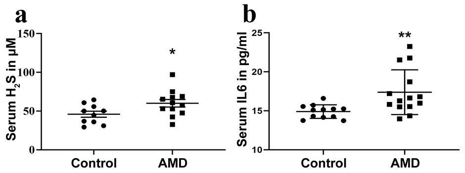
H2S in the AMD plasma samples was measured using MB assay. There was a significant increase in the levels of serum H2S with a mean of 60.08 ± 4.83 μM when compared to the control samples that showed a mean of 45.93 ± 3.92 μM (p = 0.04) (Figure 1a). The mean serum H2S levels in wet AMD was 62.8 ± 25.46 μM, (n = 5) when compared to dry AMD that showed 52.01 ± 4.77 μM, (n = 9).The AMD patients who had their anti-VEGF injections (64.06 ± 10.43 μM; n = 5) showed no statistical variations in the serum H2S levels when compared to those cases who had no such intra-vitreous injections (51.31 ± 5.139 μM; n = 9).There was a significant increase in the levels of serum IL6 in AMD patients (17.38 ± 2.87 pg/ml) when compared to controls (14.9 ± 0.87 pg/ ml, p = 0.002) (Figure 1b), whereas there was no significant difference between dry (18.13 ± 3.28 pg/ml) and wet AMD cases (16.03 ± 1.306 pg/ml).
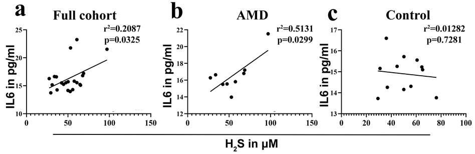
Correlation between Serum H2S and IL6
Simple linear regression correlation between serum H2S and IL6 showed a significant positive correlation (r = 0.4569; p = 0.0325) in the total study cohort of AMD and control.
Correlation between the parameters in the controls alone showed no significant correlation (r = -0.1132; p = 0.7261), whereas in AMD patients there was a significant positive correlation between the serum H2S and IL6 (r = 0.7163; p = 0.0299) (Figure 2).
Discussion
H2S plays an essential role as a gaso-transmitter similar to nitrous oxide and acts as a signaling molecule in mammals [8, 17]. Hydrogen sulphide (H2S), a potent neuromodulator, was initially thought to be a toxic gas. In humans, the concentration of H2S in serum is reported to be from 70 to 125 μM [18] and regulates various physiological cellular functions like synaptic transmission, vascular tone, inflammation, transcription, and angiogenesis and protects cells from oxidative stress. The level of H2S is crucial for the regulation of inflammatory response, wherein low dose decreases inflammation while high doses of H2S donor show controversial results. Therefore, H2S dosage is a switch to control the biphasic regulation of H2S donors on inflammation, and the generation of H2S can also be augmented by the appearance of inflammation [11]. In severe acute pancreatitis, TNFα and IL6 have been shown to induce the expression of CSE and CBS, thereby enhancing the production of H2S. The increased H2S inhibited intestinal mobility and enhanced the inflammatory response caused due to pancreatitis [19]. H2S has been reported to act as a pro-inflammatory mediator in rheumatoid arthritis and was found to be correlated with the disease activity score and tender joint count [20]. In AMD, H2S has been hypothesized to be a therapeutic target for disease management [13].In this pilot study, we find serum H2S level is significantly increased in AMD patients compared to control which is reported for the first time that needs to be validated in larger sample size. Serum IL-6 levels were also significantly increased in AMD patient samples in comparison to control. We show a positive correlation between H2S and IL6 in AMD patients and not in control. There are not many studies that associate H2S and pro-inflammatory cytokines and we speculate that H2S might induce the expression of IL-6 [19]. Also, since there was no significant difference between wet and dry AMD, H2S may be a common factor in the initiation of AMD in general and maybe a disease marker for both dry and wet AMD. Future extensive studies in larger sample size are needed to evaluate this hypothesis.
Limitations
The analysis of the H2S and IL6 were done in stored samples of the previous study. The samples were stored at -800C until analysis (no freeze thaw).
Acknowledgement
We would like to thank our statistician Mr. N. Vishwanathan for the help in the statistical analysis for the study.
Statement of Ethics
Study Approval Statement
This study protocol was reviewed and approved by Institutional Review Board (ethics committee), and the approval number is 149-2009P.
Consent to Participate Statement
Informed consent was obtained from the participants to collect blood and participate in the study.
Conflict of Interest Statement
The authors have no conflicts of interest to declare.
Funding Sources
This research was funded by Department of Biotechnology, sanction nos. BT-BRB-TF-4-2013 and BT/ PR13630/BRB/10/776/2010.
Author Contributions
OM: H2S analysis, ADM: H2S analysis verification and manuscript preparation, KAB: Sample Collection, AK: Funding, discussion for manuscript preparation and patient recruitment and sample documentation, PK: patient recruitment and clinical detail documentation SRB: Study design, funding, and manuscript editing.
Data Availability Statement
The data that support the findings of this study are not publicly available as they contain information that could compromise the privacy of research participants but are available from the corresponding author [SRB] upon reasonable request.
References
-
Jabbehdari S, Handa JT (2021) Oxidative Stress as a Therapeutic Target for the Prevention and Treatment of Early Age-related Macular Degeneration. Surv Ophthalmol 66(3): 423-440.
-
Mitchell P, Liew G, Gopinath B, Wong TY (2018) Age- related macular degeneration. The Lancet. 392(10153): 1147-1159.
-
Boyer DS, Erfurth US, Campagne MVL, Henry EC, Brittain C (2017) The pathophysiology of geographic atrophy secondary to age-related macular degeneration and the complement pathway as a therapeutic target. Retina 37(5): 819-835.
-
Frampton JE (2013) Ranibizumab: A review of its use in the treatment of neovascular age-related macular degeneration. Drugs Aging 30(5): 331-358.
-
AnandBabu K, Sen P, Angayarkanni N (2019) Oxidized LDL, homocysteine, homocysteine thiolactone and advanced glycation end products act as pro-oxidant metabolites inducing cytokine release, macrophage infiltration and pro-angiogenic effect in ARPE-19 cells. PLoS One 14(5): e0216899.
-
Kauppinen A, Paterno JJ, Blasiak J, Salminen A, Kaarniranta K (2016) Inflammation and its role in age- related macular degeneration. Cellular and Molecular Life Sciences 73(9): 1765-1786.
-
Calvert JW, Coetzee WA, Lefer DJ (2010) Novel insights into hydrogen sulfide-mediated cytoprotection. Antioxid Redox Signal 12(10): 1203-1217.
-
Abe K, Kimura H (1996) The possible role of hydrogen sulfide as an endogenous neuromodulator. J Neurosci 16(3): 1066-1071.
-
Bian J, Hu LF, Yang L, Du J, Jin H (2017) Role of Hydrogen Sulfide in Retinal Diseases.
-
Chen Z, Zhang M, Zhao Y, Xu W, Xiang F, et al. (2021) Hydrogen Sulfide Contributes to Uterine Quiescence Through Inhibition of NLRP3 Inflammasome Activation by Suppressing the TLR4/NF-κB Signalling Pathway. J Inflamm Res 14: 2753-2768.
-
Han Y, Shang Q, Yao J, Ji Y (2019) Hydrogen sulfide: a gaseous signaling molecule modulates tissue homeostasis: implications in ophthalmic diseases. cell death & disease 10: 293.
-
Salvi A, Bankhele P, Jamil J, Chitnis MK, Mbye YFN, et al. (2016) Effect of hydrogen sulfide donors on intraocular pressure in rabbits. J Ocul Pharmacol Ther 32(6): 371- 375.
-
George AK, Singh M, Homme RP, Majumder A, Sandhu HS, et al. (2018) A hypothesis for treating inflammation and oxidative stress with hydrogen sulfide during age- related macular degeneration. Int J Ophthalmol 11(5): 881-887.
-
Lindblad AS, Kassoff A, Kieval S, Mehu M, Buehler J, et al. (1999) The age-related eye disease study (AREDS): Design implications AREDS report no. 1. Control Clin Trials 20(6): 573-600.
-
Barathi S, Vadhana P, Angayarkanni N, Ramakrishnan S (2007) Estimation of hydrogen sulphide in the human lymphocytes. Indian Journal of Biochemistry and Biophysics 44(3): 179-182.
-
Ran R, Du L, Zhang X, Chen X, Fang Y, et al. (2014) Elevated hydrogen sulfide levels in vitreous body and plasma in patients with proliferative diabetic retinopathy. Retina 34(10): 2003-2009.
-
Martelli A, Testai L, Breschi MC, Blandizzi C, Virdis A, et al. (2012) Hydrogen sulphide: Novel opportunity for drug discovery. Med Res Rev 32(6): 1093-1130.
-
Karunya R, Jayaprakash KS, Gaikwad R, Sajeesh P, Ramshad K, et al. (2019) Rapid measurement of hydrogen sulphide in human blood plasma using a microfluidic method. Sci Rep 9: 3258.
-
Liu Y, Liao R, Qiang Z, Zhang C (2017) Pro-inflammatory cytokine-driven PI3K/Akt/Sp1 signalling and H 2 S production facilitates the pathogenesis of severe acute pancreatitis. Biosci Rep 37(2): BSR20160483.
-
Muniraj N, Stamp LK, Badiei A, Hegde A, Cameron V, (2017) Hydrogen sulfide acts as a pro-inflammatory mediator in rheumatic disease. Int J Rheum Dis 20(2): 182-189.
- Screening of Hospital Staff During World Glaucoma Week in a Tertiary Eye Care Centre
- Angioid Streaks with Macular Neovascularization: Clinical Insights from Two Cases
- Giant Kissing Naevus: An Oculoplastic Challenge
- Why Freedom of Vision Should Not Cost the Freedom of Feeling - LASIK in the Climate of Change
- Asymmetric Optic Nerve with Small Disc and Large Cup: A Rare and Challenging Case of Unilateral Optic Nerve Hypoplasia
- Large Angle Exotropia in a Child: A Case Report