Association of Serum 25-Hydroxyl Vitamin D Deficiency and Idiopathic Presenile Cataract -A Hospital Based Study
Background: Cataract is the leading cause of blindness in India mainly due to age related changes in the lens. Early onset cataract can be visually debilitating having high societal burden requiring early surgical intervention. Identifying modifiablerisk factors can delay or prevent early onset cataractous changes.Aim of the study: The aim of this study was to determine any association between Serum 25-Hydroxyl (25-OH ) Vitamin D Deficiency and Idiopathic Presenile CataractMaterials and Methods: The study was undertaken at Tertiary care hospital in hilly state of India. A detailed medical history was taken and presenile cataract with no identifiable cause was taken in the study group. 67 cataract and 67 controls of bothsexes were examined for 25 hydroxyl vitamin D using the electrochemiluminescence immunoassay method.Observations and Results: Mean 25-OH vitamin D level in cataract patients was 18.91 ± 12.75 ng/mL and median was 18.45 (6 – 95.98) , while in the control group, mean 25-OH vitamin D level was 21.8 ± 13 ng/mL and median was 18.95 ( 4.23 –87.2). There was no statistically significant difference among the cataracts and controls although the mean vitamin D levels were lower in patients with presenile cataract. Female preponderance was seen in the cataract group. Posterior subcapsular cataract was the most common type of morphology seen.Conclusion: In our study serum vitamin D levels were lower than reference range in the idiopathic presenile cataract patients but were not found to be statistically significant.
Abbreviation
LOCS: Lens Opacity Classification System.
Introduction
Cataract is a major cause of blindness globally and commonly occurs after 50 years of age. It is mainly due to age related degenerative process and opacification of lens. In India, it accounts for 62.6% of all cases of blindness. Cataract is one of the curable causes of blindness which is included in the VISION 2020 [1]. Presenile cataract refers to onset of cataract before the age of 50 years. Early onset cataract affects the most productive group of population and can add burden to health care needs of the country.
Several risk factors have been proposed for cataract formation in early age group such as atopy, use of corticosteroids, uveitis, diabetes mellitus and exposure to ultraviolet radiations. Some studies done in India could not attribute any risk factor to the development of presenile cataract and termed it as idiopathic [2, 3].
If we can identify a modifiable risk factor that is associated with idiopathic presenile cataract , we can prevent development of early onset cataract changes .
Vitamin D is supposed to have an anti-inflammatory role and suppress oxidative stress. It can have a protective role against cataract formation [4].
In our study, we aim to detect any association between serum vitamin D levels and idiopathic presenile cataract.
Material and Methods
The current study was conducted in the Ophthalmology Department of Tertiary care teaching hospital in hilly state of India from April 2023 to December 2023. Inclusion criteria are as follows: patients with age between 18-49 years developing cataract without a known cause or identifiable risk factor. Exclusion criteria are as follows: complicated cataract either ocular or systemic disease, congenital or developmental cataract, glaucoma, high myopia, prior ocular medication or surgery, history of tuberculosis, steroid use, use of calcium supplements or osteoporosis medications, prior trauma, autoimmune diseases, skin disorders, diabetes, cancer or cardiopulmonary disease.
The control group was enrolled from the outpatient clinic following the same criteria with no cataract detected.
Lens opacity classification system (LOCS) was used for grading of both nuclear and cortical cataracts [5]. Slit-lamp retroillumination was used to classify the type and grade of cataract. Dilated fundus examination and refraction was done. Ultrasound B scan was done in indicated cases.A sample of peripheral blood was collected in a sterile plain vacutainer from patients and healthy controls for vitamin D assay. The case group included 67 patients and the control group also included 67 participants. Proportion of gender taken in the control group was same as that of cataract group.
25 hydroxyl vitamin D levels was estimated using the electrochemiluminescence immunoassay method.
Vitamin D deficiency was taken as <20 ng/mL and severe deficiency as <10 ng/mL .Informed consent was obtained from patients and participants and the approval of the ethical committee of our institute was obtained.
The collected data was transformed into variables, coded and entered in Microsoft Excel. Data was analyzed and statistically evaluated using SPSS-PC-19 version. Quantitative data were expressed as means ± standard deviation, median, and range. Qualitative data were expressed as number and percentage. The chi-squared test was used for comparison between qualitative variables. P’ value less than 0.05 was considered statistically significant.
Results
Our study included 67 cases and 67 controls after applying the inclusion criteria .There were 32.8% (22) males and 67% (45) females in the cataract group.There were more female as compared to male in the presenile cataract group although it was not statistically significant.
Maximum number of cataract cases 65.67% (44) was seen in the age group of 41-49 years. Patients in both cases and controls were matched for age and gender with no statistical difference (Table 1).
| Cataract group (N=67) | Control group (N=67) | P value | |
|---|---|---|---|
| 18-30 | 8 (12 %) | 11 (16.4%) | 0.2 |
| 31-40 | 15 (22.38%) | 22 (32.8%) | 0.2 |
| 41-49 | 44 (65.67%) | 34 (50.7%) | 0.2 |
Table 1: Sociodemographic Profile of the Patients –Cases and Controls.
Forty- three (64%) patients had bilateral cataracts while 36% had unilateral cataract.
Mean 25-OH D level in cataract patients was 18.91 ng/ mL ± 12.75 SD and median was 18.45 ng/mL (6 – 95.98 ) . This serum 25-OH D level can be considered in deficiency category. The mean 25-OH D level in the control group was 21.8 ± 13 ng/mL and median was 18.95ng/ml (4.23 – 87.2).
Forty six (68.6%) patients in the cataract group and thirty eight ( 56.7%) patients in the control group had vitamin D deficiency ( < 20ng/ml) (Graph1).
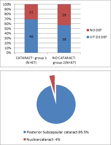
Graph 1: Vitamin D Status in Cases and Controls.
Eight (12%) patients in the cataract group and 7 ( 10.4%) patients in the control group had very severe deficiency ( < 10ng/ml).
The most common type of cataract was posterior subcapsular cataract. Sixty four (95.5%) patients had posterior subcapsular cataracts and three (4.47%) had nuclear cataract (Graph 2).
Graph 2: Type of Cataract in Patients.
No significant statistical difference was found between serum 25-OH vitamin D among cataract group and control group.
Discussion
The aim of our study was to determine any association between vitamin D deficiency and idiopathic presenile cataract in patients between 18 and 49 years in Hamirpur district.
In our study , female preponderance was seen for presenile cataract. The most common type of cataract seen in our study was posterior subcapsular cataract. 68.6% of the enrolled patients with presenile cataract had vitamin D deficiency compared to control group which had 56.7% although the difference was not statistically significant.
Cataract occurring before 50 years is becoming increasing frequent leading to cataract surgery at an earlier age. It may be associated with several factors other than genetic such as environmental factors like high ultraviolet exposure , metabolic such as diabetes mellitus, drug intake such as corticosteroids, social factors like smoke exposure and personal habits like tobacco [2]. Cataractous changes may also occur due to hypo-calcaemia when the parathyroid glands become atrophic or removed during thyroidectomy [6].
In Indian population, cataracts tend to occur earlier in life and have a higher incidence. It could be due to various causes such as genetics, environmental risk factors, nutritional deficiencies, and severe dehydration [7].
Several studies have been done in India regarding cause of presenile cataract In one study conducted in north India, the most common type of cataract identified was posterior sub capsular cataract and the major causes found were Trauma (34.46%), Uveitis (20.90%), Atopy (12.43%), Diabetes (9.60%) and idiopathic (16.38%) [8]. In a study conducted in urban area in North India, presenile cataract was observed to be significantly associated with tobacco,hy percholesterolemia,fuel exposure and lower socioeconomic status [9]. In other studies done in various parts of India, cases that presented with presenile cataract had no risk factor associated and hence were classified as idiopathic [10, 11, 12].
Few studies done have found a significant role of 25-hydroxyl vitamin D deficiency in age-related cataract patients and patients with posterior subcapsular cataract [13, 14, 15].
Visual impairment occurs early owing to the position of the opacity near the nodal point of the eye. It may have a high societal burden in terms of lost work and need for early surgical intervention.
Vitamin D is a fat-soluble steroid derivative, which plays a significant role in calcium homeostasis and bone metabolism through its actions in intestine, bone, kidney, and the parathyroid glands. It also plays an important role in pathogenesis of many immune-related diseases by regulating the production of inflammatory cytokines and immune cells [16].
Vitamin D deficiency is widespread in people regardless of their age, gender, race and geography. Vitamin D is synthesized in the skin on exposure to ultraviolet rays, still its deficiency is widely prevalent in a tropical country like India [17]. Vitamin D deficiency contributes to osteoporosis and low bone mass as well as has been linked to extra skeletal disorders such as cancer, diabetes and autoimmune disorders [18].
A recent study done indicated that low circulating levels of 25 (OH) D were associated with Age related macular degeneration and may have a protective role against cataract formation due to its anti inflammatory properties [19]. The 25 (OH) D blood test, a biomarker for Vitamin D reserves, is a low cost and widely available blood test.
In a study done in South India, only 44.8% of the study group was found to have vitamin 25 OH D levels higher than 20ng/ml [20].
The limitation of this study was it was a hospital- based study and our sample size was limited.
Conclusion
While no significant association was found, the trend of lower vitamin D levels in presenile cataract patients warrants further investigation. Furthermore studies are required to evaluate risk factors associated with development of idiopathic presenile cataract.
Financial Support and Sponsorship
Nil.
Conflicts of Interest
The authors declare no conflicts of interest.
References
-
Vision (2020) The Right to Sight – India.
-
Sreekanth B (2017) A Clinical Study on Risk Factors Cataracts in Young Adults. Int J Sci Stud 5(9): 120-124.
-
Jyothi R, Sathyan S (2017) Etiopathogenesis of presenile cataracts in Central Kerala: A cross-sectional observational study. Kerala J Ophthalmol 29: 179-183.
-
d’Hellencourt LC, Menei CMN, Bernard R, Couez D (2003) Vitamin D3 inhibits proinflammatory cytokines and nitric oxide production by the EOC13 microglial cell line. Journal of Neuroscience Research 71(4): 575–582.
-
Chylack L, Wolfe JK., Singer DM, Leske MC, Bullimore MA, et al. (1993) The lens opacities classification system III. Archives of Ophthalmology 111(6): 831–837.
-
Daba KT, Weldemichael DK, Mulugeta GA (2019) Bilateral hypocalcemic cataract after total thyroidectomy in a young woman: case report. BMC Ophthalmol 19(1): 233.
-
Nam SW, Lim DH, Cho KY, Kim HS, Kim K, et al. (2018) Risk factors of presenile nuclear cataract in health screening study. BMC Ophthalmol 18(1): 263.
-
Kumar J, Chaubey P, Singh VP, (2018) Pre-Senile Cataract: Analytical Study. IOSR Journal of Dental and Medical Sciences (IOSR-JDMS) 17(4): 6-9.
-
Das GK, Boriwal K, Chhabra P, Sahu PK, Kumar S, et al. (2019) Presenile cataract and its risk factors: A case control study. J Family Med Prim Care 8(6): 2120-2123.
-
Praveen MR, Shah GD, Vasavada AR, Mehta PG, Gilbert C, et al. (2010) A study to explore the risk factors for the early onset of cataract in India, Eye (Lond) 24(4): 686- 694.
-
Dhanya VS, Abraham M, Deepa MG (2021) Risk factors and types of presenile cataract in patients attending a Tertiary Care Hospital in Kerala- a retrospective study. International Journal of Research and Review 8: 2.
-
Mishrikotkar JP, Sethiya S, Phadke Y, Sangai S (2019) Clinical Study of Presenile Cataract in Patients Attending Tertiary Health Care Centre. International Journal of Current Medical and Applied sciences 24(2): 36-38.
-
Brown C, Akaichi F (2015) Vitamin D deficiency and posterior subcapsular cataract. Clin Ophthalmol 9: 1093-1098.
-
Abdellah MM, Mostafa EM, Salama EH, Mohamed RE (2019) Association of Serum 25-Hydroxyl Vitamin D Deficiency and Age-Related Cataract: A Case-Control Study. J Ophthalmol 2019: 9312929.
-
Rao P, Millen AE, Meyers KJ, Liu Z, Voland R, et al. (2015) The relationship between serum 25-hydroxyvitamin D levels and nuclear cataract in the carotenoid age-related eye study (CAREDS), an ancillary study of the women’s health initiative. Investigative Opthalmology & Visual Science 56(8): 4221-4230.
-
Richer SP, Pizzimenti JJ (2013) The importance of vitamin D in systemic and ocular wellness. J Optom 6(3): 124-133.
-
Nimitphong H, Holick MF (2013) Vitamin D status and sun exposure in Southeast Asia. Dermato-Endocrinology 5(1): 34-37.
-
Ritu G, Gupta A (2014) Vitamin D Deficiency in India: Prevalence, Causalities and Interventions. Nutrients 6: 729-775.
-
Millen AE, Voland R, Sondel SA, Parekh N, Horst RL, et al. (2011) Vitamin D status and early age-related macular degeneration in postmenopausal women. Archives of Ophthalmology 129(4): 481-489.
-
Mechenro J, Venugopal G, Kumar MB, Balakrishnan D, Ramakrishna BS (2018) Vitamin D status in Kancheepuram District, Tamil Nadu, India. BMC Public Health 18: 1345.
- Screening of Hospital Staff During World Glaucoma Week in a Tertiary Eye Care Centre
- Angioid Streaks with Macular Neovascularization: Clinical Insights from Two Cases
- Giant Kissing Naevus: An Oculoplastic Challenge
- Why Freedom of Vision Should Not Cost the Freedom of Feeling - LASIK in the Climate of Change
- Asymmetric Optic Nerve with Small Disc and Large Cup: A Rare and Challenging Case of Unilateral Optic Nerve Hypoplasia
- Large Angle Exotropia in a Child: A Case Report