Evaluation of ACP Derma Barrier Cream in the Healing Process of Induced Wounds in Dogs
The treatment of skin lesions comes up against the difficulty of the treatment due to the animal's temperament, causing stress and reluctance to administer medication, which may lead the tutor to interrupt the treatment. Knowing that coconut water has therapeutic properties that benefit tissue regeneration, we aimed to evaluate the healing activity of ACP Derma barrier cream, a coconut water-based product, to measure the degree of repair of the wound and to compare the evolution of the healing process, through macroscopic evaluations of the wounds. Five dogs were used in which 3 circular incisions were made on their backs. Wounds were treated with the 3 ointments (ACP, CMR® and Vetaglós®) and were macroscopically evaluated in the periods of 4, 7, 10 and 14 days of treatment. There were significant reductions in the wound area up to the 14th day, resulting in a satisfactory healing of the ointments used. ACP Derma was shown to be superior in terms of adherence to the lesion and showed effective potential for wound healing in dogs, equivalent to treatment with commercial ointments, making it a more cost-effective therapeutic option.
Introduction
The study of healing and treatment of skin lesions is extremely important in the veterinarian medicine field due to the high numbers of animals with lesions of different types and origins constantly admitted for veterinary care [1]. The combination of the knowledge of this process with the particularities of each patient helps to create a healing therapy, which results in the tissue repair and in the reestablishment of the animal’s homeostasis [2]. Any failure during the repair process can result in chronic injuries or scarring, which increases the financial expense of the tutors due to constant treatments and considerable medical costs, affecting quality of life of patients. Great efforts have been made to develop new therapies for wound repair [3].
Countless researches and several methods are being developed and applied to accelerate the healing process. The interest in the use of herbal medicines is increasing [4]. Among them, coconut water powder (ACP) has already been used in clinical studies that evaluate the effects of skin healing in Wistar rats [5]. The author concluded that the ACP treatment enhanced the tissue repair process, promoting the development of collagen fibers. It was evaluated the use of ACP-based biofilms (ACP-501) as a support in the treatment of oral affections in patients with head and neck cancer [6].
Based on previous pre-clinical and clinical studies which consisted in one of the most important steps to ensure the efficacy and safety of a new product, it was proposed the development of a coconut water powder-based bioemulsion (ACP-502) to use in treatment of diabetic foot as a wound protector and healing inducer. Such work proved the effective potential in the healing of diabetic foot ulcers, contributing to a new, more accessible therapeutic approach in treatment of wounds in diabetic patients. The treatment interfered in the clinical score of the affected patients regarding the mean healing time, the risks of complications and permanent sequelae. Therefore, the approach was considered a viable, easy-to-use and lower-cost option [7].
The search for a way to advance and improve the healing process of skin wounds in dogs comes up through observation of the long-time period of clinic admission of these animals, to treat extensive skin wounds. A product which can reduce the healing time improves the treatment, since most long-term protocols can cause stress, difficulties to administer medications to the animal. Also, it can lead the tutor to decide to stop the treatment, due to the long time needed and other adversities on the way.
The difficulty of treatment can increase due to the animal’s temperament, along with the consistency of ointments of common use. All of it delays the healing time. Few experimental studies to evaluate skin healing using dogs are found in the literature, most likely due to the difficulties in handling these species in an experimental environment. The research for new therapies to enhance this process is fundamental.
Based on previous results and on the non-identification of clinical or experimental studies which have evaluated the coconut water in the healing process in dog’s wounds, the aim of this work was to evaluate the healing activity of the herbal formulation ACP Derma cream barrier (coconut water powder-based) compared to the traditionally used ointments: CMR® e Vetaglós®.
Materials and Methods
Ethical Aspects
The project was submitted and approved by the Committee on Ethics in the Use of Animals of the institution under process number 11251322/2019. The study was carried out according to the Ethical Principals of Animal Experimentation.
Animals
Five street dogs (Canis lupus familiaris) were rescued from the urban perimeter a large city. All animals were females, mixed breed, weighing from 4.5 kg to 12.4 kg, and ages ranging from 5 months to 1 year and a half. These animals were placed in a kennel, where they had been through individual care, access to water and super premium commercial dog food until the end of the experiment.
The dogs were dewormed and an ectoparasiticide was administrated. Blood was collected to perform leishmaniasis test and blood count (all were negative for leishmania). The procedures were carried out at a veterinary clinic; in the same city the dogs were rescued.
The animals that presented hematological alteration, such as anemia and hemoparasitosis, were treated and underwent 15 days of adaptation process. The animals were carefully observed to check their general health, especially the condition of nutrition and cleanliness, as well as any aspects that could interfere with the full course of the experiment. At the end of the process, all animals were spayed, vaccinated against rabies and adopted by responsible tutors.
Induced Wounds
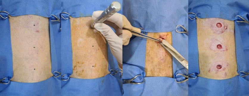
The bitches were submitted to injectable dissociative anesthesia, using the association of 125 μg/m2 dexmedetomidine, 0.2 mg/kg methadone, 0.3 mg/kg IM midazolam, and after 15 minutes, ketamine 2 mg/kg IV. After arranging the animals in the prone position, a trichotomy was performed on the thoracic spine region of each animal, followed by antisepsis with 0.2% povidone iodine.
The incision areas were marked on the back of the animals using a marker brush, keeping 5 cm between the lesions, in three locations in the thoracic sagittal midline, proximal, middle and distal third. After demarcation, circular incisions were made in the delimited areas with the aid of a metal biopsy Punch Keyes of 1 cm in diameter, with a cutting blade on its lower edge, piercing the skin and the subcutaneous tissue with the aid of a scalpel. Surgical scissors and anatomic forceps; Thus, it was possible to perform the removal of the skin fragment and consequently exposing the thoracolumbar fascia (Figure 1). The same instrument was used to induce all wounds, being properly sterilized for use in the next animal. The full depth of the punch was used, standardizing the size of the wound, and paying attention to the fact that all layers were removed, leaving only the underlying muscle.
Treatment
The animals remained in individual kennel. They were submitted to the treatment, initiating the ointment application soon after the wound induction, following the sequence: Thoracic lesion of the proximal third: CMR; Thoracic lesion of the middle third: VETAGLÓS; Thoracic lesion of the distal third: ACP.
Post-surgical analgesia was carried out using Tramadol 2 mg/kg and Dipyrone 25 mg/kg, both every 8 h PO, for 3 days. The animals used an Elizabethan collar to avoid licking the surgical wound throughout the treatment.
The topical application of a layer of the respective substances was enough to completely cover the wound areas, without the need for bandaging. The application was done once a day, always at the same time, for 14 days and by the same researcher.
Wound Evaluation
Surgical wounds were daily observed to evaluate the qualitative parameters of the main macroscopic characteristics, regarding the presence and intensity of exudate, wound bed color, presence and color of scabs (yes or no). They were considered healed when covered by macroscopically distinguishable epithelium cells were.
The presence of edema was reported in absent, mild, moderate and severe scores.
All animals had their wounds photographed using a smartphone with a high-resolution digital camera (64 megapixels), positioned 30 cm away and parallel to the wound. The calibration marker (quick response code [QR]) was positioned close to the wound, and a photograph was taken only after QR the code recognition by the wound measurement app Imito Wound (Figure 2). This approach avoids underestimating or overestimating of the wound area estimate.
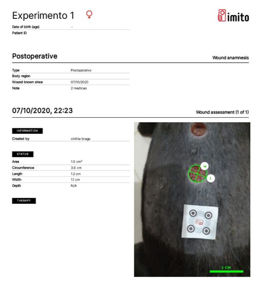
The image was positioned and scaled on the smartphone screen to occupy the entire surface. The configuration of the area in Imito app was obtained by tracing the circumference with a point-to-point line. With the amplified image, the distance between the points becomes smaller, which improves the contour of the wound edge. The application automatically calculated width, circumference and area with digital planimetry, on days 4, 7, 10 and 14 of the healing process. Wound closure was evaluated by measuring the area of the wound retraction on the days previously mentioned.
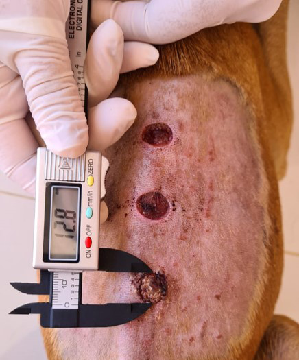
The wound reduction analysis was performed with a digital caliper (Figure 3) to evaluate the linear transversal and horizontal measurement of the lesion and subsequent area calculation, equivalent to an ellipse. Where a: transversal measure and b: horizontal measure. The area reduction was followed through the days; A= π x a x b/4.
The mean ulcer repair rate (RRm) was also calculated, which denotes how many mm2 the ulcer area decreased in a given time interval (between t1 and t2), being expressed in mm2 per day and defined by the following quotient: RRm = - A (t2) – A (t1)/ t2-t1; where A(t1) and A(t2) are the ulcer areas at times t1 and t2, respectively, where t2 > t1. Mean repair rates were calculated among days RR4-7, RR4-14, RR7-10, RR7-14 and RR10-14.
Statistical Analysis
Data were submitted to Shapiro-Wilk and Bartlet tests to investigate the normal distribution of residuals and homoscedasticity, respectively. Repair rate data between days 4 and 7 (RR4-7), 4 and 10 (RR4-10), 4 and 14 (RR4-14), 7 and 10 (RR7-10), 7 and 14 (RR7 -14) and, finally, 10 and 14 (RR10-14) were analyzed in a completely randomized design with treatment effect (ACP x CMR x VET) through analysis of variance (ANOVA). Application wound area (AWA), caliper wound area (CWA) and wound circumferences (WC) were analyzed in a randomized block design (animals) in a split-plot scheme (4, 7, 10 and 14 days). When a statistical difference was observed, the Student-Newman-Keuls test (SNK) was applied to compare the means. Data were expressed as mean and standard error of the mean (SEM). The significance level considered was 5% (P < 0.05). All experiments were performed using the R software (Team, 2020).
Results
During the inflammatory period, the presence of edema was observed until the 4th day in animal 2, being intense in the wound treated with CMR and moderate in the wound treated with Vetaglós. In animal 3, the wound treated with Vetaglós presented mild edema. On the 7th day of treatment, animal 2 presented mild edema in the wound treated with CMR. In both animals, the wounds treated with ACP did not show edema, as well as in the other animals in any of the wounds.
Twenty percent of the wounds treated with CMR presented edema until the 7th day of treatment, while 40% of the wounds treated with Vetaglós presented edema until the 4th day. On the 10th and 14th day of treatment, none of the wounds presented edema in any of the treatments (Table 1).
| D4 | D7 | D10 | D14 | |
|---|---|---|---|---|
| CMR | 20% | 20% | 0% | 0% |
| VETAGLÓS | 40% | 0% | 0% | 0% |
| ACP | 0% | 0% | 0% | 0% |
Table 1: Relative frequency (%) regarding the presence of edema in induced wounds treated with CMR, Vetaglós and ACP ointments.
The exudate characteristics of the inflammatory phase were also monitored during the experiment. On the first postoperative day, serosanguineous exudate was observed in animal 1 in all wounds, moderate in the ones treated with CRM and Vetaglós wound and mild in ACP. In animal 2, the serosanguineous exudate was more intense in the wound treated with CMR, moderate in Vetaglós and absent in the wound treated with ACP. Animal 3 did not present exudate in any of the wounds. Animal 4 presented moderate serous exudate in wounds treated with CMR and Vetaglós and mild in wounds treated with ACP.
On the 4th day of treatment, only animal 2 still had serosanguineous exudate in wounds treated with CMR and Vetaglós in a moderate and mild intensity, respectively. The wound treated with ACP, as well as the other wounds in the other animals, did not present exudate.
In all animals the wounds did not presented infection with the presence of purulent drainage (Table 2).
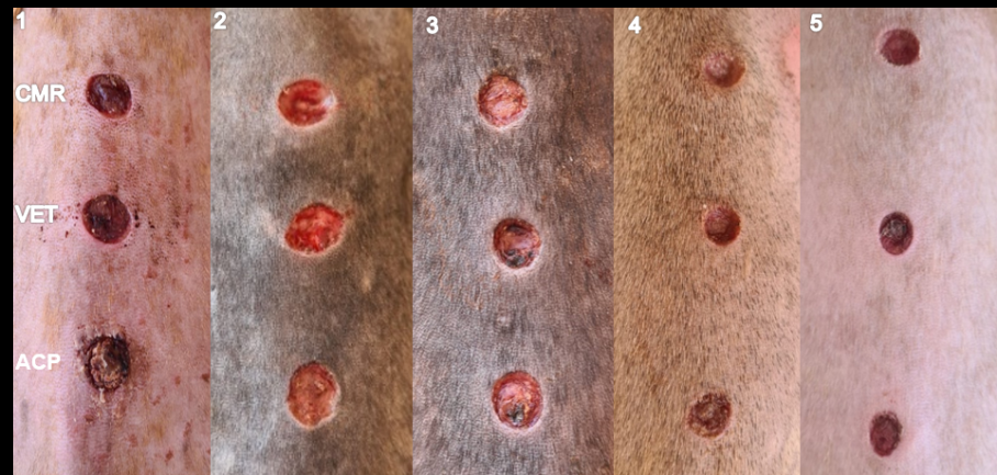
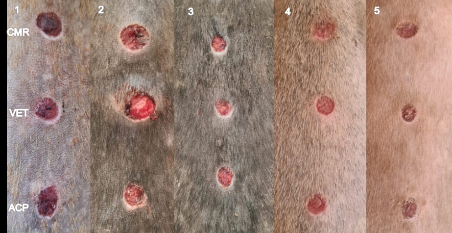
Regarding the presence of scabs and wound coloration, on the 4th day of evaluation (Figure 4), animal 1 had dry, reddish-colored wounds that were treated with CMR and Vetaglós and in the one treated with ACP it showed onset of retraction and intense yellowish scab. Animal 2 presented moist and reddish wounds, treated with CMR and Vetaglós. The lesion treated with ACP was dry and with a slightly yellowish scab. Animal 3 had all wounds dry, reddish and crusted. Only the lesion treated with CMR showed partial retraction. In animal 4, all wounds were dry, reddish and crusted, but the scab was more evident in the wound treated with ACP. Animal 5 had dry, reddish and crusted wounds. 100% of wounds treated with ACP were dry and crusted, while CMR and Vetaglós still had moist and no scabs.
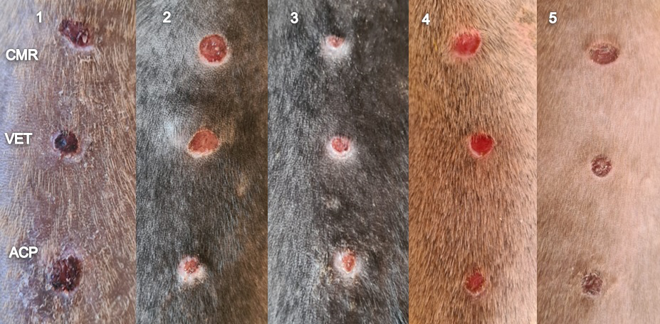
On the 7th day (Figure 5), the wound treated with Vetaglós in animal 2 had moisture and still did not have scabs, unlike the other animals.
| Treatment | Exudate intensity (%) | |||
|---|---|---|---|---|
| Intense | Moderate | Discreet | Absent | |
| 1st day | ||||
| CMR | 20% | 40% | 0% | 40% |
| VETAGLÓS | 0% | 60% | 0% | 40% |
| ACP | 0% | 0% | 40% | 60% |
| 4th day | ||||
| CMR | 0% | 20% | 0% | 80% |
| VETAGLÓS | 0% | 0% | 20% | 80% |
| ACP | 0% | 0% | 0% | 100% |
Table 2: Relative frequency (%) referring to the intensity of exudate (serosanguineous or serous) in induced wounds treated with
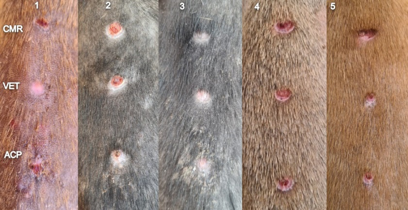
On the 10th day (Figure 6), all wounds had scabs. In animal 1, the crusts were thicker and presented dark coloration in all wounds. In the other animals, the lesions were more reddish. All wounds were dry and had no purulent drainage.
On the 14th day (Figure 7), all wounds were healed in all animals. In animal 1, the wound treated with Vetaglós no longer had scabs, as all the wounds in animal 3.
Repair rates are shown in Table 3. ACP, CMR and VET did not significantly influence RR4-7, RR4-14, RR7-10, RR7- 14 and RR10-14. However, RR4-10 varied in function of treatments (P < 0.05). ACP differed statistically from CMR and was similar to VET. VET and CMR did not differ.
| Treatment | Repair rate | |||||
|---|---|---|---|---|---|---|
| Days 4-7 | Days 4-10 | Days 4-14 | Days 7-10 | Days 7-14 | Days 10-14 | |
| ACP | 0.13±0.02 | 0.15±0.02 a | 0.12±0.02 | 0.16±0.02 | 0.12±0.01 | 0.08±0.02 |
| CMR | 0.03±0.04 | 0.06±0.02 b | 0.06±0.01 | 0.09±0.01 | 0.09±0.01 | 0.08±0.02 |
| VET | 0.08±0.02 | 0.11±0.02 ab | 0.09±0.01 | 0.14±0.05 | 0.08±0.01 | 0.08±0.01 |
| p-value# | 0.11 | 0.02 | 0.06 | 0.26 | 0.3 | 0.98 |
Table 3: Mean±standard error of the mean and p-value of repair rates between days 4, 7, 10 and 14 for ACP, CMR and VET.
The mean values for AWA and WC and the effects of days and treatments are shown in Table 4. For AWA, there was an interaction among treatments and days. ACP and VET showed differences between day 4 and 7. CMR only started to reduce between day 7 and 10. ACP decreased until day 14. On the other hand, VET and CMR stabilized on day 10. On
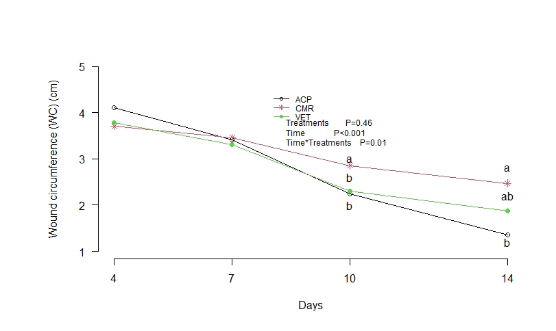
day 4, ACP showed the highest value. On the other days, there was no difference among treatments. For WC, ACP showed a significant reduction between days 4 and 7. CMR and VET reduced WC between days 7 and 10, stabilizing thereafter. On the other hand, ACP showed decreasing values until day 14. Treatments did not differ on days 4 and 7. On days 10 and 14, CMR had WC higher than ACP (Figure 8).
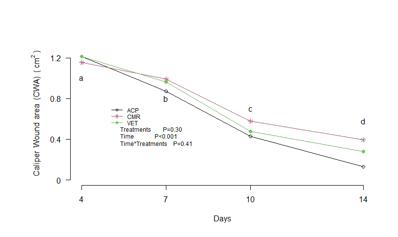
Treatments did not differ for CWA (C = 0.30). Differently, the days influenced significantly. There was a significant reduction from days 4 to 14 (Figure 9).
| Application wound area (cm2) | Days | |||
|---|---|---|---|---|
| Treatment | 4 | 7 | 10 | 14 |
| ACP | 1.3±0.2 Aa | 0.9±0.1 Ab | 0.4±0.1 Ac | 0.1±0.1 Ad |
| CMR | 1.0±0.1 Ba | 0.9±0.1 Aa | 0.6±0.1 Ab | 0.4±0.1 Ab |
| VET | 1.1±0.1 Ba | 0.9±0.1 Ab | 0.4±0.1 Ac | 0.3±0.1 Ac |
| Sources of variation | Ointment | Day | P x D | |
| p-value # | 0.94 | <0.001 | 0.02 | |
| Circumference. cm | Days | |||
| Treatment | 4 | 7 | 10 | 14 |
| ACP | 4.1±0.3 Aa | 3.4±0.2 Ab | 2.2±0.4 Bc | 1.3±0.2 Bd |
| CMR | 3.7±0.2 Aa | 3.4±0.3 Aa | 2.8±0.4 Ab | 2.5±0.3 Ab |
| VET | 3.8±0.2 Aa | 3.3±0.3 Aa | 2.3±0.4 Bb | 1.8±0.3 ABb |
| Sources of variation | Ointment | Day | P x D | |
| p-value | 0.46 | <0.001 | 0.02 | |
| a, b, c, d indicate difference between days in the same treatment. | ||||
| A, B indicate difference between treatments on the same day. | ||||
| *SEM: Standard error of the mean. # Significance levels of the F-test of the analysis of variance. |
Table 4: Mean±standard error of the mean and p-value of the application wound area (cm2), circumference (cm) and caliper wound ar
Discussion
During the literature review, no studies were found with the aim to evaluate the healing action of powdered coconut water ointment in dogs comparing to allopathic (Vetaglós®) and homeopathic (CMR®) ointments commercially used in the veterinary field as a wound healing for dogs and cats. No experimental study with this purpose was identified in the studied species.
ACP Derma Barrier Cream is composed by powdered coconut water and vegetable oils (coconut oil, moringa oil, linseed oil). When topically applied, the substances of vegetable oils (triglycerides, phospholipids and antioxidants) can act synergistically through several mechanisms: production of skin barrier homeostasis, antioxidative activities, anti-inflammatory properties, direct or indirect antimicrobial activities (upregulation of antimicrobial peptides), wound healing [8].
Signs of inflammation, such as edema, hyperemia and the presence of exudate are expected findings after a wound is made and can be minimized by agents that favor healing [9].
In wounds treated with the ACP Derma barrier cream, there was an important role in the inflammatory phase (until the 4th day), reducing edema and presenting an absence of exudate, promoting an earlier entry into the proliferative phase. At the beginning of the proliferation phase (4-7 days), it was observed scab formation and the beginning of retraction was more evident when compared to the other treatments.
Up to the fourth day after the operation, 20% of the wounds treated with CRM and 40% of the wounds treated with Vetaglós presented edema, while none was present in the lesions treated with ACP. The absence of edema may be associated with the presence of oleic and linoleic acids present in the ACP barrier cream. Showed that the wounds treated with n-9 and n-6 presented less edema in the first 48 hours when compared to the control group [10].
On day 7, 20% of wounds treated with CMR still had edema. The results differed from the study carried out, comparing herbal medicines of topical use in the skin healing process in horses. It was observed that in the inflammatory phase of the healing process, wounds treated with calendula had less swollen edges and serous, smooth and thin scabs, compared to the other groups of the treatment [11]. Nonetheless, the results are in accordance with the literature, which states that the occurring edema in the region of the wounds can last up to seven days, and it is a consequence of the increase of the vascular permeability that develops in the inflammatory phase of the wound, as a protective tissue response [12].
The initial phase of healing, the inflammatory phase, is fundamental for the repair process. Without inflammation there is no repair. There is an increase in capillary permeability and consequent migration of cells (leukocytes, lymphocytes, erythrocytes) to the wound, which, with the accumulation of plasma, constitute the inflammatory exudate [13]. This process was observed in this work where exudate was present on the first day of treatment with varying intensity in all of them. On the 4th day, only the ACP treatment showed absence of exudate. Similar result was previously presented where no exudates were seen in the macroscopic observation carried out in the three groups of treatments in rats (amorphous and crystalline-aqueous, amorphous and crystalline-gel and triglyceride freeze-dried coconut water) [5].
the observation of scab formation on a skin wound favors the repair process. The dryness of the superficial scab also helps in the wound contraction process [5]. During dehydration the lesion decreases in size and takes tissue adhered to it. In this study, on the 4th day of treatment, all wounds treated with ACP had scabs, while in the other treatments there was moist and no scabs.
Using the temporal progression from a macroscopic point of view, under evaluation of the wound area treated with ACP, it could be observed that, on the 4th day, there was an expansion of the ulcerated area when compared to the initial area produced and standardized by the punch, in agreement with the results from Magalhães [5] where the group treated with Amorphous and crystalline-aqueous coconut water and amorphous and crystalline-gel freeze-dried coconut water also showed expansion.
In this study, the mean repair rate of wounds treated with ACP had a higher percentage on all days compared, except on days 10-14, where results were similar to the others. Notwithstanding, treatments did not significantly influence the repair rate. The RRM 4-10, on the other hand, varied according to the treatments (P=0.02), where wounds treated with ACP (RRM=0.15) differed statistically from those treated with CMR (RRM=0.06) but they were similar to Vetaglós (RRM=0.11). The evolution of scar repair at the beginning of the proliferative phase (4-10 days) was more evident in wounds treated with ACP, possibly due to the presence of biopolymers composed of glycoside residues that show piezoelectricity compared to collagen, maintaining the high level of moisture in the wound bed. The concentration of water in the wound bed determines the migration of keratinocytes from other cells because the neovascularization process induces an increase of oxygen intake. The fibroblasts, along with other molecules, support the matrix architecture wth the ability to synthesize not only collagen, but also aminoglycans [14]. Studies show that freeze-dried coconut water significantly increases the deposition of collagen in healing process [5].
Despite that on the 14th day the mean area of wounds treated with ACP was lower (0.1) when compared to CMR (0.4) and Vetaglós (0.3), this study did not find statistically significant data, regarding the differences in the size of the area of treated wounds. Nonetheless, the area of wounds treated with ACP decreased until day 14, while the other treatments stabilized on day 10. However, on day 14, the circumference of lesions not treated with ACP (1.3) was statistically different (P=0.02) from the lesions treated with CMR (2.5) and Vetaglós (1.8). The last two were similar. The circumference of wounds treated with ACP showed values always decreasing from day 4 to 14, when compared to the other ointments that stabilized the circumference values on days 7 and 10. The model presented in this work showed greater repair from a macroscopic clinical point of view, contributing in a way so that the healed area of the ACP was smaller on the 14th day of treatment, however not statistically different from the others. With significant reductions, the ACP treatment had a superior performance on the 14th day, regarding the size of the circumference, being statistically different and smaller than the others.
It is necessary to emphasize that in the composition of coconut water is possible to find the presence of some fatty acids such as linoleic, oleic and linolenic. Also, vitamin C, among other elements that have already shown, in studies, an influence on wound healing. Furthermore, coconut water is rich in phytohormones (indole-acetic acid), which are cytokines of plant growth active in promoting cell division. They are also involved in cell growth and differentiation in other physiological processes. Coconut water also contains other compounds that show cytokine-like activities which are purine derivatives (diphenylurea), having biological activities considered excellent for cells [15].
In addition to essential fatty acids, vitamins A also contribute to the tissue repair process. In his study, the author describes that vitamin A acted in the stimulation of fibroblasts, collagen deposition and formation of connective tissue [16]. It is fundamental to the proliferation phase, important in the formation of granulation tissue. Vitamin A and its derivatives, called retinoids, regulate cell proliferation and differentiation, playing an important role in dermatological treatment. Ascorbic acid (vitamin C), when applied topically, promotes good tissue repair in the initial phases of the healing process, both qualitatively and quantitatively [17].
Another important agent in the healing process is urea. It has a moisturizing effect and water retention capacity in the epidermal barrier [18]. It is a widely used moisturizing agent due to its ability to retain the skin and may have helped to maintain the moisture in the wound, preventing it from becoming dry. The presence of vitamins A and urea in Vetglós ointment and vitamin C in ACP may have contributed to the satisfatory healing response of the treated wounds.
Evaluating the healing effect of CMR ointment in Wistar rats, it was observed that the ointment had great angiogenic potential, with a shorter healing time when compared to the extract of Bidens pilosa L., honey and an allopathic ointment [19]. It was showed the angiogenic effect of Calendula officinalis, which occurs due to the presence of triterpenes and steroids in its composition, not due to inflammation, as occurs in other healing products [20]. Histologically, in an experiment carried out, it was observed that animals treated with Bellis perenis extract presented more grouped and intertwined collagen fibers when compared to animals in the control group, which ensured a better aesthetic appearance at the location of the wound [21]. In this study, despite not showing good adhesion to the lesion surface, the homeopathic CMR ointment also had a satisfactory healing effect. Vetaglós and ACP ointments showed excellent adhesion to the wound surface when compared to CMR. ACP had better consistency, facilitating handling and, consequently, treatment.
Although there are no repellent compounds in the formulation of the ointments and the barrier cream is rich in sugars, due to the powdered coconut water, no attraction of insects to the wounds was observed, even in the rainy period, which predisposes to greater proliferation of these. It can also be considered that none of the treatments in this experiment induced hypersensitivity. The study presented here did not use any substance to infect the wounds. On the other hand, treatments were carried out in order to keep the wound bed free from contamination. In none of the treatments was observed purulent drainage or infection.
As one of the components of ACP and coconut oil, lauric acid and its derivative monolaurin has shown antimicrobial activity by disintegrating the membrane of lipid-coated bacteria, including Staphylococcus aureus and Staphylococcus epidermidis [22]. The antimicrobial and healing properties of sunflower seed oil (containing linoleic acid) in rats was studied, comparing it to a group treated with clostebol+neomycin, treating wounds topically with these substances, in rats inoculated with 0.1mL of Staphylococcus aureus. Overall results indicated significant antimicrobial activity, anti-inflammatory and wound healing properties [23]. Lauric acid and linoleic acid in ACP ointment probably also built up an antimicrobial influence during the treatments, showing satisfactory results when compared to Vetaglós ointment, which has antimicrobial substances (Gentamicin Sulfate, Sulfanilamide, Sulfadiazine) in its composition. As for the CMR ointment, despite not having antibiotics in its composition, it was reported an in vitro study a considerable action of Myristica sebifera on bacterial strains isolated from suppurative lesions [24].
In this study, there were significant reductions in the wound area until the 14th day, resulting in a satisfactory healing by the ointments used for the treatment, resulting in the development of granulation tissue, conforming to the literature [25].
Conclusions
According to the experimental model proposed in this study, all treatments presented healing potential. In view of the results, it is evident that the ACP Derma barrier cream, based on powdered coconut water, showed effective potential for wound healing in dogs, being equivalent to the treatment with commercial allopathic (Vetaglós) and homeopathic (CMR) ointments, products of common use.
The consistency of ACP Derma proved to be superior regarding the adherence to the surface of the lesion. The use of ACP Derma barrier cream to treat wounds in dogs resulted in a better cost-effectiveness. Also, it offered a therapeutic option that brought benefits regarding the feasibility of application and the market price.
Authors Contributions
We certify that all authors have participated sufficiently in the intellectual content, conception and design of this work, of the analysis and interpretation of the data, as well as the writing of the manuscript, to take public responsibility for it and have agreed to have our name listed as a contributor. Cinthia de Sousa Braga was responsible for investigation, formal analysis, validation, data curation, visualization, writing (original draft). Lúcia Danel Machado da Silva was responsible for conceptualization, methodology, project administration, supervision, writing (original draft, review and editing).
Acknowledgements
- Conflict Of Interest: The authors have stated that there are no competing interests. None of the authors has financial or personal relationships that may influence or distort the content of the article.
- Funding Information: This research did not receive any specific grant from funding agencies in the public, commercial, or not-for-profit sectors.
- Data Availability Statement: The data that support the findings of this study are available from the corresponding author upon reasonable request.
References
-
Nazaret, Thuanny Lopes (2018) Study to evaluate the action and efficacy of the formulation of ketanserine tartrate (0.345) and asiaticoside (0.20), applied topically, in the healing process of cutaneous lesions in dogs. UNESP Institutional Repository.
-
Oliveira IVPM, Dias RVC (2012) Wound healing: stages and influencing factors. Acta Veterinaria Brasilica 6(4): 267-271.
-
Su L, Zheng J, Wang Y, Wei Zhang, Dahai Hu (2019) Emerging progress on the mechanism and technology in wound repair. Biomedicine & Pharmacotherapy 117: 109191.
-
Martelli A, AndradeTAM, Santos GMT (2018) Perspectives on the use of herbal medicines in tissue healing: systematic review. Archives of Health Investigation 7(8): 344-350.
-
Magalhães MSF (2007) Evaluation of the effects of dersani**® and of** liophylized coconut water on the cutaneous model of second-intention wound healing in wister rats. PhD Thesis.
-
Santos ES, Moreira ACO, Salgueiro CM, et al. (2015) Uso de biofilme à base de produtos naturais no tratamento da osteorradionecrose de cabeça e pescoço. In: XV Encontro de Pós-Graduação e Pesquisa da UNIFOR, Fortaleza.
-
Moura AVLR (2017) Avaliação da cicatrização de feridas em pé diabético tratadas com bioemulsão à base de **água** de coco: Estudo clínico fase II, Master degree thesis, 2017.
-
Lin T-K, Zhong L, Santiago JL (2018) Anti-inflammatory and skin barrier repair effects of topical application of some plant oils. Int J Mol Sci 19(1): 70.
-
Oliveira JE, Martins DL, Dias MPR, Tiago Luis Eilers Treichel, Tales Dias do Prado (2020) Macroscopic evaluation of the healing of skin wounds treated with pequi tree leaf extract (_Caryocar brasiliense_). Braz J Develop 6(4): 17649-17659.
-
Cardoso CRB, Souza MA, Ferro EAV, Sílvio Favoreto Jr, Janethe Deolina Oliviera Pena (2004) Influence of topical administration of n‐3 and n‐6 essential and n‐9 nonessential fatty acids on the healing of cutaneous wounds, Wound Repair Regen 12(2): 235-243.
-
Martins PS, Alves ALG, Hussni CA, Sequeira JL, Nicoletti JLM, et al. (2003) Comparação entre fitoterápicos de uso tópico na cicatrização de pele em equinos. Archives of Veterinary Science 8(2): 1-7.
-
Hosgood G (2013) Biologia da Cicatrizaç**ão** de Feridas. In: Williams, J, et al. Bsava Manual de Feridas Em Cães e Gatos. (2 Edn.) São Paulo: Roca cap 1: 1-14.
-
Garros IC, Campos ACL, Tâmbara EM, Tenório SB, Torres OJ, et al. (2006) Extract from _Passiflora edulis_ on the healing of open wounds in rats: morphometric and histological study. Acta Cirúrgica Brasileira 21(3): 55-65.
-
Nunes JF, Salgueiro CCM (2011) Strategies to improve the reproductive efficiency of goats in Brazil. Small Ruminant Research 98(3): 176-184.
-
Metivier JR. Dormência e germinação (1979) In: FERRI MG., et al. Fisiologia vegetal. São Paulo: Editora da Universidade de São Paulo 2: 343-392.
-
De Nardi AB, Rodaski S, Sousa RS, DLK Baudy, JHT Castro (2004) Secondary Wound Healing In Dermoepidermal Wounds Treated With Essential Fatty Acids, Vitamins A And E, Soy Lecithin And Polyvinylpyrrolidone Iodine In Dogs. Archives of Veterinary Science 9(1). **17. Ávila** VJB, Duarte GP, Machado IG (2004) Topical application of ascorbic acid in ulcer provoked on the tongue of guinea pigs (Cavia porcellus) histological study Revista da Faculdade de Odontologia-UPF 9(2): 27-32.
-
Addor FASA, Schalka S, Pereira VMC, Folino, Bruno Brandão (2009) The skin moisturizing effects of different concentrations of urea: a clinical and corneometry study. Surg Cosmet Dermatol 1(1): 5-9.
-
Santos CEC, Carvalho MGS, Costa BE, WG Ferreira Junior, CC Lima, et al. (2020) Effect of Bidens pilosa L. extract, honey and homeopathic and allopathic ointments on the healing of skin wounds in Wistar rats. Arq Bras Med Vet Zootec 72(4):1286-1294.
-
Parente LML, Andrade MA, Brito LAB, Veridiana Maria Brianezi Dignani de Moura, Marina Pacheco Miguel, et al. (2011) Angiogenic activity of Calendula officinalis flowers L. in rats. Acta Cir Bras 26(1): 19-24.
-
Karakaş FP, Karakaş A, Boran Ç, Arzu Uçar Türker, Funda Nuray Yalçin, et al. (2012) The evaluation of topical administration of Bellis perennis fraction on circular excision wound healing in Wistar albino rats. Pharm Biol 50(8): 1031-1037.
-
Preuss HG, Echard B, Enig M, Brook I, Elliott TB (2005) Minimum inhibitory concentrations of herbal essential oils and monolaurin for gram-positive and gram- negative bacteria. Mol Cell Biochem 272(2): 29-34.
-
RodriguesKL, Cardoso CC, Caputo LR, Carvalho JC, Fiorini JE, et al. (2004) Cicatrizing and antimicrobial properties of an ozonised oil from sunflower seeds. Inflammopharmacology 12(3): 261-270.
-
Shakthidharan V (2011) Anti-microbial action of _Myristica sebifera_ against isolates from suppurative infections an in-vitro study (PhD Thesis).
-
Mandelbaum SH, Di Santis ÉP, Mandelbaum MHSA (2003) Cicatrization: current concepts and auxiliary resources-Part I. An bras Dermatol Rio de Janeiro 78(4): 393-410.
- The Digital Stethoscope: Harnessing AI in Veterinary Medicine Without Losing Our Healing Touch
- Meningoencephalomyelitis of Unknown Etiology: Short-Term Effect of Two Treatment Protocols on Cerebrospinal Fluid
- Safety and Efficacy of the HomeoPet Cough in Domestic Pets –A Clinical and Correction Analysis Based Upon User Response Survey
- Non Human Animals Responses to Social Loss
- Owner Reported Clinical Outcomes of a Homeopathic Proprietary Preparation for the Treatment of Upper Respiratory and Nasal Disorders in Companion Animals
- Effects and Diagnostic Approach of Ultrasound in Veterinary Practice: A Systematic Review