Seroepidemiological Survey of Lumpy Skin Disease among Cattle in El Gezira State (Central Sudan)
Lumpy skin disease (LSD) is an infectious viral disease that affects cattle, buffalos and selected wild animals. A cross-sectional study was conducted in El Gezira State, Central Sudan to determine the seroprevalence and risk factors of LSD in cattle in seven localities. A total of 197 serum samples were collected randomly from apparently healthy cattle during the period between April – May, 2021 and tested serologically with a commercial ELISA kit. The overall true seroprevalence of lumpy skin disease virus (LSDV) was 24.6% (50/198). Univariate analysis showed that there was a significant difference (p < 0.05) in seropositivity to LSD between age groups, sex as well as breed. Multivariate analysis revealed that only two independent risk factors (age and breed) were statistically significant (p < 0.05). It is apparent from the present study that LSDV infection is quite prevalent among cattle in El Gezira State, Central Sudan. To the best of the author’s knowledge, this is the first seroepidemiological study of LSDV infection in Central Sudan. The authors recommend that the concerned authorities expend more efforts to control this economically important disease in Sudan.
Introduction
Lumpy skin disease (LSD) is an infectious viral disease that affects cattle, buffalos and some wild animals which was first reported in 1929 in Zambia [1]. The causative agent called Neethling virus is a double-stranded DNA virus that is a member of the genus Capripoxvirus that belongs to the Poxviridae family. It is transmitted mechanically between hosts by arthropod vectors [2]. The disease is characterized by different symptoms generally including high fever, significant weight loss and development of nodules in the skin and internal organs. Severe symptoms may lead to animal death [3]. Morbidity and mortality depend on the health and breed of the cattle, the insect vector involved in the transmission, and the isolation of the virus [2]. The disease is considered as a major economic risk to the cattle industry due to decreased milk production rates, weakness and reduction of animal weight, sterility of bulls and abortion of pregnant cows [4].
Control and prevention of lumpy skin disease virus (LSDV) is undertaken by the slaughter of infected animals, quarantine, livestock movement, vector control and vaccination [5]. Globally the disease spread largely in Africa in sub-Saharan countries [6]. LSDV was also reported in Egypt [7], India [1], Middle Eastern European and Asian regions and in South-East Europe, the Balkans and the Caucasus [1]. In Sudan LSD was first observed in 1971 in the west and the causative agent was isolated and confirmed [8]. Searching the available literature has revealed that no studies have been conducted on lumpy skin disease in El Gezira State, Central Sudan. This study was thus carried out to determine the seroprevalence and risk factors of LSD in cattle in seven localities of the state.
Materials and Methods
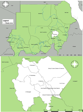
Study area: This study was conducted in seven localities of El Gezira State. The state lies between latitudes 14° 13.872’ N and longitudes 33° 21.582’ E in the central region of the country. It is boarded to the north by Khartoum State, to the south by Sinnar State, to the west by White Nile State and to the east by Gedarif State (Figure 1). The climate is dry with a short rainy season from July to September.
Study design: A cross-sectional study was conducted to determine the prevalence of Immunoglobulin G (IgG) antibodies against LSDV in cattle using enzyme-linked immunosorbent assay (ELISA). Sample size for this survey was estimated using the formula n=z2PQ/ L2 [9], where 𝑛 was the required number of individuals to be examined; z was a constant = 1.96; P is a known or estimated prevalence; Q= (1-P); 𝐿 is the allowable error. The estimated number using this formula assuming the 15% prevalence rate was 195. A total number of 198 serum samples were included in this survey.
Sample collection: Sample collection was conducted according to the animal welfare code of Sudan (Ethical number: AWCS220245). Collection of blood from animals was performed by qualified veterinarians following proper physical restraint of animals to ensure both personnel and animal safety. A total of 198 blood samples (129 crossbred and 69 indigenous animals were collected randomly from apparently healthy animals during the period between April – May, 2021. Samples were collected from 7 different localities in El Gezira State including El managil, Madani, Gazira East, Elkamleen, Elhasahesa, Twenty-fourth Elgorashi and Gazira south. Serum was separated and frozen in -20oC until tested. Risk factors analyzed in this study included age, sex, breed, locality and water source.
Serological Screening Using Elisa: The sera samples were examined for antibodies against the Capripox viruses (CPV) using the Capripox double antigen ELISA (ID.Vet Innovatitive Diagnostics, France) according to the manufacturer’s instructions. The optical densities (OD) were estimated using a microplate ELISA reader (Stat Fax 4200, USA) at 450 nm wavelength. Sera with ratios of sample to positive control antibody (S/P%) of titers ≥ 30% were evaluated positively.
Statistical Analysis. Statistical package for Social Science (SPSS) software (IBM version 20.0, USA) was employed for the analysis of the results. Logistic regression was applied to assess the association between various risk factors and the prevalence of LSD. A multivariable model for the outcome variable was constructed using logistic regression analysis and chi-square. A p-value of ≤ 0.05 indicated a significant association. The true prevalence was determined according to Stevenson formula [10]: True prevalence=(AP+Sp -1)/(Se +Sp-1) The apparent seroprevalence (AP) was modified for specificity (Sp) and sensitivity (Se) for the ELISA assay with 99.7% and 91%, respectively.
Results
Using ELISA, the overall true seroprevalence of LSDV- specific IgG was 24.6% (50/198, 95%CI, 0.520-0.539)
- among cattle in El Gezira State. As for various localities, the highest prevalence of LSDV seropositivity was recorded in
- Twenty-fourth Elgorashi (34.8%), Elkamleen (30.6%) and
- Madani (29.3%), while the lowest prevalence was recorded in the Gezira East locality (15%) (Table 1). The variation in seroprevalence among seven localities was statistically insignificant (p-value = 0.523).
- Locality
- No of tested animals
- No of positive
- True prevalence (%)
- 95%CI p- value
- Elmanagil
- 31
- 7
- 22
- Madani
- 30
- 9
- 29.3
- Gezira East
- 32
- 5
- 15
- Elkamleen
- 32
- 10
- 30.6
- Elhasahesa
- 32
- 6
- 18.1
- 24 Elgorashi
- 31
- 11
- 34.8
- Gezira south
- 10
- 2
- 19.4
- Total
- 198
- 50
- 24.6
Table 1: Univariate analysis for the association of origin of collected samples (locality) and seropositivity for Lumpy skin disease
Univariant logistic regression on the other hand, showed that there was a significant difference (p-value < 0.05) between age groups, sex, breed and seropositivity to LSD. The highest seropositive rate was noted in cattle aged > 6 years old (38.81%). The gender which showed significant association with seroprevalence of LSD revealed that the Risk factor Groups Animals tested Animals affected (%) p-value highest seroprevalence rate was detected to be in female (28.93%) compared to male (10.26%) animals. Moreover, the breed showed a significant difference, and the highest seropositivity rate was noted in local breed (37.68%) compared to the crossbreed (18.60%) (Table 2).
< 3 80 10 (12.5%) Age (years)
0.001* 3-6 51 14 (27.45%) > 6 67 26 (38.81%)
Sex Male 39 4 (10.26%) 0.016* Female 159 46 (28.93%)
Breed Cross 129 24 (18.60%) 0.003* Local 69 26 (37.68%)
South Gezira 10 2 (20%)
Madani 30 9 (30%)
Managil 31 7 (22.6%)
24 Gurash 31 11(35.5)
Hasahesa 32 6 (18.75%)
Kamlin 32 10 (31.25%)
East Gezira 32 5 (15.63%)
Barraled 73 14 (19.18%)
Water Source
0.318 Canar 30 9 (30%)
Turaa 95 27 (28.42%)
*significantly different, with a p-value < 0.05 Table 2: The Univariable association between potential risk factors and LSDV seropositivity among cattle in El Gezira State, Sudan using the chi-square test.
Multivariate analysis showed that only two independent risk factors were statistically significant (p-value < 0.05). Older cattle (> 6 years) were three times more likely to be infected with LSD (OR = 3.128, CI = 1.155 – 8.470, p-value = Risk factor Animals tested Animals positive (%) p-value Odds ratio 95% CI Lower - Upper Age (years) < 3 80 10 (12.5) Ref - -
3- 6 51 14 (27.5) 0.123 2.285 0.798 – 6.538
> 6 67 26 (38.8) 0.025* 3.128 1.155 – 8.470
Sex
Male 39 4 (10.3) Ref - -
Female 159 46 (28.9) 0.477 1.62 0.429 – 6.126
Breed
Cross 129 24 (18.6) Ref - -
Local 69 26 (37.7) 0.016* 2.353 1.176 – 4.706
0.025). The local breed was two times more probable to be infected compared to cross breed (OR = 2.353, CI = 1.176 – 4.706, p-value = 0.016) (Table 3).
* p-value < 0.05 is significantly different Table 3: Multivariate analysis using logistic regression model for significant association (p < 0.05) of risk factors and LSDV seropositivity among cattle in El Gezira State, Sudan.
Discussion
Lumpy skin disease is a transboundary viral disease. The first cases of LSVD reported in Sudan were in 1971 from an outbreak that occurred in the west of the country [8]. A following outbreak was reported in Khartoum State in 1989 among imported Friesian cattle. The cases were confirmed by histopathological, electron microscopical findings and virus neutralization [11]. Recently an outbreak of LSD among cattle was reported in the Butana area, Eastern Sudan. The cases were diagnosed using serological, virological and molecular techniques [12].
In the present study, the overall true seroprevalence rate of LSDV in cattle was 24.6% which is lower than that reported in Red Sea State using serum neutralization test (SNT) (57%) and Agar gel immunodiffusion (AGID) test (68%) during the period between March, 2003 to August, 2005 [13]. This may be due to the samples which were collected during two active outbreaks in Red Sea State. Similar findings however were reported in Egypt (24%) [14] and in Ethiopia 26.5% by Molla et al. [15] in central and north-western parts of Ethiopia. Albeit, the seroprevalence of LSD in this study was much higher than those described in Western Wollega, Ethiopia (6.4%) [16], in north-eastern Ethiopia (7.4%) [17] and in Uganda (8.7%) [3]. The variation in seroprevalence rates may be referred to the climatic diversity of the study areas, sampling time, the difference in the sample population, the activity of biting insects and the test method applied for the survey [18, 15]
The LSDV-specific antibodies revealed among cattle proposed natural infection as LSDV vaccination is not applied in the study area. In addition, screened animals were aged > 6 months, thus maternal immunity was no longer existing.
In the current study certain factors may have an effect on the seroepidemiology of LSDV infection in El Gezira State, Sudan. Our study indicates that LSD seropositivity increased significantly with age groups. This is in line with the results obtained by Abera et al. [15/16] and Selim et al. [19] who reported a positive association between LSDV infection and age groups. This is may be attributed to repeated exposure of older cattle to biting flies’ seasons compared to calves and young cattle. Furthermore, young animals are often kept indoors protecting them from the vector-borne diseases [20]. A significant association (p-value = 0.016) was also observed between LSDV seropositivity and cattle breed. The local breed was more likely to infection compared to crossbreed. In contrast, other investigators noted that exotic imported breeds are at more risk of LSDV infection than indigenous breeds [11, 16, 19, 21]. This may be due to the high number of screened crossbreed (n=129) samples compared to the local breed (n= 69) examined in our study. In the present study, an insignificant association (p-value = 0.477) was detected between gender and LSDV seropositivity that was more prevalent in females (28.9%) compared to males (10.3%). This in accordance with the results obtained by Selim, et al. [19] in Egypt. Other investigators however reported high LSD prevalence in males compared to females. They attributed the difference to certain factors such as tiredness besides the existence of numerous abrasion on the skin in the draft male that may attract the carrier flies leading to infection transmission [22, 23].
Conclusions
It is concluded that LSDV antibodies are prevalent in El Gezira State, Sudan. Breed and age-groups had a significant effect on the seropositivity of LSDV infection. Further surveillance of LSD at the country level is important in order to estimate its economic impact on animal industry in Sudan. In addition, the understanding of shepherds about risk factors that associated with LSD must be refined to better control of the disease in Sudan. Finally, more efforts are required on the part of veterinary authorities to control LSD which can be mainly achieved by vaccination besides other control measures.
Data Availability
Data used to support the findings of this study are available from the corresponding author upon request.
Conflict of Interest
The authors declare that there is no conflict of interest regarding the publication of this paper.
Funding Statement
This work was supported by Kassala Veterinary Research Laboratory, Animal Resources Research Corporation (ARRC), Khartoum, Sudan.
Acknowledgements
The authors wish to thank Dr. Selma K. Ahmed for providing the map of the study area.
References
-
Kumar N, Chander Y, Kumar R, Khandelwal N, Riyesh Y, et al. (2021) Isolation and characterization of lumpy skin disease virus from cattle in India. PLoS ONE 16(1): e0241022.
-
Mulatu E , Feyisa A (2018) Review: Lumpy Skin Disease, Journal of Veterinary Science and Technology 9(3): 535.
-
Ochwo S, Vander Waal K, Munsey A, Nkamwesiga J, Ndekezi C, et al. (2019) Seroprevalence and risk factors for lumpy skin disease virus seropositivity in cattle in Uganda. BMC Veterinary Research, pp: 236.
-
Abdullatif RB, Allawe AB (2021) Investigation of Lumpy Skin Disease Virus in Baghdad City. Indian Journal of Forensic Medicine and Toxicology, pp: 2121-2125.
-
OIE (2012) Lumpy Skin Disease: Aetiology Epidemiology, Diagnosis, Prevention and Control References Infect Dis Poverty.
-
OIE (2017). Lumpy skin Disease, OIE Terrestrial Manual.
-
House JA, Wilson TM, El Nakashly S, Karim I, Ismail I, et al. (1990) The isolation of lumpy skin disease virus and bovine herpesvirus- from cattle in Egypt. J Vet Diagn Invest 2(2): 111-115.
-
Ali BH, Obbaid HM (1977) Investigation of the first outbreaks of Lumpy Skin Disease in the Sudan. British Veterinary Journal 133(2): 184-189.
-
Martin SW, Meek AH, Willeberg P (1987) Veterinary Epidemiology Principles and Methods. Lowa States University Press.
-
Stevenson M (2008) An introduction to veterinary epidemiology. Massey University Palmerston North.
-
Khalafalla A, Gaffar Elamin M, Abbas Z (1993) Lumpy skin disease: Observations on the recent outbreaks of the disease in the Sudan. Rev Elev Med Vet Pays Trop 46(4): 548-550.
-
Hussien M, Osman A, Bakri E, Elhassan A, Elmahi M, et al. (2022) Serological, virological and molecular diagnosis of an outbreak of lumpy skin disease among cattle in Butana area, Eastern Sudan. Vet Med Sci 8(3): 1180- 1186.
-
Elsaim N (2007) Prevalence of lumpy skin disease in cattle in Red Sea State, Sudan. MSc Thesis, University of Khartoum, Sudan 7: 1022.
-
Mona AE , Selim A, and Abd Elmoneim A (2019) Prevalence and molecular characterization of Lumpy Skin Disease in cattle. Benha Veterinary Medical Journal, pp: 144-147.
-
Molla W, Frankena K, Gari G, Kidane M, Shegu D, et al. (2018) Seroprevalence and risk factors of lumpy skin disease in Ethiopia. Preventive Veterinary Medicine, pp: 99-104.
-
Abera Z, Degefu H, Gari G, Kidane M (2015) Sero- prevalence of lumpy skin disease in selected districts of West Wollega zone, Ethiopia. BMC Vet Res, pp: 135.
-
Hailu B, Tolosa, Gari G, Teklue T, Beyene B (2014) Estimated prevalence and risk factors associated with clinical Lumpy skin disease in north-eastern Ethiopia. Preventive Veterinary Medicine 115(2): 64-68.
-
Issimov A, Kutumbetov L, Orynbayev M, Khairullin B, Myrzakhmetova B, et al. (2020) Mechanical Transmission of Lumpy Skin Disease Virus by Stomoxys Spp (Stomoxys Calsitrans, Stomoxys Sitiens, Stomoxys Indica). Diptera: Animals (Basel) 10(3): 477.
-
Selim A, Manaa E, Khater H (2021) Seroprevalence and risk factors for lumpy skin disease in cattle in Northern Egypt. Trop Anim Health Prod 53(3): 350.
-
Troyo A, Calderón-Arguedas O, Fuller D, Solano M, Avendaño A, et al. (2008) Seasonal profiles of Aedes aegypti (Diptera: Culicidae) larval habitats in an urban area of Costa Rica with a history of mosquito control. J Vector Ecol 33(1): 76-88.
-
Babiuk S, Bowden T, Parkyn G, Dalman B, Manning L, et al. (2008) Quantification of lumpy skin disease virus following experimental infection in cattle. Transbound Emerg Dis 55(7): 299-307.
-
Radostits O, Gay C, Hinchcliff K, Constable P (2006) Veterinary Medicine E-Book: A textbook of the diseases of cattle, horses, sheep, pigs and goats, Elsevier Health Sciences.
-
Gari G, Grosbois V, Waret-Szkuta A, Babiuk S, Jacquiet P, et al. (2012) Lumpy skin disease in Ethiopia: Seroprevalence study across different agro-climate zones. Acta Trop 123(2): 101-106.
- The Digital Stethoscope: Harnessing AI in Veterinary Medicine Without Losing Our Healing Touch
- Meningoencephalomyelitis of Unknown Etiology: Short-Term Effect of Two Treatment Protocols on Cerebrospinal Fluid
- Safety and Efficacy of the HomeoPet Cough in Domestic Pets –A Clinical and Correction Analysis Based Upon User Response Survey
- Non Human Animals Responses to Social Loss
- Owner Reported Clinical Outcomes of a Homeopathic Proprietary Preparation for the Treatment of Upper Respiratory and Nasal Disorders in Companion Animals
- Effects and Diagnostic Approach of Ultrasound in Veterinary Practice: A Systematic Review