Mixed Canine Mammary Gland Tumour in a 10 Year Old Lhasa Apso Bitch Histopathological valuation and Surgical Management
Mixed mammary gland tumors are the most frequent canine mammary gland neoplasms with a high incidence rate among females, however, there is paucity of studies that describe their clinic-pathological presentations and management approaches. This case evaluated the clinic-pathological data and management of mixed mammary gland tumors. A 10-year-old Lhasa Apso bitch weighing 11.4kg was referred to the University of Jos Veterinary Teaching Hospital with a ventral abdominal mass. Clinical examination revealed an enlarged left inguinal mammary gland with two palpable masses. The inguinal region was aseptically prepared for surgery and anesthesia achieved by the administration of 1% atropine sulphate (Atropine® - Shanxi Shuguang Pharmaceutical Co., Ltd., Qixian, China) and Chlorpromazine as preanesthetics and 50mg/mL Ketamine hydrochloride (Jawa ketamine® - Swiss Parenterals Ltd, Gujarat, India) + 2% Xylazine (Alfasan, Woerden-Holland) as induction and maintenance anesthetics. The glandular, connective/fibrous and fatty tissues of the mammary gland were carefully dissected to achieve the complete excision of the encapsulated mammary tumour masses before the reconstruction of the resulting defect. Postoperatively, penicillin and streptomycin, piroxicam, vitamin B complex, dexamethasone, vincristine sulphate and ivermectin were administered. The excised masses histologically presented numerous tubules with some containing mucin. Also there were the presence of acini with neoplastic epithelial cells and myoepithelial cells, cartilage and bony trabeculae. Surgical debulking with adjunctive immuno-chemotherapy was curative for the mixed mammary gland tumours in this bitch.
Introduction
Mixed tumors are the most frequent mammary gland neoplasms in bitches [1]. They are characterized by epithelial, myoepithelial, and mesenchymal components often with the potential to undergo malignant transformation [1, 2]. Microscopically, benign epithelial (ductal and/or acinar cells), myoepithelial, and mesenchymal elements, along with the presence of cartilage and/or bone are signature components of benign mixed tumors [2]. Carcinomas are present in mixed tumors but they are usually characterized by the presence of malignant epithelial and benign mesenchymal components, sometimes represented by variable presence of cartilage, bone and adipose tissue. However, mammary carcino- sarcomas are neoplastic in nature with both malignant epithelial and mesenchymal components [2]. Prognosis of mammary gland tumour is influenced by several factors including age, histological type and grade, clinical stage, tumor size and biological behavior including mitotic index, regional or distant metastases, micro-vascular density, and molecular markers with a single nucleotide polymorphism in exon 9 of BRCA1 and exon 24 of BRCA2 significantly associated with canine mammary glands tumour [3, 4]. Surgical management is the first option for all bitches with mammary tumors [5]. Adjuvant chemotherapy is indicated for patients with regional or distant metastases and for those with mammary neoplasms with poor prognoses [3].
Available cancer management modalities include: chemotherapy, immunotherapy, biological therapy, radiotherapy, endocrine therapy, surgical excision and their combinations. Combining antiangiogenic strategies and antineoplastic chemotherapeutic protocols with surgical intervention may benefit bitches with malignant neoplasms [4, 6]. Thus, the management of mixed mammary gland tumour in this 10-year-old bitch will invaluably contribute to the knowledge available to clinicians for effective management in bitches and by extension, in humans since many similar features exist between human mammary/ salivary and canine mammary gland mixed tumours [7].
Case Management
History and Clinical Examination
A 10-year-old Lhasa Apso bitch weighing 11.4kg was referred to the University of Jos veterinary teaching hospital with a diagnosis of a ventral abdominal mammary gland tumour from the referring Veterinarian. History revealed that the mass was first noticed about a year ago with gradual size increment. Clinical examination revealed rectal temperature, pulse rates and respiratory rates of 38.4ºC, 76.0 beats/ minute and 40.0 cycles/minute respectively. Upon further physical examination, the left inguinal mammary gland was enlarged with two palpable masses felt.
Management
Following clinical examination, blood sample and fine needle aspirate of the swelling were collected and sent to Clinical pathology laboratory complete haemogram and for exfoliative cytology following the methods first described by Robinson, et al. [8], respectively. The inguinal region was prepared for aseptic surgery by clipping the hair around the area, scrubbing using chlorhexidine gluconate (Savlon®) and application of mild antiseptic solution. Anesthesia was achieved by the administration of 1% atropine sulphate (Atropine® - Shanxi Shuguang Pharmaceutical Co., Ltd., Qixian, China) at 0.02 mg/kg and Chlorpromazine 25mg/ kg as preanesthetics and 50mg/mL Ketamine hydrochloride (Jawa ketamine® - Swiss Parenterals Ltd, Gujarat, India) at 12mg/kg + 2% Xylazine 1mg/kg (Alfasan, Woerden-Holland) at 2mg/kg as induction and maintenance anesthetics. The bitch was fastened unto the surgical table on dorsal recumbency and given a square fashion draping, exposing the proposed surgical site. The glandular, connective/fibrous and fatty tissues of the mammary gland were carefully dissected to assess and doubly ligate the caudal epigastric vessels before a complete excision of the encapsulated mammary tumour mass was done with a scalpel blade while ensuring to incorporate a 1cm margin of healthy tissue. The surrounding healthy tissues were sutured and a subcuticular suture placed using size 0 vicryl® to approximate the skin edges before the application of horizontal mattress sutures using size 2 silk for skin apposition (Figure 1).
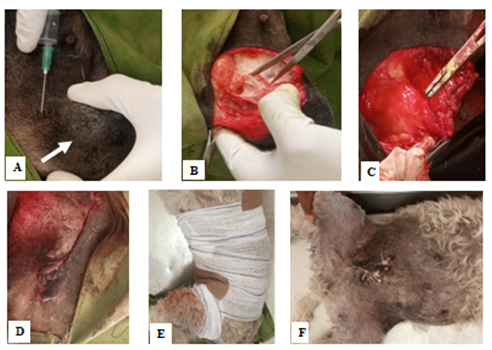
Post-operatively, Inj. 200,000iu penicillin 12,000iu/ kg and 200mg streptomycin 20mg/kg IM x 5/7, Inj. 10mg piroxicam 0.3mg/kg IM x 2/7 E.O.D, Inj. vitamin B complex IM x 2/7, and Inj. 2mg dexamethasone 0.3mg/kg IM x 2/7 were administered. Elizabethan collar was placed over the dog’s neck and physical activities reduced while diet was improved until healing was achieved. Two masses weighing 139.6 grams were removed from the mammary gland and sent for histological evaluation (Figure 2). The sutures were removed on day-10 following surgery and the bitch was administered with vincristine sulphate 0.025mg/kg slow IV and ivermectin 0.6mg/kg SC once weekly ϰ 2/52 before being discharged following normal vitals and haemogram (Table 1).
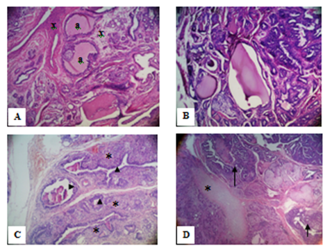
• Presenting numerous tubules with some containing mucin (a) and increased connective tissue stroma (x) HE, 100ϰ, (B) numerous tubules with some containing mucin (a) and dense cellularity (m), HE, 100ϰ, (C) acini with neoplastic epithelial cells (arrow head) and myoepithelial cells (asterisk). HE, 100ϰ, (D) cartilage (star) and bone trabeculae (arrow). HE, 40ϰ.
| Parameters | Patient’s Values | Normal Range | |
|---|---|---|---|
| Pre-surgery | Post-surgery | ||
| Packed Cell Volume (%) | 45 | 44 | 37-55 |
| Haemoglobin (g/dL) | 13.6 | 13.4 | 12.00 -18.00 |
| Plasma Protein (g/dL) | 10 | 7.2 | 6 – 7.5 |
| Plasma Colour | Clear | Clear | Clear |
| Total leucocyte (x109/L) | 8.4 | 8.8 | 6 – 17 |
| Seg. Neutrophils (x109/L) | 7.81 | 8.22 | 3.6 – 13.09 |
| Band Neutrophils(x109/L) | 0 | 0.03 | 0 – 0.85 |
| Lymphocyte (x109/L) | 0.59 | 0.47 | 0.72 – 5.1 |
| Monocytes (x109/L) | 0 | 0 | 0.18 – 1.7 |
| Eosinophils (x109/L) | 0 | 0.08 | 0.12 – 1.7 |
| Basophils (x109/L) | 0 | 0 | Rare |
Table 1: Haemogram of 10-Year-old Lhasa Apso with Mixed Canine Mammary Gland Tumour.
Follow-up re-examination of the site at 3-month interval until a period of 1-year with no evidence of tumour re-growth was indicative of complete remission.
Local infiltration of the clipped and aseptically prepared surgical site with lidocaine and adrenaline following a rectangular draping with exposed surgical field (A), firm mass on the left inguinal mammary gland (white arrow), dissection of the exposed mammary tissues and tumour mass (B), suturing the mammary gland tissues (C) approximated skin edges by subcuticular sutures (D), lightly bandaged surgical site (E) and healed surgical site 4 weeks post-surgery. Discussion Mammary gland tumours, like other tumours are the result of mutation inactivating tumour suppressor genes and DNA repair genes, thus enabling abnormal cell proliferation. Mammary tumours are the most frequent neoplasia in female dogs constituting a serious problem in veterinary medicine [1]. Risk factors could be non-modifiable such as gender, age, genetic mutation, personal history, family history and modifiable which include; not being physically active, obesity, hormones, reproductive history and alcohol entities. Diagnosis of mixed mammary gland tumour was reached by the cytological appearance similar to reports by Dolka, et al. [9] in which lobular hyperplasia cholesterol clefts and high cellularity were observed. The case supports the findings of Sorenmo, et al. [5] that surgical excision is usually the first option in mammary tumour with an immediate esthetically pleasing appearance which gets better as the surgical wound heal by primary intention.
Histologically, the mammary tumour presented numerous tubules with some containing mucin and increased connective tissue stroma with visible dense cellularity. Also the presence of acini with neoplastic epithelial cells and myoepithelial cells, cartilage and bony trabeculae signified the mixed nature of the mammary tumour in this case. These findings corroborates the reports by Cassali, et al. [10] where they reported canine mixed mammary gland tumour to present a complex histological pattern consisting of epithelial and mesenchymal component. Goldschmidt, et al. [11] and Sharma and Farooq [12] reported that the mesenchymal component consists of cartilage and/ or bone, mostly in combination with adipose tissue. Also, they reported epithelial component of the neoplasm to be malignant while the mesenchymal component is often benign. Malignant mixed mammary tumour (Carcinosarcoma) is composed of cells that morphologically resemble epithelial cells, luminal epithelium and/or myoepithelial and have various types of differentiation including adeno, squamous, mucinous and anaplastic features [12]. Benign mixed tumours are characterized by benign proliferation of cells that are morphologically similar to epithelial components and mesenchymal cells that produce cartilage and bone [2, 12]. Malignant types of canine mammary tumours are more frequent than benign mammary tumours. Due to vast histological diversity of canine mammary tumours their diagnosis is difficult and provides little prognostic information [13, 14, 15].
Conclusion
Canine mammary tumours attract special interest because of their similarities with human breast cancer. Mixed tumours are uncommon lesions in the human breast, but they are found most frequently in the mammary gland of the female dogs and in the human salivary glands. Canine mixed mammary gland tumours could be effectively managed by early surgical excision with adjunctive chemotherapy using vincristine sulphate at 0.025mg/kg patient’s body weight.
References
-
Cassali GD, Bertagnolli CA, Ferreira E, Gamba CO, Araujo K, et al. (2012) Canine mammary mixed tumours a review. Vet Med Int 274608: 1-7.
-
Misdorp W (1999) Histological classification of the mammary tumors of the dog and the cat. In: S. Series (Ed.), WHO international histological classification of tumors of domestic animals. Washington, DC: AFIP.
-
Cassali GD, Lavalle GE, Nardi AB, Ferreira E, Bertagnolli AC, et al. (2014) Consensus for the Diagnosis Prognosis and Treatment of Canine Mammary Tumors. Brazilian Journal of Veterinary Pathology 4(2): 153-180.
-
Nunes FC, Damasceno KA, De Campos CB, Bertagnolli AC, Lavalle GE, et al. (2019) Mixed Tumors of the Canine Mammary Glands Evaluation of Prognostic Factors Treatment and Overall Survival. Vet Anim Sci 7: 100039.
-
Sorenmo KU, Worley D, Goldschmidt MH (2013) Tumors of the mammary gland. In Withrow SJ, et al. (Eds.) Withrow and MacEwen’s Small animal clinical oncology. 5th (Edn.), St. Louis: Saunders Elsevier. pp: 538-556.
-
Tkaczyk Wlizlo A, Smiech A, Kowal K, Rozanska D, Slaska B (2023) Histopathological Evaluation of Canine Mammary Gland Tumours: A study of 92 cases. Medycyna Weterynaryjna 79(7): 356-363.
-
Genelhu MCLS, Cardoso SV, Gobbi H, Cassali GD (2007) a comparative study between mixed type tumours from human salivary and canine mammary glands. BMC Cancer 7: 218-227.
-
Robinson IA, McKee G, Nicholson A, Arcy JD, Jackson PA, et al. (1994) Prognostic value of cytological grading of fine-needle aspirates from breast carcinomas. Lancet 343(8903): 947-949.
-
Dolka I, Czopowicz M, Gruk Jurka A, Wojtkowska A, Sapierzynski R, et al. (2018) Diagnostic Efficacy of Smear Cytology And Robinson’s Cytological Grading of Canine Mammary Tumors with Respect to Histopathology Cytomorphometry Metastases and Overall Survival. PLoS One 13(1): e0191595.
-
Cassali G, Damasceno K, Bertagnolli A, Estrela-Lima A, Lavalle G, et al. (2017) Consensus Regarding the Diagnosis Prognosis and Treatment of Canine Mammary Tumors Benign Mixed Tumors Carcinomas in Mixed Tumors and Carcinosarcomas. Brazilian Journal of Veterinary Pathology 10: 87-99.
-
Goldschmidt M, Pena L, Rasotto R, Zappulli V (2011) Classification and Grading of Canine Mammary Tumors. Vet Pathol 48(1): 117-131.
-
Sharma A, Farooq SU (2022) Canine Mammary Tumours Advances in Classification Grading and Expression of Biological Markers. International Journal of Bio resource and Stress Management 13(12): 1496-1503.
-
Misdorp W, Cotchin E, Hampe JF, Jabara AG, Von Sandersleben J (1972) Canine malignant mammary tumours: II. Adenocarcinomas, solid carcinomas and spindle cell carcinomas. Vet Pathol 9(6): 447-470.
-
Olayemi FO, Azeez IO, Ogunyemi A, Ighagbon FO (2009) Study on Erythrocyte Values on the Nigerian Indigenous Dog. Folia Veterinaria 53(2): 65-67.
-
Avazi DO, Awasum CA, Hassan AZ, Ayo JO, Aluwong T, et al. (2019) Evaluation of levels of interleukin-6 interleukin-8 and some haematologic parameters of dogs with cutaneous wounds. Cytokine 113: 128-138.
- The Digital Stethoscope: Harnessing AI in Veterinary Medicine Without Losing Our Healing Touch
- Meningoencephalomyelitis of Unknown Etiology: Short-Term Effect of Two Treatment Protocols on Cerebrospinal Fluid
- Safety and Efficacy of the HomeoPet Cough in Domestic Pets –A Clinical and Correction Analysis Based Upon User Response Survey
- Non Human Animals Responses to Social Loss
- Owner Reported Clinical Outcomes of a Homeopathic Proprietary Preparation for the Treatment of Upper Respiratory and Nasal Disorders in Companion Animals
- Effects and Diagnostic Approach of Ultrasound in Veterinary Practice: A Systematic Review