Diagnostic and Therapeutic Aspects of Mucosal Leishmaniasis
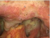
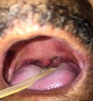
Leishmaniasis is caused by protozoa of the genus Leishmania, with the presentation restricted to the mucosa being infrequent. Although the nasal mucosa is the main site affected in this form of the disease, it is also possible the involvement of the lips, mouth, pharynx and larynx. The lesions are characteristically ulcerative-vegetative, with granulation tissue formation. Patients usually complain of pain, dysphagia and odynophagia. Differential diagnosis should include cancer, infectious diseases and granulomatous diseases. We present a case of a 64-year-old male patient, coming from an endemic area for American Tegumentary Leishmaniasis (ATL), with a chief complaint of persistent dysphagia and nasal obstruction for 6 months. The lesion was ulcerative with a purulent infiltration into the soft palate and uvula. After excluding other diseases, ATL was suggested as a hypothesis, having been requested serology and biopsy of the lesions. Was started the treatment with pentavalent antimony and the patient presented regression of the lesions in 30 days, with no other complications.
Introduction
Another study, Clinico-Audio-Radiological and The American Tegumentary Leishmaniasis (ATL) is a disease caused by protozoa of the genus Leishmania, and initially presenting in the cutaneous surface, which can or cannot be followed by mucosal involvement. Mucosal lesions can occur during the activity of the skin lesion or even years after their resolution. The presentation with mucosal involvement is rare and is difficult to diagnose because several diseases have similar clinical picture. However, ATL is not always considered in the differential diagnosis [1, 2, 3]. Differential diagnoses include skin infectious diseases such as those caused by fungi and bacteria (tertiary syphilis, leprosy, and paracoccidioidomycosis), granulomatous diseases such as sarcoidosis, blastomycosis, and Wegener's granulomatosis, as well as cancerous lesions such as squamous cell carcinoma and, lymphomas [1, 2, 4, 5].
A 64- year-old Male farmer presented to our clinic from an endemic area for Leishmaniasis, because of his persistent dysphagia and nasal obstruction over the past six months. Physical examination revealed ulceration and a purulent infiltration into the soft palate and uvula associated with edema and local purulent discharge. Biopsy revealed unspecific granulomatous reaction, inflammatory infiltrate, without direct visualization of leishmania parasite. Based on clinical and epidemiological findings, we chose to start treatment with pentavalent antimony parenteral, 20mg/kg/day. The lesions regressed in 4 weeks, leaving only residual adhesions in nasal cavities.
Discussion
The epidemiological situation of American Tegumentary Leishmaniasis shows that its prevalence and incidence are high in Brazil, as in other countries in the Americas, Europe, Africa and Asia. Epidemiological data from the World Health Organization (WHO) shows a global prevalence of 12 million people and an incidence of approximately 400,000 cases/year. In Brazil, the ATL is considered an endemic disease and is prevalent in adults and males, although it can affect individuals of both genders and all ages. The protozoan species most commonly found in Brazil and the Americas are Leishmania brasiliensis, L. guyanensis and L. amazonensis [4, 6]. Those species, also called New World species, can cause crippling injuries with formidable esthetic and psychological consequences in the affected individual [1, 3, 5]. The clinical presentation of ATL happens due to the interaction of parasite species and host immune response is mediated by humoral and cell response [1]. Usually, the clinical presentation of ATL with mucosal involvement is the occurrence of lesions in the nasal cavity and may be associated with the oral cavity, pharynx and larynx [2, 3, 4, 7]. The occurrence of isolated mucosal lesions is rare and, in Brazil, only 4.4% of the reported cases have this presentation [8]. When there is the oropharynx’s involvement the hard palate is often involved, which can be disseminated to the soft palate, uvula and pharynx. Nasal obstruction symptoms may occur due to the expansion of infiltrative process in the nasopharynx region. Are frequent symptoms of oropharynx involvement: dysphagia, odynophagia, dysphonia, coughing, drooling and oral wounds [3]. When there is mucosal involvement the treatment is more difficult and the occurrence of relapses is common [1, 9]. The WHO considers the clinical manifestations associated with a positive parasitological test confirmative of the diagnosis of ATL, but points out that serologic tests have limited value. The conduct’s manual of ATL followed by this country [10] correlate the clinical and epidemiological diagnosis to be made on the occurrence of typical lesions, especially when an individual resides or comes from endemic regions to the ATLHowever, diagnostic confirmation by parasitological methods is desired and should be attempted whenever possible. The mucosal form of the disease, usually by having an atypical presentation, can lead to delays in diagnosis and wrong management of the disease [5], which one, could present with decreased effectiveness of therapy because of the late institution and the occurrence of functional and esthetic sequelae [4]. The mucosal form of ATL has an important intrinsic limitation, represented by the difficulty in obtaining appropriate biological samples for the achievement of parasitological tests [1, 7]. Even the direct investigation of the parasite has low sensitivity, around 48% [1], with articles presenting a variation range of 15- 70% [7], being lower with the chronicity of injury, mainly if the symptoms has initiated more than 3 months. Also, in the histological analysis the parasite’s visualization is difficult and the classic finding consists of nonspecific granulomatous reaction associated with inflammatory infiltrate rich in lymphocytes [7]. There is no consensus on the ideal way to obtain material for direct detection of parasite at mucosal lesions, but it is recommended to obtain samples from the edge of the lesion, which usually present with tumescent and hyperemic aspect, for the ulcerated skin lesions [4]. Although the patient in question present evolution of symptoms for six months, featuring chronicity and reducing the chances of parasite visualization, the biopsy of oropharynx’s lesion with parasitological histopathologic investigation is necessary in an attempt to confirm the diagnosis, specially due to the atypical presentation of the patient’s clinical condition. More accurate tests of molecular biology, such as the technique of Polymerase Chain Reaction (PCR), have very high costs and are only available in large urban centers [7] making it difficult to carry out this investigation in this patient. Faced with the difficulty to visualize the parasite at parasitological examinations, indirect methods of identifying the presence of the disease has been used and the search for immunoglobulins of the IgG, IgA, IgM and IgE classes is being studied as a diagnostic alternative to direct parasitological examinations in ATL [1, 11]. Serological testing by immunofluorescence, ELISA and Western blot are some of the many tests that can be used. The sensitivity and specificity of these depend on the technique used and the presentation of the disease [7]. Zeyrek, et al. [12] point out that serological tests are of great value for the diagnosis of visceral leishmaniasis, but have limited importance for the mucocutaneous leishimaniasis (MCL) form. Still, in their assay, the authors demonstrated that, in their targed population consisted by individuals from highly endemic area for ATL and with typical skin lesions, serologic tests make up an important diagnostic tool, reporting sensitivity of 78.4% and specificity of 69.3% for IgG searched by ELISA’s technique. Sarkari, et al. [13] corroborated the results found by Zeyrek and his team, showing a sensitivity of 83.6% and specificity of 62.7% for the diagnosis of ATL in patients with cutaneous presentation by the total IgG analysis and 84.7% sensitivity and 54.3% specificity by IgM analysis by ELISA’s technique, but weren’t found no similar reports in patients with atypical clinical condition without cutaneous involvement. Study conducted by Souza, et al. [11] evaluated the antibody profile in serum samples from patients (n=37) with clinical diagnosis confirmed or compatible with mucocutaneous leishmaniasis treated at a hospital in Uberlândia/MG/Brazil. A percentage of 86.5% of these patients had predominant cutaneous manifestations and disease’s time evolution ranged from 1 month to 10 years. The authors found 94.6% of reagent samples for IgG and 21.5% for IgM, even with most of the samples showing little time evolution of the lesions. They also rated the avidity of these antibodies in an attempt to correlate with disease duration, but it wasn’t found a pattern in the answers, which shows the complexity of the immune response in ATL. Immunological tests are indirect tests that visualize the late cellular response of the host, and the Montenegro’s Skin Test its one of the available tests. It has sensitivity close to 90% and specificity around 75%, and when there is mucosal involvement the patient usually has a strong positive reaction [6]. Although it is a low cost and good accessibility test, the applicability in endemic areas for Chagas disease, tuberculosis and leprosy becomes impracticable due to cases of false positive by cross-reaction, besides also having technical difficulties that is inherent to the method [7]. Being the patient from an endemic region for the three mentioned pathologies, the choice for this method to diagnostic aid would not be appropriate. The diagnosis of ATL is difficult to be realized, either by no suspicion of the disease or lack of additional tests with good accuracy, and there is no gold standard for diagnosis. To be a neglected disease that affects specially the least developed countries and the population of low social and economical level, there is little interest in developing new parasite detection techniques, as well as adjust the cost-benefit relation to the reality of the most affected population, which means that often the laboratory research is not accessible to all suspected cases and makes it impossible to wait for diagnostic confirmation to initiate the treatment [7, 14]. Considering the difficulty of obtaining a diagnosis of sure, can be made a presumptive diagnosis by the junction of the clinical history, epidemiological history and laboratory data, and the absence of one of these factors does not exclude the ATL. The patient described presented clinical complaints of dysphagia and nasal obstruction for 6 months. Before being evaluated in this service, he had already made medical attendance for a period in the city of São Paulo/SP, where tests to exclude other pathologies were requested. The results were negative and the patient was still with undefined diagnosis. Evaluating the patient, was observed on physical examination the presence of lesions in ulcerative-infiltrative type at oropharynx region of the soft palate and uvula, and associated with edema and purulent discharge. The history showed important epidemiology for ATL and this diagnosis was suggested. Serology was requested for leishmaniasis and realized biopsy of the lesion. Serology showed positive result for IgG and biopsy with histopathological analysis shows no parasite view. Even without the identification of the parasite, it was decided for the institution of treatment due to clinical condition that, although not specific, is compatible with the mucosal form of ATL associated with the origin of endemic region and serologic analysis reagent that is suggestive of pathology. The treatment of MCL is still no consensus and their choice may change according to the patient's profile, the species involved and the clinical presentation of the disease. Although several drugs with better toxicity profile has been tested and validated for specific populations and species of the parasite, the drug of choice for treating MCL presentation of ATL recommended by PAHO/WHO is still pentavalent antimony. The potential toxicity to the kidneys, liver, heart, pancreas and haematopoietic system limits the use in some populations such as liver disease, kidney disease and patients with important arrhythmias [9]. The recommended dose is 20mg/kg/day administered intramuscularly or intravenously for, at least, 4 weeks [15], with studies showing cure rate between 30 and 90% [16]. In the event of recurrence pentavalent antimony can be repeated, at a dose of 10 to 15mg/kg 12/12hs for another 4 weeks or can be used pentamidine 4mg/kg intramuscularly three times a week until the disappearing of the lesions [15]. Amphotericin B and pentamidine are second-line treatments recommended in the case of contraindications to the use of pentavalent antimony [14, 17]. Two Brazilian assays tested the use of lower doses of pentavalent antimony in the treatment of mucosal form of ATL. Oliveira-Neto, et al. [9] used a dose of 5mg/kg/day intramuscularly for 30 days in patients with mild to moderate severity mucosal form, and if the patient does not present improvement or complete regression of the lesions, the same scheme was repeated for 15 days. The authors observed efficacy of 91.4% with treatment and was not observed resistance to the use of higher doses in cases where there was no response to initial treatment. In another study, Oliveira-Neto & Mattos [18] tested low doses of pentavalent antimony in two patients resistant to treatment (antimony in high doses, amphotericin B and/or pentamidine). The treatment schedule consisted in an ampoule of antimony 405mg intramuscularly three times a week for 10 to 12 weeks. No side effects were observed and after the application time the lesions of patients were cured. It was also not observed the occurrence of relapse during the accompaniment period (9 months to 1 year). The authors suggested that the treatment with high doses of the drug for short periods could be substituted by lower doses and longer time, which would reduce the side effects and acceptance of the treatment by the patients. Others medications e combined therapy are being researched in order to increase the cure rates of ATL and/or reduce relapses and adverse effects caused by the drugs currently in use. Randomized, double-blind and controlled assay made by Machado, et al. [19] compared the efficacy of a combination of pentavalent antimonial with pentoxifylline (test group) to a combination of antimonial and placebo (control group) in patients who had severe mucosal form of ATL. Patients who used the combined antimony-pentoxifylline showed curing time of mucosal lesions less than patients in the control group. The test group also achieved higher cure rate, of 100%, while the control group had a cure rate of 58% with a treatment cycle. Patients were followed for 2 years and no relapse was observed during this period. Hodiamont, et al. [20] recommend the use of combination antimony and pentoxifylline in the treatment of ATL while waiting for more assays about the effectiveness of other drugs. Studies realized with the drug Miltefosine showed good results in patients in Colombia, suggesting that this drug could be an option in the treatment of mucosal ATL, but more randomized controlled studies should be conducted to indicate this medication as a routine [21]. Studies using azoles, specifically itraconazole, in the treatment of MCL presented discrepant and insufficient results to recommend the drug as an alternative treatment in LTA with mucosal presentation [14, 16]. Local treatment by infiltration of the drug, the use of ointments, thermo- therapies and surgery to remove the lesion has not been validated for mucosal forms of the disease, but they are options when there is only skin involvement with minor injuries and a few number of lesion plus a low risk of progression to the mucocutaneous form of the disease, or when the patient has contraindications to systemic treatment [16, 20, 21].
Conclusion
American Tegumentary Leishmaniasis is a disease that can have multiple presentations, with the mucosal form being the rarest one. Because of its atypical clinical picture, diagnosis of mucosal form is more difficult, especially because various diseases, benign and malignant, makes differential diagnosis and can be confused with the ALT. The laboratory tests limitations, such as low sensitivity of direct research and poor accessibility to more accurate methods, make the epidemiological context of greater importance in the diagnosis and treatment of disease.
References
-
Zajtchuk JT, Casler JD, Netto EM, Grogl M, Neafie RC, et al. (1989) Mucosal leishmaniasis in Brazil. Laryngoscope 99(9): 925-939.
-
Lessa MM, Lessa HA, Castro TWN, Oliveira A, Scherifer A, et al. (2007) Leishmaniose mucosa: aspectos clínicos e epidemiológicos. Rev Bras Otorrinolaringol 73(6): 843-847.
-
Neto FXP, Rodrigues AC, Silva LL, Palheta ACP, Rodrigues LG, et al. (2008) Otorhinolaryngologic Manifestations Relating American Tegumentary Leishmaniasis: Literature Review. Int Arch Otorhinolaryngol 12(4): 531-537.
-
Mota LAA, Miranda RR (2011) Manifestações dermatológicas e otorrinolaringológicas na Leishmaniose. Arquivos Int Otorrinolaringol 15(3): 376-381.
-
Nadler C, Enk CD, Leon GT, Samuni Y, Maly A, et al. (2014) Diagnosis and management of oral leishmaniasis--case series and literature review. J Oral Maxillofac Surg 72(5): 927-934.
-
Grant A, Spraggs PD, Grant HR, Bryceson AD (1994) Laryngeal leishmaniasis. J Laryngol Otol 108(12): 1086-1088.
-
Gomes CM, Paula NA, Morais OO, Soares KA, Roselino AM, et al. (2014) Complementary exams in the diagnosis of American tegumentary leishmaniasis. An Bras Dermatol 89(5): 701-709.
-
(2014) Informe Epidemiológico das Américas. Organização Pan-Americana da Saúde, Organização Mundial da Saúde. Informe Leishmanioses (3).
-
Oliveira-Neto MP, Mattos M, Pirmez C, Fernandes O, Goncalves-Costa SC, et al. (2000) Mucosal leishmaniasis ("espundia") responsive to low dose of N-methyl glucamine (Glucantime) in Rio de Janeiro, Brazil. Rev Inst Med Trop São Paulo 42(6): 321-325.
-
(2010) Brasil Ministério da Saúde Secretaria de Vigilância em Saúde Manual de Vigilância da Leishmaniose Tegumentar Brasilia.
-
Souza MA, Silva AG, Afonso Cardoso SR, Favoreto JS, Ferreira MS (2005) Perfil de isotipos de imunoglobulinas e subclasses de IgG na leishmaniose tegumentar americana. Rev Soc Bras Med Trop 38(2): 137-141.
-
Zeyrek FY, Korkmaz M, Ozbel Y (2007) Serodiagnosis of anthroponotic cutaneous leishmaniasis (ACL) caused by Leishmania tropica in Sanliurfa Province Turkey where ACL is Highly Endemic. Clin Vaccine Immunol 14(11): 1409-1415.
-
Sarkari B, Ashrafmansouri M, Hatam G, Habibi P, Abdolahi Khabisi S (2014) Performance of an ELISA and indirect immunofluorescence assay in serological diagnosis of zoonotic cutaneous leishmaniasis in iran. Interdiscip Perspect Infect Dis.
-
Lima EB, Porto C, Motta JOC, Sampaio RNR (2007) Tratamento da Leishmaniose Tegumentar Americana / Treatment of american cutaneous leishmaniasis. An Bras Dermatol 82(2): 111-124.
-
(2004) Organización Panamericana de la Salud. Guía para el tratamiento de lasc enfermedades infecciosas.
-
Monge-Maillo B, López-Vélez R (2013) Therapeutic options for old world cutaneous leishmaniasis and new world cutaneous and mucocutaneous leishmaniasis. Drugs 73(17): 1889-1920.
-
Lessa HA, Machado P, Lima F, Cruz AA, Bacellar O, et al. (2001) Successful treatment of refractory mucosal leishmaniasis with pentoxifylline plus antimony. Am J Trop Med Hyg 65(2): 87-89.
-
de Oliveira-Neto MP, Mattos Mda S (2006) Successful therapeutic response of resistant cases of mucocutaneous leishmaniasis to a very low dose of antimony. Rev Soc Bras Med Trop 39(4): 376-378.
-
Machado PR, Lessa H, Lessa M, Guimarães LH, Bang H, et al. (2007) Oral pentoxifylline combined with pentavalent antimony: a randomized trial for mucosal leishmaniasis. Clin Infect Dis 44(6): 788-793.
-
Hodiamont CJ, Kager PA, Bart A, de Vries HJ, van Thiel PP, et al. (2014) Species-directed therapy for leishmaniasis in returning travellers: a comprehensive guide. PLoS Negl Trop Dis 8(5): e2832.
-
Blum J, Desjeux P, Schwartz E, Beck B, Hatz C (2004) Treatment of cutaneous leishmaniasis among travellers. J Antimicrob Chemother 53(2): 158-166.
- 4th Branchial Cleft Sinus Anomaly Presenting as Recurrent Thyroid Abscess in A Child: A Case Report
- Parotid Duct Injury Repaired Using an Angiocatheter Stent: A Case Report
- Organization and Functionality of the Referral and Counter-Referral System for ENT Disorders in District Hospitals of N’Djamena, Chad: A Cross-Sectional Analytical Study
- Facial Metastases from a Gastrointestinal Stromal Tumor: A Case Report
- Panorama of Ent Cancers and Literature Review: Epidemiological Profile and Therapeutic Management
- Could Antimicrobial Resistance Prove to Be Both a Threat and an Opportunity for us?