Tracheal Myiasis: Case Report
Myiasis is known to exist since immemorial time. Physician harnessed it to treat infected wounds especially in pre-antibiotic era. We report a case of tracheal myiasis in a tracheostomized patient of post chemo radiotherapy for a laryngeal carcinoma. This was successfully treated by manual removal of maggots. Myiasis may prove fatal as it can lead to total airway obstruction if left untreated.
Introduction
Myiasis, a not so common entity in tropics, is an opportunistic parasitic infestation of human and animals, which are caused by house fly larvae (maggots). This can be classified as accidental, facultative (opportunistic) or obligate [1]. Many factors are responsible for myiasis such as low socioeconomic status, immune compromised state, mental retardation and unhygienic living conditions. The misery is generally associated with traumatic wounds and erosive or ulcerative lesions of skin and mucosa [2]. Myiasis is usually accidental and most often results in subcutaneous infestation (furuncular myiasis). Many species of dipterous flies among the genera chrysomyia have been reported to be the most important obligatory myiasis producers among human and animals [3]. Severity of myiasis depends on location of infestation, size of wound and degree of tissue inflammation. We report a case of a post chemoradiotherapy for a laryngeal carcinoma larynx who had tracheostomy with crawling maggots in trachea.
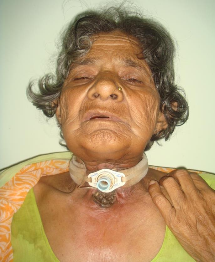
A 54 years old female patient (Figure 1) belonging to rural background with low socioeconomic as well as low hygienic status, had carcinoma of the right hemilarynx with neck metastases, for which she underwent tracheostomy and full course of chemoradiotherapy. Four and half months later, she presented with tickling and crawling sensation just below the tracheostomy tube which was associated with foul smelling and serosanguinous discharge. After 24 hours maggots were noted below the tracheal stoma. The procedure was explained to the patient and the treatment started after the informed consent was obtained. The maggots were removed manually with forceps after application of topical liquid paraffin application. Subsequently 0 degree nasal endoscope was passed through the tracheostome until no maggot was left behind. Daily stoma cleaning, dressing and change of tracheostomy tube were done. The patient was discharged with clean and healthy tracheal stoma and she is on regular follow up.
Discussion
The utilization of larvae for wound healing has been well documented across over centuries in different cultures especially the China [4]. The beneficial effect of using larvae in wounds was first reported by Pare in 1557 while treating battle wounds in Napoleon’s army [5]. The first clinical application of maggots therapy was performed during the American civil war [6]. Later, Baer refined the technique by using sterile maggots for management of wound infection. The therapy became more popular and was widely used for the treatment of chronic or infected wounds across America and Europe during the 1930s, before the antibiotic era [7]. Female flies are attracted to odoriferous suppurating lesions and open wounds, the eggs are deposited on the unbroken, soft skin of various parts of the body that are contaminated by blood or mucus discharge [1]. These eggs can be transferred to other sites by patient’s own fingers due to itching and poor hygienic habits. On hatching, the maggots penetrate deep into the tissue aided by their sharp oral hooks and anchoring inter- segmental spines which scrape away the tissue and lacerate the fine blood vessels, while feeding. During feeding on necrotic or living tissue the caudal ends of the maggots with their blackish peritremes remains visible at the surface of the lesion, enabling the larvae to breathe [3]. Infestation is commonly implicated to larvae of Chryosomyia bezziana, which is a cause of obligatory myasis on human and animal tissue especially in the nose, ear, face, gums and serous cavities. The predisposing factors include poor hygienic condition, especially with underlying diseases like atrophic rhinitis, leprosy, diabetes with purulent sinusitis, midline granuloma, malignancy and syphilis involving nose [1, 8]. Progressive necrosis of muscles continues which is associated with larval growth and invasion until a large cavernous lesion is formed, where the larvae aggregate and remain active. Hemorrhage from the lesion may be severe and surrounding tissue becomes tense, edematous and emits characteristic pungent odor. Sometimes myiasis cause severe pain, however, our patient didn’t complain of pain as the radiotherapy caused radio necrosis and subsequent fibrosis that destroyed sensory nerve endings of skin of the neck. Further maggots destroying nerve endings during the process of invasion have also been reported [1]. Myiasis patient should be treated with utmost sympathy and not with disdain. Simply apply turpentine oil, chloroform or liquid paraffin and remove the maggots manually [9]. Broad spectrum antibiotics should be given to treat the infection. General hygiene and sanitation conditions should be improved to avoid reinfection by parasite for better prognosis of such patients. We have emphasize the importance of simple manual removal of maggots from tracheostomised patient as otherwise it may prove dangerous in a tracheostomized patient.
References
-
Gopalakrishnan S, Srinivasan R, Saxena SK, Shanmugapriya J (2008) Myiasis in different types of carcinoma cases in Southern India. Indian J Med Microbiol 26(2): 189-192.
-
Masoodi M, Hosseini K (2003) The respiratory and allergic manifestations of human myiasis caused by larva sheep bot fly (Oestrus ovis): A report of 33 pharyngeal cases from southern Iran. Ann Trop Med Parasitol 97(1): 75-81.
-
Lee HL, Young YK (1991) Human aural myiasis. Southeast Asian J Trop Med Public Health 22(2): 274- 75.
-
Chan DC, Fong DH, Leung JY, Patil NG, Leung GK (2007) Maggot debridement therapy in chronic wound care. Hong kong Med J 13(5): 382-386.
-
Pare A (1952) The battle of S Quintin (1557) In: Keynes G (Eds.) The Apologie and treatise of Ambrose Pare, Chicago: The University of Chicago Press 68-70.
-
Mumcuoglu KY (2001) Clinical application for maggots in wound care. Am J Clin Dermatol 2001; 2(4): 219-227.
-
Beasley WD, Hirst G (2004) Making a meal of MRSA- the role of biosurgery in hospital-acquired infection. J Hosp Infect 56(1): 6-9.
-
Schiff TA (1993) Furuncular cutaneous myiasis caused by cuterebra larva. J Am Acad Dermatol 28(2): 261-263.
-
Singh I, Gathwala G, Yadav SPS, Wig U (1991) Ocular Myiasis. Indian Paediatr 28: 1524-1525.
- 4th Branchial Cleft Sinus Anomaly Presenting as Recurrent Thyroid Abscess in A Child: A Case Report
- Parotid Duct Injury Repaired Using an Angiocatheter Stent: A Case Report
- Organization and Functionality of the Referral and Counter-Referral System for ENT Disorders in District Hospitals of N’Djamena, Chad: A Cross-Sectional Analytical Study
- Facial Metastases from a Gastrointestinal Stromal Tumor: A Case Report
- Panorama of Ent Cancers and Literature Review: Epidemiological Profile and Therapeutic Management
- Could Antimicrobial Resistance Prove to Be Both a Threat and an Opportunity for us?