Tuberculous Otitis Media with Facial Paralysis-Case Report and Review of Literature
Tuberculous otitis media is characterised by non-specific symptoms. Its diagnosis is confirmed with histological and gene amplification tests. We present a case report of a patient presenting with an ear discharge and facial nerve paralysis who was treated with facial nerve decompression and anti-tubercular treatment. The objective of this report is to create awareness about tuberculous otitis media and that it should always be considered in the differential diagnosis of chronic otitis media.
Introduction
Tuberculosis remains an insidious disease worldwide. A report in 2017 by the World Health Organization estimated that tuberculosis affects 10 million people each year [1]. India accounts for 1/4th of the global TB burden (2.79 million) and 40 % of the Indian population has latent TB [2, 3]. In addition, significant data on mortality associated with M. tuberculosis infection revealed 4.8 lakh deaths annually. Extra-pulmonary TB refers to involvement of organs other than the lungs e.g., pleura, lymph nodes, abdomen, genitourinary tract, skin, joints and bones or meninges [4].The TB India 2017 report estimated 1,01,434 extra pulmonary TB cases [2]. Tuberculous otitis media (TOM) occurs secondary to direct transmission from these organs and accounts for 0.05-0.9% of all cases of chronic otitis media [5]. Here we describe a case of tuberculosis of the middle ear in an adult patient, with no previous history of TB, whose diagnosis was made with histological and molecular biological testing.
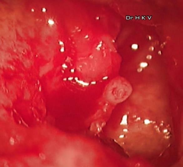
A 48-year-old woman presented with a history of intermittent right sided- ear discharge since 2 years, which was painless, scanty, purulent and blood stained, along with decreased hearing. Her most recent episode began two months ago. She also developed ipsilateral facial weakness since 1 week. Otoscopic examination of the right ear revealed a large central perforation of the tympanic membrane with granulation in the middle ear. Also evident was a polyp, appearing to originate from the attic. Facial nerve function on the right side was House Brackmann Grade IV. Ear swab for culture and sensitivity showed no bacterial overgrowth. Her pure tone audiogram showed right severe mixed hearing loss HRCT Temporal Bone was suggestive of chronic otitis media with mastoiditis and dehiscence of the horizontal and vertical segments of the facial canal. The chest X-ray was normal. A right modified radical mastoidectomy with a type III tympanoplasty was performed. Intraoperatively, granulation tissue, which was pale and greyish was removed along with the polyp and sent for histopathology. On drilling it was noticed that bone chipped off with much ease. This raised our suspicions, to consider TOM as a diagnosis. The malleus and incus were found eroded, thereby removed. The stapes was seen intact. The horizontal and vertical segments of the facial nerve were decompressed. The histopathology report of the middle ear granulation revealed chronic granulomatous inflammation with Langerhan giant cells along with acid fast bacilli identified on the Ziehl-Neelson stain, confirming tuberculous otitis media. Also, the polymerase chain reaction testing for middle ear mucosa was positive for Mycobacterium tuberculosis.
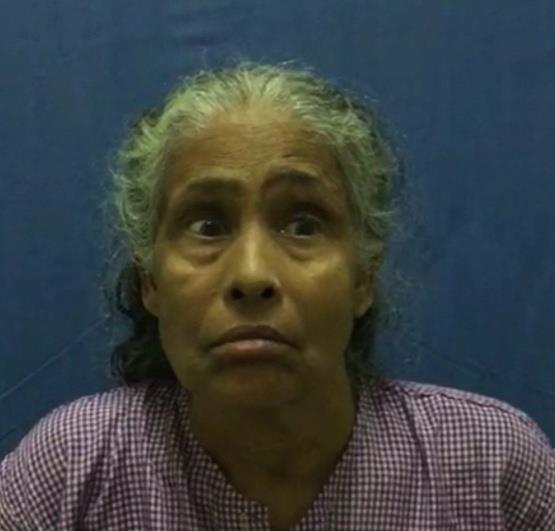
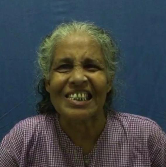
Appropriate anti-tubercular treatment was started with the standard DOTS category 1, which includes a 4 drug regimen in the first two months (Isoniazid, Rifampicin, Pyrazinamide and Ethambutol), followed by a 2 drug regimen for four months (Isoniazid and Rifampicin). Along with her anti-Koch’s treatment, she followed-up in the ENT OPD every 2 weeks, during which we kept a watchful eye for any development of granulation tissue or ear discharge. The patient had an un eventful recovery. The facial paralysis improved and was completely resolved by 3 months post-operative (Figures 1-3).

Discussion
Tuberculosis otitis media, is a relatively uncommon form of otitis caused by Mycobacterium tuberculosis. Mills study has mentioned that incidence of TOM has reduced from 3-5% to 0.04-0.09 % since the last century [6].This is attributed to the advent of antibiotics. In India, there are studies which corroborate this decline in incidence. In 1991, Grewal DS, et al. [5] conducted a 15- year study reporting incidence to be 0.05-0.9%. A more recent study done in 2013 by Deenadayal DD, et al. [7] estimated incidence to be 0.02%7. The 3 postulated mechanisms of pathogenesis are aspiration of mucus through the Eustachian tube, hematogenous transmission from other tuberculous foci and direct implantation through the external auditory canal via a tympanic membrane perforation [8]. It is characterised by painless, odourless otorrhoea; multiple perforations that tend to coalesce into a total perforation; pale granulomatosis; severe hearing loss which is disproportionate to other symptoms [9]. Vertigo, bony necrosis and facial paralysis are present to varying degrees and do not have a characteristic pattern [10]. Although being described as painless, Pusalkar, et al. [11] suggest the presence of a dull ache due to granulation tissue in the mastoid cavity. A review of literature by Skolnik, et al. [12] states that multiple perforations are rarely seen and only 0.16% of patients develop facial nerve palsy.
Therefore, anti-tuberculosis treatment is often delayed as a diagnosis is made only by direct smears, culture, histological and molecular biological testing and these usually require surgical sampling [8, 13]. Smears of ear discharge have been found positive for AFB in 0-20% cases and cultures are positive in 5-44% of cases [14, 15]. This lack in sensitivity is perhaps owed to frequent use of antibiotic ear drops [16]. Histology findings reveal Langerhan Giant cells, granulomas and caseation necrosis [17]. Polymerase chain reaction (PCR) amplification allows detection of Mycobacterium tuberculosis [18]. These 2 tests prove to be cardinal tools for a definitive diagnosis of TOM. Surgery is required to make a diagnosis using histology and PCR testing. It also allows for reconstruction of the middle ear and mastoid, as well as restoration or salvage of hearing.
Conclusion
To conclude, as symptoms of TOM are unspecific, it should be suspected as a differential diagnosis of chronic otitis media. Its latency, low sensitivity culture tests and rapidly progressive symptoms calls upon the ENT
surgeon to use a more aggressive approach in making a TOM diagnosis, for a disease that can be treated.
References
-
World Health Organization (2017) Global tuberculosis control 2017. Geneva: WHO.
-
TB India (2017) Revised National Tuberculosis Control Programme. Annual Status Report Central TB Division-New Delhi.
-
Mahmood T (2016) 40% of India’s population play host to the TB bacillus as a latent TB.
-
Odetoyinbo O (1988) Early diagnosis of tuberculous otitis media. Journal of Laryngology and Otology 102(2): 133-135.
-
Grewal DS, Baser B, Shahani RN, Khanna S (1991) Tuberculous otitis media presenting as complications: Report of 18 cases. Auris Nasus Larynx 18(3): 199-208.
-
Mills RP (1997) Management of chronic suppurative otitis media. In: Kerr AG, Booth JB, eds. Scott- Brown’s Otolaryngology (otology) 6th (Edn.), London: Butterworth 3.
-
Deenadayal DS, Kumar BN, Bommakanti V, Sameeri KL (2016) Tuberculous Otitis Media— A Rare Entity or a Missed Diagnosis. Int J Otolaryngol Head & Neck Surg 5(2): 65-72.
-
Windle-Taylor PC, Bailey CM (1980) Tuberculous otitis media: A series of 22 patients. Laryngoscope 90: 1039-1044.
-
Textbook of Pediatric Otorhinolaryngology-Head and Neck Surgery. Volume 1.Otology and Rhinology. Chris de Souza, James A Stankiewicz, Phillip K Pelliteri Chapter 9:106.
-
Hwang GH, Jung JY, Yum G, Choi J (2013) Tuberculous Otitis Media with Facial Paralysis Combined with Labyrinthitis. Korean J Audiol 17(1): 27-29.
-
Plester D, Pusalkar A, Steinbach E (1980) Middle ear tuberculosis. The Journal of Laryngology and Otology 94(12): 1415-1421.
-
Skolnik PR, Nadol JB, Baker AS (1986) Tuberculosis of the middle ear: review of the literature with an instructive case report. Rev Infect Dis 8(3): 403-410.
-
Luca Bruschini, Annalisa Ciabotti, Stefano Berrettini (2016) Chronic Tuberculous Otomastoiditis: A Case Report. J Int Adv Otol 12(2): 219-221.
-
Yaniv E (1987) Tuberculous otitis: An underdiagnosed disease. Am J Otolaryngol 8(6): 356- 360.
-
Kirsch CM, Wehner JH, Jensen WA (1995) Tuberculous otitis media. South Med J 88(3): 363- 366.
-
De Paep K, Offeciers FE, Van de Heyning P, Claes J, Marquet J (1989) Tuberculosis in the middle ear: 5 case reports. Acta Oto-Rhino-Laryngologica Belgica 43(4): 321-326.
-
Cho YS, Lee HS, Kimetal SW (2006) Tuberculous Otitis Media: a clinical and radiologic analysis of 52 patients. Laryngoscope 116(6): 921-927.
-
Bhalla Rk, Jones TM, Rothburn MM, Swift AC (2001) Tuberculous otitis media—a diagnostic dilemma. Auris Nasus Larynx 28(3): 241-243.
- 4th Branchial Cleft Sinus Anomaly Presenting as Recurrent Thyroid Abscess in A Child: A Case Report
- Parotid Duct Injury Repaired Using an Angiocatheter Stent: A Case Report
- Organization and Functionality of the Referral and Counter-Referral System for ENT Disorders in District Hospitals of N’Djamena, Chad: A Cross-Sectional Analytical Study
- Facial Metastases from a Gastrointestinal Stromal Tumor: A Case Report
- Panorama of Ent Cancers and Literature Review: Epidemiological Profile and Therapeutic Management
- Could Antimicrobial Resistance Prove to Be Both a Threat and an Opportunity for us?