Epistaxis in Pregnancy-A Rare Case Report
Introduction: Pyogenic granuloma is a benign lesion of unknown etiology that occurs in the skin and mucous membrane. It has been reported as a common lesion in oral cavity and less commonly in nasal cavity. Although its etiology is unclear, pyogenic granuloma is commonly associated with pregnancy, oral contraceptives and trauma. Case: A 22 year old female patient with 7 months of amenorrhoea came to ENT opd with complaints of epistaxis and swelling in left nasal cavity ultimately diagnosed as pyogenic granuloma. The patient was treated with endoscopic excision of nasal mass under general anesthesia. The management of pregnant female with such lesion was challenging, so we had to operate her. Even radiation exposure was a risk while radioimaging before surgery. Conclusion: Pyogenic granuloma is rapidly growing lesion that should be considered in the differential diagnosis of gravid patient with oral and nasal mass. Early excision of mass helped in avoidance of further complications such as fetal distress, low birth weight and reduces maternal mortality and morbidity.
Introduction
Pyogenic granuloma (also known as a “eruptive hemangioma”, “granulation tissue-type hemangioma”, “granuloma gravidarum”, “lobular capillary hemangioma”, “pregnancy tumor”, and “tumor of pregnancy” [1, 2] is a vascular lesion that occurs on both mucosa and skin, and appears as an overgrowth of tissue due irritation, physical trauma, hormonal factors [3, 4]. Lobular capillary hemangioma is microscopic proliferation of capillaries with lobular structure. Oral mucosa is the most common site. It is rapidly growing benign lesion. It was first described as human botryomycosis by Poncet and Dor in 1897. Causes of epistaxis: hereditary telangiectasia, trauma, neoplastic like juvenile nasoangiofibroma, capillary and cavernous hemangioma, hormonal like endometriosis, vicarious menstruation, systemic causes like hypertension (most common).
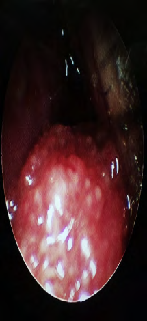
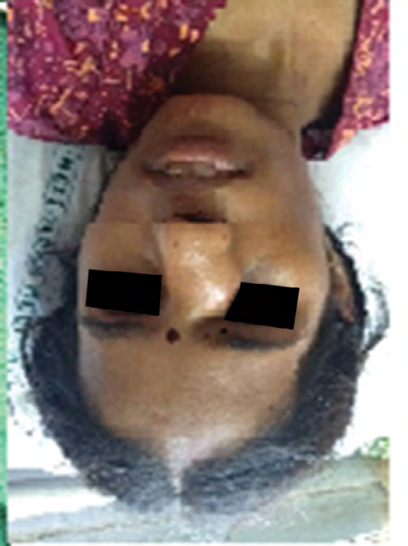
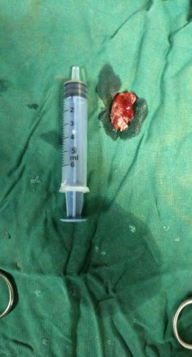
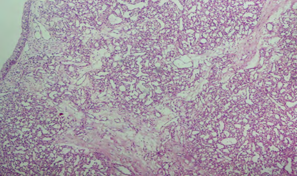
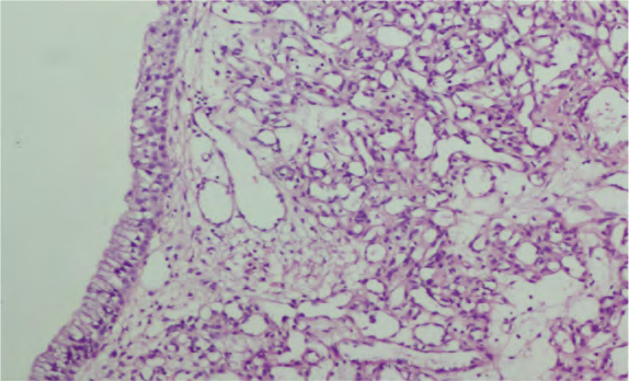
A 22 year old female patient came to our opd with 7 months of amenorrhoea with compalints of nasal bleeding and nasal obstruction since 2 months. There was no history of trauma or any recent infection. On anterior rhinoscopy, there was a mass in left nasal cavity which was smaller initially then rapidly increased in size to fill the left nasal cavity completely causing obstruction of left nasal cavity. It used to bleed on touch or otherwise also. On diagnostic nasal endoscopy, revealed a reddish hemorrhagic polypoid mass in left nasal cavity arising from nasal septum from postero-superior part. Patient was otherwise healthy. Her blood parameters were within normal limits. Bleeding was so profuse that we had to excise this nasal mass by endoscopic approach transnasally and excised mass sent for histopathology. Lesion composed of proliferation of small capillaries sized vessels lined by endothelium along with anastomizing channels. Areas of hemorrhagic and necrosis are seen.
Imp: Capillary Hemangioma
CT-PNS: Soft tissue lesion density inleft anterior nasal cavity with extension to adjacent maxillary region it is causing bony destruction of left nasal bone with blockage anteriorly (Figures 1-5).
Histopathology
Discussion
Pyogenic granuloma is acquired vascular tumour of benign etiology. It can occur at any age. It is more common in females than males of 20-30 years of age (3-5 times). In pregnant females it is more common in second and third trimester. It is presented as unilateral epitaxis, nasal obstruction with or without facial pain, headache. Our patient has similar complaints (left nasal obstruction, epistaxis). But these symptoms are not specific for pyogenic granuloma. Any mass in nasal cavity may present with similar complaints. Differential diagnosis are nasal polyp, menigocoele, sarcoidosis, hemangiosarcoma, wegener’s granulomatosis [5]. As pyogenic granuloma in pregnancy is due to hormonal changes, we can wait and watch till it regress es by itself after delivery. But if bleeding is so profuse then it might require intervention such as electrodessication (cauterisation) or excision by endoscopic approach. In our patient, we waited till 1 month; but symptoms were not relieved with conservative line of management ultimately we had to excise her nasal mass endoscopically and sent it for histopathological examination Prognosis is excellent; It may recur in 16% of the patients. There are least chances of its malignanr transformation.
Conclusion
Removal of pyogenic granuloma is indicated to treat any bleeding, discomfort, cosmetic distress, and diagnostic uncertainty. A number of malignant tumors may clinically mimic pyogenic granuloma, making histopathologic confirmation important if the presentation is atypical.
References
-
William J, Timothy B, Dirk E (2006) Andrews’ Diseases of the Skin: Clinical Dermatology. Saunders Elsevier.
-
Ronald RP, Jean BL, Joseph JL (2007) Dermatology: 2-Volume Set. St Louis: Mosby.
-
Jafarzadeh H, Sanatkhani M, Mohtasham N (2006) Oral pyogenic granuloma: a review. J Oral Sci 48(4): 167-175.
-
Puxeddu R, Berlucchi M, Ledda GP, Parodo G, Farina D, et al. (2006) Lobular capillary hemangioma of the nasal cavity: a retrospective study on 40 patients. Am J Rhinol 20(4): 480-484.
-
Simo R, de Carpentier J, Rejali D, Gunawardena WJ (1998) Pediatric pyogenic granuloma presenting as a unilateral nasal polyp. Rhinology 36(3): 136-138.
- 4th Branchial Cleft Sinus Anomaly Presenting as Recurrent Thyroid Abscess in A Child: A Case Report
- Parotid Duct Injury Repaired Using an Angiocatheter Stent: A Case Report
- Organization and Functionality of the Referral and Counter-Referral System for ENT Disorders in District Hospitals of N’Djamena, Chad: A Cross-Sectional Analytical Study
- Facial Metastases from a Gastrointestinal Stromal Tumor: A Case Report
- Panorama of Ent Cancers and Literature Review: Epidemiological Profile and Therapeutic Management
- Could Antimicrobial Resistance Prove to Be Both a Threat and an Opportunity for us?