Dermal Pleomorphic Sarcoma, A Rare Neoplasm
The European Commission, in its surveillance program for rare cancers, has defined a rare neoplasm as one with an incidence of 6 per 100,000 people per year. Within this definition, dermal pleomorphic sarcoma is found. Being a tumor whose origin remains unknown and without knowing the cell that originates it, the diagnosis is one of exclusion. For the diagnosis, the support of an expert pathologist and the use of immunohistochemistry and genomic techniques are suggested. The clinical case of a patient with this pathology is presented, which, due to its low prevalence, is important for dissemination among health professionals for a correct diagnosis.
Clinical Case
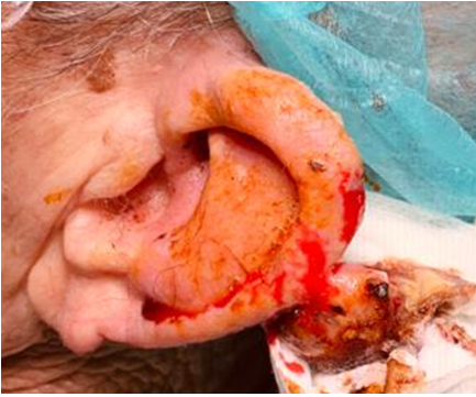
An 87-year-old male presented with a solid, bleeding, ulcerated lesion in the left auricle of three months’ duration, approximately 2 cm in its largest diameter (Figure 1). As important antecedents, the patient refers to unprotected sun exposure since youth.
The patient undergoes surgical resection, and the following pathology report is obtained.
Dermal Pleomorphic Sarcoma
- total tumor size: 2 X 1.5 cm.
- level of infiltration: reticular dermis.
- ulceration: yes.
- tumor necrosis: yes.
- mitotic index: 30 x 10 CGA.
- vascular invasion: no.
- perineural invasion: no.
- tumor-free cartilage. Immunohistochemical study
- positive for CD10 and vimentin The patient is currently asymptomatic three months after surgical resection. Since there was no presence of vascular or perineural invasion and the cartilage was free of tumor cells, the prognosis is expected to be favorable and timely follow-up will be performed.
Developing
In 2007, the epidemiology and genetics research program of the National Cancer Institute and the National Institute of Rare Diseases determined that a rare neoplasm is one with an incidence of less than 15 cases per 100,000 people or a report of less than 40,000 cases new per year in the United States. Other agencies such as the European Commission, in its surveillance program for rare cancers, have defined a rare neoplasm as one with an incidence of 6 per 100,000 people per year [1]. Within the rare neoplasms, undifferentiated pleomorphic sarcomas located of the the head and neck are located.
They are malignant tumors that originate from mesenchymal cells of the extremities in 50%, of the trunk or peritoneum in 40% and in 10% of the head and neck [2].
Reported international incidence rates for this condition range from 1.8 to 5 cases per 100,000 per year.
These cancers are more frequent in patients with chromosomal mutations and in some of the following syndromes: Gardner syndrome, Grolin syndrome, Tuberous sclerosis, Von Recklinghausen disease and Werner syndrome, with exposure to previous radiotherapy increases the risk of sporadic presentation, in the case of lymphangiosarcoma, the presence of chronic lymphedema and for hepatic angiosarcomas, exposure to chemical products such as vinyl chloride and arsenic are risk factors [3].
Undifferentiated pleomorphic sarcoma, formerly known as malignant fibrous histiocytoma, has highly variable characteristics, and no clear line of differentiation can be found using immunohistochemical techniques. When it begins in the skin, it is called dermal pleomorphic sarcoma, and is considered a tumor with histological characteristics common to atypical fibroxanthoma but with a worse prognosis [4]. Due to the confusion that has historically been created in the nomenclature of these tumors and the update carried out by the World Health Organization in 2013, there are few large series of this pathology from which guidelines and conclusions can be drawn for the proper management of this tumor.
Dermal pleomorphic sarcoma is located in the dermis and is made up of two populations of cells in different proportions: atypical spindle cells and pleomorphic epithelioid cells [5].
It usually appears in elderly patients and is characteristically located in areas of skin exposed to the sun, manifesting as an exophytic, asymmetric, commonly ulcerated and bleeding lesion, of rapid growth and medium size. Clinically it would be indistinguishable from atypical fibroxanthoma, so that it is usually not suspected on physical examination and is usually diagnosed as epidermoid carcinoma [4, 6].
As it is a tumor whose origin remains unknown and without knowing the cell that originates it, the diagnosis is one of exclusion.
The diagnosis of this neoplasm is carried out following the steps that would be used to diagnose any other neoplasm and is based primarily on the histological study to define its treatment [6]. The intervention of an expert pathologist is always suggested, since histological studies still are subject to many errors, which reach 25 to 40% in routine conditions, mainly due to the lack of experience of the professionals given the low prevalence of these diseases and the high biological complexity of the tumors, for which it is necessary to use of immunohistochemistry and genomic techniques, in addition to the use of decision support systems that use the extraction of information from image analysis, graph analysis, real-time updating that is, the support of digital pathology to make diagnoses in collaboration with others pathologists [7].
Usually it is not necessary to order preoperative imaging tests. In cases where deep infiltration is suspected, a computed tomography will be requested to rule out bone involvement or an MRI to study deep soft tissue involvement.
The histological criteria that define dermal pleomorphic sarcoma and differentiate it from atypical fibroxanthoma are: infiltration of subcutaneous cellular tissue, perineural or perivascular infiltration, and the presence of necrosis, these findings being sufficient to diagnose pleomorphic sarcoma [8].
In relation to immunohistochemistry, dermal pleomorphic sarcoma does not have a characteristic marker, although it is positive for vimentin, CD10, CD99 and actin, all of which are nonspecific and only have an indicative value, however, it is very important and more useful for diagnosis, ask for other markers that are negative for this sarcoma and positive in other tumors with which a differential diagnosis must be made [9].
Taking into account that dermal pleomorphic sarcoma is a recently defined tumor, there are no consensus guidelines for its management. Its treatment is surgical and it is usually curative. The factor that most correlates with the possibility of recurrence is the presence of positive surgical margins [4].
Radiotherapy is reserved for inoperable cases or for those in which palliative treatment is decided. In cases with metastasis, chemotherapy with adriamycin or ifosfamide would be used [5].
Factors associated with a poor prognosis include: patients older than 60 years of age, tumors greater than 5 cm in diameter, histological characteristics of high grade malignancy, and margins with tumor presence after resection [10].
Some published series report a recurrence of 20-28% of cases and the development of metastases in 10-20% of patients, especially to the skin, lung and lymph nodes [4, 5].
Therefore, follow-up is recommended by physical examination of the skin and lymph node territories every 3 months in the first year and subsequently every 6 months until completing 5 years of follow-up and depending on the case, the patient could be reviewed once a year until 10 years after the intervention [11].
Conclusion
It is considered an interesting case for disclosure among health professionals due to its low prevalence, classified within rare neoplasms by international institutions, seeking to broaden diagnostic horizons within health professionals.
References
-
Gatta G, Capocaccia R, Trama A, Martínez-García C (2010) The burden of rare cancers in Europe. Adv Exp Med Biol 686: 285-303.
-
Alorjani MS, Matalka II, Alfaqih MA, Jahmani RA, Alsinglawi BS, et al. (2022) Soft Tissue Sarcomas: A 16- Year Experience of a Tertiary Referral Hospital in North Jordan. Medicina (Kaunas) 58(2): 198.
-
Singer S, Maki RG, O’Sullivan B (2011) Soft tissue sarcoma. In: DeVita VT Jr, Lawrence TS, Rosenberg SA: Cancer: Principles and Practice of Oncology. 9th ed. Lippincott Williams & Wilkins pp: 1533-1577.
-
Tardío JC, Pinedo F, Aramburu JA, Suárez Massa D, Pampín A, et al. (2016) Pleomorphic dermal sarcoma: a more aggressive neoplasm than previously estimated. J Cutan Pathol 43(2): 101-112.
-
Miller K, Goodlad JR, Brenn T (2012) Pleomorphic dermal sarcoma: adverse histologic features predict aggressive behavior and allow distinction from atypical fibroxanthoma. Am J Surg Pathol 36(9): 1317-1326.
-
(2002) PDQ Cancer Information Summaries. Bethesda (MD): National Cancer Institute (US).
-
Sidiropoulos K, Glotsos D, Kostopoulos S, Ravazoula P, Kalatzis I, et al. (2012) Real time decision support system for diagnosis of rare cancers, trained in parallel, on a graphics processing unit. Comput Biol Med 42(4): 376-386.
-
McCalmont TH (2012) Correction and clarification regarding AFX and pleomorphic dermal sarcoma. J Cutan Pathol 39(1): 8.
-
Hanlon A, Stasko T, Christiansen D, Cyrus N, Galan A (2017) LN2, CD10, and Ezrin Do Not Distinguish Between Atypical Fibroxanthoma and Undifferentiated Pleomorphic Sarcoma or Predict Clinical Outcome. Dermatol Surg 43(3): 431-436.
-
Trovik LH, Ovrebo K, Almquist M, Haugland HK, Rissler P, et al. (2014) Adjuvant radiotherapy in retroperitoneal sarcomas. A Scandinavian Sarcoma Group study of 97 patients. Acta Oncol 53(9): 1165-1172.
-
Rios Vinuela E, Serra-Guillen C, Llombart B, Requena C, Nagore E, et al. (2020) Pleomorphic dermal sarcoma: a retrospective study of 16 cases in a dermato-oncology centre and a review of the literature. Eur J Dermatol 30(5): 545-553.
- 4th Branchial Cleft Sinus Anomaly Presenting as Recurrent Thyroid Abscess in A Child: A Case Report
- Parotid Duct Injury Repaired Using an Angiocatheter Stent: A Case Report
- Organization and Functionality of the Referral and Counter-Referral System for ENT Disorders in District Hospitals of N’Djamena, Chad: A Cross-Sectional Analytical Study
- Facial Metastases from a Gastrointestinal Stromal Tumor: A Case Report
- Panorama of Ent Cancers and Literature Review: Epidemiological Profile and Therapeutic Management
- Could Antimicrobial Resistance Prove to Be Both a Threat and an Opportunity for us?