Isolation and Biochemical Identification of Enterococcus Faecalis on Moribund Nile Tilapia (Oreochromis Niloticus L)
The study was able to isolate five colonies of presumed Enterococcus spp. from moribund Nile tilapia (Oreochromis niloticus L.) using selective medium. Of the 5 bacterial isolates, three were identified as E. faecalis and the remaining were Enterococcus spp. using the Remel RapIDTM STR System. The commensal Enterococcus spp. can also cause diseases on Nile tilapia.
Introduction
Enterococci are Gram-positive, catalase-negative, non- spore forming and facultative anaerobic bacteria that can occur either as single cocci or in chains [1]. Enterococci are considered commensals of the gastrointestinal tract of a variety of organisms, including humans. They are found in a number of environments, due to dissemination in animal excrement and environmental persistence [2].
Among commercially important fish species, enterococci have been reported worldwide in yellowtail (Seriola spp.), eels (Anguilla japonica), menhaden (Brevoortia patronus), striped mullet (Mugil cephalus) and striped bass (Morone saxatilis) [3]. Among the dozens of different enterococcal species, Enterococcus faecalis is the main cause of human enterococcal infections [4].
For a long time, enterococci were assumed non- pathogenic since they were normally existed in humans and animal flora. Nevertheless, causing severe diseases and high lethality under certain conditions was revealed that they have notable virulence factors. In several studies conducted, enterococci possess some properties that might relate to virulency [5]. Aggregation substance (AS) is one of the most accentuated among these factors. The importance of AS were demonstrated in epidemiological [6] and experimental studies [7, 8].
Materials and Methods
Collection and Dissection of Tilapia Samples
Ten pieces of moribund Nile tilapia reared in ponds were collected from a private farm. All samples exhibited signs of bacterial infection such as sloughing of scales, lesion, fin rot, bulging of eyes, corneal opacity, body deformities and/or abnormal body coloration. The samples were immediately transported in the laboratory for dissection to reveal the internal organs. The liver of all samples was removed and homogenized in disinfected mortar and pestle.
Isolation of Enterococcus spp
Two series of 10-fold dilutions (10-1 and 10-2) of liver homogenate was made in Phosphate Buffered Saline (PBS). One hundred microliters (100 µL) of the diluted sample was plated onto a selective medium, Edwards Medium. The plates were incubated for 18 to 24 h at 35 to 37°C. The colonies of Enterococcus spp. appeared black in the medium. Five of the black colonies were streaked to Trypticase Soy Agar (TSA) slants.
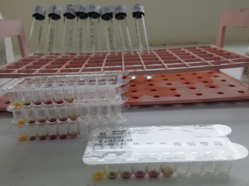
Biochemical Characterization and Identification of the Enterococcus spp. Isolates RapID STR System is comprised of RapID STR Panels and RapID STR Reagent. Each RapID STR Panel has several reaction cavities molded into the periphery of a plastic disposable tray. Reaction cavities contain dehydrated reactants and the tray allows simultaneous inoculation of each cavity with a predetermined amount of inoculum. A suspension of the test organism in RapID Inoculation Fluid was used as the inoculum which rehydrates and initiates test reactions. After incubation of the panel, each test cavity was examined for reactivity by noting the development of a color. In some cases, reagents were added to the test cavities to provide a color change. The resulting pattern of positive and negative test scores was used as the basis for identification of the test isolate by comparison of results to reactivity patterns stored in the Electronic RapID Compendium (ERIC™) database or by use of the RapID STR Differential Chart.
Results and Discussion
Clinical Signs of Bacterial Infection
All of the collected tilapia samples manifested clinical signs of bacterial infection such as sloughing of scales, lesion, fin rot, bulging of eyes, corneal opacity, body deformities and/or abnormal body coloration (Figure 1). The dominant clinical sign was abnormal coloration (50%) e.g. reddening and/or blackening of body parts. Two of the samples showed watery intestine and liver upon dissection (Table 1).
| Fish Samples | Clinical Signs of Bacterial Infection | |||||||
|---|---|---|---|---|---|---|---|---|
| Sloughing of scales | Lesion | Fin rot | Bulging of eyes | Corneal opacity | Body deformities | Abnormal body coloration | Watery internal organs | |
| 1 | X | X | X | X | X | X | X | |
| 2 | X | |||||||
| 3 | X | |||||||
| 4 | X | |||||||
| 5 | X | |||||||
| 6 | X | X | ||||||
| 7 | X | |||||||
| 8 | X | |||||||
| 9 | X | X | X | X | X | X | X | |
| 10 | X |
Table 1: External and internal signs of bacterial infection manifested by the collected tilapia samples. Note: X denotes the pres
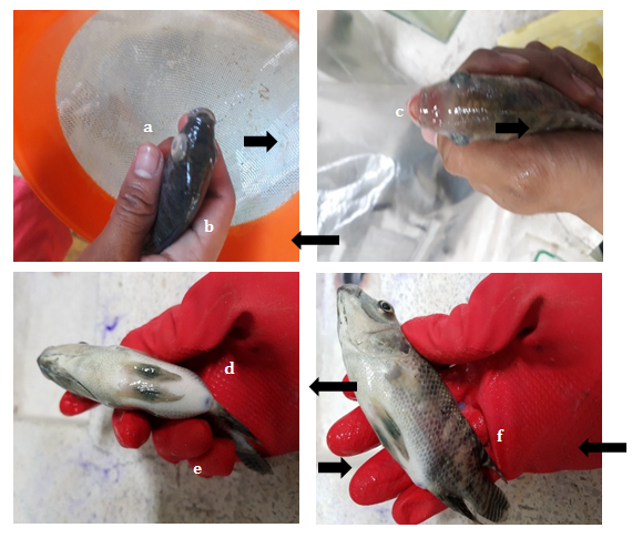
It was proven in the study of Madrid, et al. [9] that the commensal E. faecalis can cause diseases or even 40% mortality to cultured tilapia at a density of 108 cells/mL. The gross signs of the enterococossis have appeared as early as day 2 post-inoculation (PI) and began to cause death after day 5 PI. The infected groups showed pronounced clinical signs of bacterial infection such as uni-eye exopthalmia and eye opacity, reddening on the base fin and nasal area, protruded belly and ulcerative lesions. Ulcerations of the body surfaces were reported by Austin & Austin [10] in tilapia as a characteristic of septicemia. Behaviorally, the infected tilapia exhibited loss of appetite and erratic swimming. Observed internally was the accumulation of fluid in the peritoneal cavity. Presence of hemorrhages was also observed on the necropsied tilapia. According to Perera, et al. [11] hemorrhaging of internal organs or other gross dermal and epidermal lesions was common in diseased fish infected with Streptococcal group. According to the report of Plumb, the clinical signs and pathological manifestations of E. faecalis are similar to streptococcosis which exhibits exopthalmia, muscular hemorrhages, acute bronchitis, superlative inflammation in the eyes and necrosis of the spleen and kidney.
The pathogenesis of the bacterium is of two mechanisms namely the host inflammatory cascade or by direct damage as a result of secreted toxins or proteases [12]. Enterococcal cytolysin and two proteases, a zinc metalloprotease (gelatinase) and a serine protease are secreted factors that contribute to the severity of disease [4]. In addition to secreted proteins, E. faecalis and E. faecium can produce a toxic oxygen metabolite that results to cell or organ damage [13].
Isolation of Enterococcus spp.
Colonies of Enterococcus spp. appeared black in Edwards Medium because of the presence of aesculin in the medium. In aesculin-negative isolate such as Streptococcus spp., the colonies appeared blue. Aside from Enterococcus spp., aesculin also aids in the identification of Listeria spp., Aerococcus spp. and Leuconostoc spp. (drugs.com). Biochemical Characterization and Identification of the Enterococcus spp. Isolates Remel RapIDTM STR System (Figure 2) is a qualitative micromethod employing conventional and chromogenic substrates for the identification of medically important streptococci and related organisms which have been isolated from human clinical specimens. The tests used in RapID STR System are based on microbial degradation of specific substrates detected by various indicator systems. The RapID STR System is intended to aid in the identification of Lancefield groups A,B,C,D, and G streptococci, viridans streptococci, and Streptococcus pneumoniae, Enterococcus spp., Aerococcus spp., Gemella spp., Leuconostoc spp., Pediococcus spp., Weisella confusa, and Listeria monocytogenes [14, 15, 16, 17].
Of the 5 bacterial isolates, three were identified as E. faecalis (isolates 1, 2 and 5) and the remaining were Enterococcus spp, (isolates 3 and 4) (Table 2). This identification system was also used in the study of Abuseliana, et al. [18] with 99.81% identification report for all the Streptococcus spp. isolates.
| Biochemical Tests | |||||||||||||||
|---|---|---|---|---|---|---|---|---|---|---|---|---|---|---|---|
| Isolates | ARG | ESC | MNL | SBL | RAF | INU | GAL | GLU | NAG | PO 4 | TYR | HPR | LYS | PYR | Remarks |
| 1 | + | + | + | + | - | - | - | + | + | - | - | - | + | + | Enterococcus faecalis |
| 2 | + | + | + | + | - | - | - | + | + | - | - | - | + | + | Enterococcus faecalis |
| 3 | + | + | - | - | - | + | - | + | - | - | - | - | + | + | Enterococcus spp. |
| 4 | + | + | - | - | - | + | + | + | - | - | - | - | + | + | Enterococcus spp. |
| 5 | + | + | + | + | - | - | - | + | + | - | - | - | + | + | Enterococcus faecalis |
Conclusion
Through biochemical characterization using the Remel RapIDTM STR System, the five bacterial isolates from moribund Nile tilapia were identified as E. faecalis and Enterococcus spp. The commensal Enterococcus spp. can also cause diseases on Nile tilapia.
References
-
Fisher K, Phillips C (2009) The ecology, epidemiology and virulence of _Enterococcus._ Microbiology 155: 1749- 1757.
-
Gilmore MS, Courvalin DBP, Dunny GM, Murray BE, Rice LB (2002) The Enterococci: Pathogenesis, molecular biology, and antibiotic resistance. ASM Press, Washington DC.
-
Kitao T (1993) Bacterial diseases of fish. In: Inglis V, Roberts RJ, Bromage NR (Eds.), Blackwell, Oxford, Pasteurellosis, pp: 159-165.
-
Jett BD, Huycke MM, Gilmore MS (1994) Virulence of enterococci. Clininical Microbiology Reviews 7: 462-478.
-
Mundy LM, Sahm DF, Gilmore M (2000) Relationships between enterococcal virulence and antimicrobial resistance. Clinical Microbiology Reviews 13: 513-522.
-
Elsner HA, Soottka I, Mack D, Claussen M, Laufs R, et al. (2000). Virulence factors of Enterococcus faecalis and Enterococcus faecium blood culture isolates. European Journal of Clinical Microbiology and Infectious Diseases 19: 39-42.
-
Olmested S, Dunny G, Erlandsen S, Wells C (1994) A plasmid-encoded surface protein on Enterococcus faecalis augments its internalization by cultured intestinal epiethelial cells. Journal of Infectious Diseases 170(6): 1549-1556.
-
Schlievert PM, Gahr PJ, Assimacopoulos AP, Dinges MM, Stoehr JA, et al. (1998) Aggregation and binding substances enhance pathogenicity in rabbit models of _Enterococcus faecalis_ endocarditis. Infection and Immunity 66: 218-223.
-
Madrid FGN, Reyes AT, Doctolero JS (2020) Effect of UV light exposure on antibiotic susceptibility and pathogenicity of _Enterococcus faecalis_ to Nile tilapia (_Oreochromis niloticus_). Undergraduate thesis, Central Luzon State University, Philippines.
-
Austin B, Austin DA (1987) Bacterial fish pathogens. Disease in farmed and wild fish. Ellis Horwood, Chichester, UK.
-
Perera PR, Johnson SK, Collins M, Lewis D (1994) _Streptococcus iniae_ associated with mortality of _Tilapia_ _nilotica_ x _T. aureus_ hybrids. Journal of Aquatic Animal Health 6(4): 335-340.
-
Hancock LE, Gilmore MS (2006) Pathogenicity of enterococci. In: Fishcetti, (Ed.), ASM press.
-
Huycke MM (2002) Physiology of Enterococci. The Enterococci: Pathogenesis, molecular biology and antibiotic resistance.
-
Lennette EH, Ballows WJ, Hausler WJ, Truant JP (1980) Manual of Clinical Microbiology, 3rd (Edn.), ASM, Washington DC.
-
Balows A, Hausler WJ, Herrmann KL, Isenberg HD, Shadomy HJ (1991) Manual of Clinical Microbiology, 5th (Edn.), ASM, Washington DC.
-
Berlutti F, Thaller MC, Schippa S, Pantanella F, Rompei R (1993) International Journal of Systematic Bacateriology 43: 63-68.
-
Holt JG, Krieg NR, Sneath PH, Staley JT, Williams ST (1994) Bergey’s Manual of Determinative Bacteriology, 9th (Edn.), Williams and Wilkins, Baltimore.
-
Abuseliana AF, Daud HHM, Aziz SA, Bejo SK, Alsaid M (2011) Pathogenicity of _Streptococcus agalactiae_ isolated from a fish in Selangor to juvenile red tilapia (_Oreochromis_ sp.). Journal of Animal and Veterinary Studies 10(7): 914-919.
- Gallic and Citric Acid Present in the Peels of Tropical Fruits as an Alternative in the Fight against Cancer
- Treating the Forehead Lines with Combination of Forehead and Glabellar Botulinum Toxin Among Japanese Patients
- Clinical Evaluation of Patients Suffering from Breast Cancer & Determination of Treatment Therapies and Better Strategies Related to Breast Cancer
- Medieval Recipes by Al-Zahrāwī for Heart Palpitations Treatment
- Etiology and Prescription Errors of Myocardial Infarction in Different Health Care Systems of Azad Kashmir
- Early Diagnosis and Multidisciplinary Management of Turner Syndrome: A Paediatric Case Study