Survey of Protozoan Parasites in the Mediterranean Horse Macherel (Trachurus mediterraneus) from Zliten Coast, Libya
This study was conducted to estimate the prevalence, mean abundance, and mean intensity of common protozoa infection on Mediterranean horse macherel (Trachurus mediterraneus) from Zliten coast, Libya. A total of 32 specimens of Mediterranean horse macherel species were collected randomly. The fish were transported immediately alive to the laboratory in the Department of Fish Biology and Fish Culture, College of Marine Resources, Asmarya University, where they were maintained alive in well aerated glass aquaria. The gills, fins, and skin were examined for ectoparasitic protozoa using a light microscope. Among the examined fish, the high desnsity of parasitic infectious in internal organ was observed in kideny (0.375), liver (0.5), blood and spleen (0.25), respectively. Seven species of parasitic protozoan namely Haemogregarina spp., Trypanosoma spp., Cryptopia spp., Microsporidia spp., Henneguya spp., Thelohania spp. and Ichthyophthirius multifiliis were identified in Mediterranean horse macherel (Trachurus mediterraneus) from Zliten coast.
Introduction
Protozoa’s are single-celled organisms, many of which are free-living in the aquatic environment. Typically, no intermediate host is required for the parasite to reproduce [1, 2, 3]. Protozoa are one of the major sectors of fish parasites that have been long neglected because of its inherent difficulty in studying compared to other larger parasites. They can build up to very high numbers when fish are crowded causing weight loss, debilitation, and mortality. Among protozoa, ecto-and endoparasitic protozoa occupy an important sector as it is one of the hazardous threats to fish health [4, 5, 6].
These parasites attack the fish, causing massive destruction of skin and gill epithelium. Even moderate infection of these organisms on small fish may prove a fatal disease, since the infection may cause the fish to stop feeding [6, 7, 8, 9, 10]. Some fish parasites would develop in humans if the fish is eaten raw, but none would be harmful if the fish is thoroughly cooked. All reports of people being infested with fish parasites were because of ingestion of raw fish or insufficiently cooked fish [11]. Most fish especially in the wild population are likely to be infested with parasites, but in the great majority of cases, no significant harm to the host may be ensued or identified. Thus, there are only few reports of parasites causing mortality or serious damage to the fish populations, but this may be largely because such effects go unnoticed [12]. Fishermen or consumers often observe parasites in wild fish only when they are so obvious as to lead to rejection of fish [13].
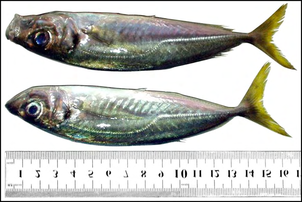
Mediterranean horse macherel (Trachurus mediterraneus), an important cultivable fish species, is found usually near the bottom, at times also in surface waters; pelagic and migratory in large schools. They feed on other fishes especially sardines, anchovies, etc. and small crustaceans [14]. It can be caught by various gears such as seines and fixed nets. The contribution of Trachurus mediterraneus to local fisheries differs in each sea. In the Black Sea, this species makes up 54% of the total catch (2,919 t), whereas it makes up 39% in the Marmara Sea (562 t), 4% in the Aegean Sea (247 t) and 3% in the northeastern Mediterranean Sea. There is currently no knowledge of horse mackerel stock structure among fishing areas of the eastern Mediterranean Sea [14]. The present study was conducted to investigae general survey and density of internal protozoan parasites on Mediterranean horse macherel (Trachurus mediterraneus) in the coastal area in Zliten city.
Materials and Methods
Samples Collection
A total of 32 specimens of fish species namely Mediterranean horse macherel (Trachurus mediterraneus) were collected randomly. Fishes were collected by fishermen by using cast net and gill nets, during August 2016 from Zliten coastal area (Lat 32°30’ N; Long. 14°43’ E) Modern harbor facility 156 km east of Tripoli. The fish were transported immediately alive to the laboratory in the Department of Fish Biology and Fish Culture, College of Marine resources, Asmarya University, where they were maintained alive in well aerated glass aquaria.
Internal Examination
Blood Smear: Firstly the fishes were brought out of water and the tails was cut by sharp scissor to obtain blood from caudal vain or artery with pressure. A drop of blood (0.5 ml) was placed on the edge of microscope slide and touched with another slide (spreader) at 45 angle and moved quickly forward to make smear, then the smear was left to dry. Liver, Kidney and Spleen Smear: The routine dissection method was adopted, as a ventral incision was made from the anus to the pectoral region and another vertical from the anus to the lateral line. The side flap was lifted and the internal organ exposed. The operculum was removed to expose the gill [15]. Then samples were taken from: liver, kidney and spleen and after that dried from blood with filter paper to absorb the fluids and blood. Then impresser smear was done by press the tissue on different places on glass slide by forceps. The smears were dried, fixed in methanol for 10 min, and then stained with 5% Giemsa’s solution in phosphate buffer (pH 7.3) for 30 min. Smears were then examined using light microscope fitted with an oil immersion lens. Photography: Kruss optronic microscope fitted with camera (BEL,Eurkm 10.0) was used to photograph the parasites. The parasites were identified using images from websites such www.fishbase.se and by making their sketches as observed on the microscope and compared with the pictorial guide on fish parasites.
Results
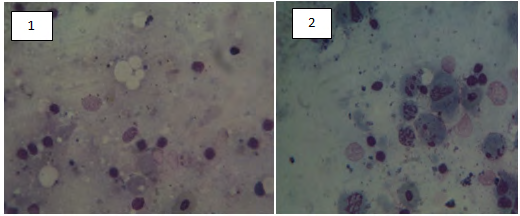
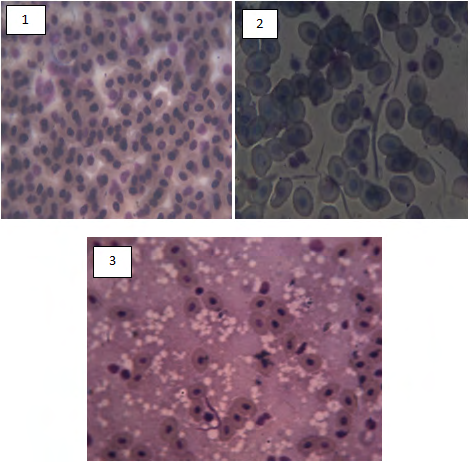
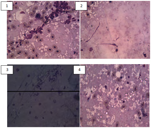
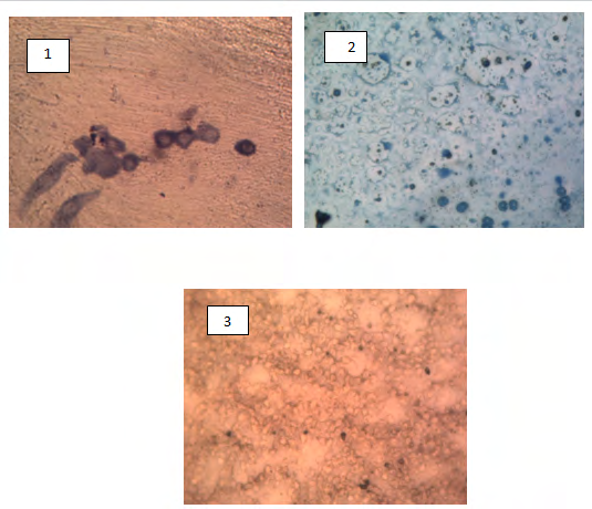
A total of 32 fish of Trachurus mediterraneus were collected and inspected for parasites from Zliten coastal area. The present study showed the existence of different stages species of external and internal protozoans (Figures 1-5). The distribution of the parasites, their location on the fish host body, mean density of infection are summarized in Table 1.
| Site of infection | ||||
|---|---|---|---|---|
| Parasites | Blood | Liver | Spleen | Kidney |
| Haemogregarina spp. | + | + | + | - |
| Trypanosoma spp. | + | - | - | - |
| Cryptopia spp. | - | + | - | - |
| Microsporidia spp. | - | + | - | + |
| Henneguya spp. | - | + | - | - |
| Thelohania spp. | - | - | + | - |
| Ichthyophthirius multifiliis | - | - | - | + |
| Unidentified protozoa. | - | - | - | + |
| Total | 2 | 4 | 2 | 3 |
| Density | 0.25 | 0.5 | 0.25 | 0.375 |
Table 1: List of parasites species found in the internal organs.
Discussion
Microscopic examination of fresh-mounted scraps of fish’s skin, gills blood and impresser smears from liver, kidney, and spleen, revealed high density of external and internal parasites. According to Roberts, et al. [12] parasite of fish can either be external or internal. Parasitic infections often give an indication of the quality of water, since parasites generally increase in abundance and diversity in more polluted waters and are the most diverse and common pathogens the aqua culturist may likely encounter, and parasitic diseases are very common in fish all over the world and are of particular importance in the tropics, also the study recorded flagellated protozoan from external and internal organs. Eiras [16] reported that flagellate protozoans are mainly characterized by the presence of one or more flagella for movement. The majority of them are ectoparasites while others can be found parasitizing internal organs, such as dinoflagellates as Amyloodinium ocellatum, Piscinoodinium pillulare, Trypanosoma and Ichthyobodo are the main representatives, under a microscope, it can be observed three forms of the dinoflagellate from the scraps of fish: pear-shaped, banana- shaped and the mature rounded parasite of brownish color [17, 18], although in high infestations different developmental stages can be found. Special care must be taken on the mature trophonts that could be confused with Ichthyophthirius multifiliis by an inexperienced person. Amyloodinium comprises dinoflagellates of varied shape depending on the life stage. The causative agent of velvet disease Amyloodinium ocellatum is ubiquitous, affects marine farmed fish and may provoke important outbreak mortalities and economical losses in aquaculture systems [6, 9, 10, 19, 20, 21], also haemoflagellate Trypanosoma was identified. This is ubiquitous throughout freshwater and marine environments and may cause problems in aquaculture. Baker [22] reported that Natural infection with trypanosomes may be very common where their leech vector are abundant. These species were: Apiosoma piscicola, A. conica, Scopulata epibranchial, Vorticella spp_., Ambiphrya ameiuri, Amphileptus_ spp., Chilodonella hexasticha, Tetrahymena corlissi, Trypanosoma mansouri, Trypanosoma syanophilum, Trypanosoma spp. and Cryptobia can be found parasitizing either the gills or skin of marine and freshwater fishes around the world and the majority of them are ecto commensals. However, some species are pathogenic for young fish [23, 24]. Also the result showed that Haemogregarina sp. in blood, liver and spleen. These groups comprise several blood protozoan parasites [25, 26]. They are commonly found in both erythrocytes and leukocytes of marine fishes [27], fishes are likely to act as intermediate hosts, while leeches or gnathiid isopods are probably the definitive ones, also the result appear Microsporidia in kidney and liver and this agree with finding of Morrison and Sprague [28] who reported that fish seem to survive infections, in spite of the presence of huge Xenomata often pressing on and displacing important organs, while infection by some _Microsporidia_ns undoubtedly has a morbid effect on the fish [29, 30].
Internal parasites include Myxozoa are parasites that are widely dispersed in native and pond-reared fish populations. Most notorious is the whirling disease of trout, manifested by skeletal deformities, which is also claimed to have been introduced with rainbow trout into South Africa [30]. Trypanosome is haemoflagellate parasite ubiquitous throughout freshwater and marine environments and may cause problems in aquaculture. Members of Trypanosome present a slender body, elongated, cylinder-shaped with more or less thin extremities, free flagellum, undulating membrane besides the nucleus and kinetoplast and volute granules disposed generally in the middle of the body [31].
Conclusion
This research based on study microscopic examination of fresh-mounted scraps of fish’s skin, gills blood and impresser smears from liver, kidney, and spleen, revealed high density of external and internal parasites. Seven species of parasitic protozoan namely Haemogregarina spp., Trypanosoma spp., Cryptopia spp., Microsporidia spp., Henneguya spp., Thelohania spp. and Ichthyophthirius multifiliis were identified in Mediterranean horse macherel (Trachurus mediterraneus) collected from Zliten coast.
Declaration of Competing Interest
There is no conflict of interest to declare between the authors of this manuscript.
Acknowledgement
The authors are grateful to the head Department of Fish Biology and Fish Culture, College of Marine Resources, Asmarya University, Zliten, Libya for providing necessary laboratory facilities and also to head department of Fisheries and Wildlife Science, Sudan University of Sciene & Technology.
References
-
Iwamoto M, Ayers T, Mohn BE, Swerdlow DL (2012) Epidemiology of seafood-Associated in fections in the United States. Clinical microbialogy Rev 23(2): 399-411.
-
Marino S, Cilfone NA, Mattila JT, Linderman JJ, Flynn JL, et al. (2014) Macrophage polarization drives granuloma outcome during Mycobacterium tuberculosis infection. Infect Immun 83(1): 324-328.
-
Ryan U, Hijjawi N, Feng Y, Xiao L (2019) Giardia: An under-reported foodborne parasite. International Journal of Parasitololgy 49(1): 1-11.
-
Papera l (1996) Parasitical disease of fish in Africa up data C F A Technical paper, Rome FAO.
-
Coupe A, Howe L, Burrows E, Sine A, Pita A, et al. (2018) First report of Toxoplasma gondii sporulated oocysts and Giardia duodenalis in commercial green-lipped mussels (Perna canaliculus) in New Zealand. Parasitoloical Research 117(5): 1453-1463.
-
Moratal S, Ayuela MAD, Cardells J, Marco Hirs JN, Puigcercos S, et al. (2020) Potential Risk of Three Zoonotic Protozoa (_Cryptosporidium_ spp., _Giardia_ _duodenalis_ and _Toxoplasma gondii_) Transmission from Fish Consumption. Foods 9(12): 1913.
-
Enayat A (2011) Review of some ecto-and endo protozoan parasites infecting Sarotherodon galilaeus and Tilapia Zillii from Damietta Branch of River Nile, Egypt. Journal of American Science 7: 1-3.
-
Reynolds K (2017) Waterborne. AMA J ethics 19(10): 1036-1042.
-
Buchmann K (2015) Impact and control of protozoan parasites in maricultured fishes. Parasitology 142(1): 168-177.
-
Marino AMF, Giunta RP, Salvaggio A, Castello A, Alfonzetti T, et al. (2019) Toxoplasma gondii in edible fishes captured in the Mediterranean basin. Zoonoses Public Health 66(7): 826-834.
-
FAO (2005) Libayn Arab Jamahiriya fisery Couny Profile, food and Agriculture Organization of the United Nations, Italy.
-
Roberts RJ (2001) Fish Pathology. 3rd (Edn.), Elsevier Health Sciences, New York, USA, pp: 492.
-
Roberts RJ (1995) Parasitology of Teleosts. 1st (Edn.), In: Fish Pathology, London, UK, pp: 23.
-
Vaniz WFS, Carangidae W, Bauchot ML, Hureau JC, Nielsen J, et al. (1986) Fishes of the north-eastern Atlantic and the Mediterranean. In: Whitehead PJP, et al. (Eds.), The Quarterly Review of Biology 61(2): 815-844.
-
Buke D (1980) Some histological techniques applicable to fish tissues. In: Lionel E Mowdely (Ed.), Disease of fish. Symposia of zoological society of Lon. No. 30 Acad. Press. Acadimic Research, pp: 153-189.
-
Eiras JC (1994) Elements of Ichthyoparasitology. Porto: Fundacao Engenheiro Antônio de Almeida, pp: 339.
-
Martin ML, Moraes JRE, Andrade PM, Schalch SHC, Moraes FR (2001) Infection in cultivated freshwater fish from the northeast region of Sao Paulo State, Brazil. Parasitological and pathological aspects. Brazil Journal of Biology 5: 6-11
-
Foin AA (2005) Parasites et parasitoses des poissons dornement deau douce: aide au diagnostic et propositions de traitement [Tese]. Maisons-Alfort: Ecole Nationale Veterinaire dalfort 7: 18-23.
-
Levy MG, Poore MF, Colorni A, Noga EJ, Vandersea MW, et al. (2007) A highly specific PCR assay for detecting the fish ectoparasite Amyloodinium ocellatum. Dis Aquat Organ 73(3): 219-226.
-
Pereira JC, Abrantes I, Martins I, Barata J, Frias P, et al. (2011) Ecological and morphological features of occurrences in cultivated gilthead seabream L.: a case study. Amyloodinium ocellatum _Sparus aurata_. Aquaculture 310(3-4): 289-297.
-
Moreira CB, Hashimoto GSO, Rombenso AN, Candiotto FB, Martins ML, et al. (2013) Outbreak of mortality among cage-reared cobia (Rachycentron canadum) associated with parasitism. Rev Bras Parasitol Vet 22(4): 588-591.
-
Baker JR (1960) Trypanosomes and dactylosomes from the blood of freshwater fishes in East Africa. Parasitology 50: 515-526.
-
Kuperman BI, Matey VE, Barlow SB (2002) Flagellate Cryptobia branchialis (Bodonida: Kinetoplastida), ectoparasite of tilapia from the Salton Sea. Hydrobiologia 5: 44-53.
-
Paiva MJR, Felizardo NN, Luque JL (2005) Parasitological and hematological analysis of Nile tilapia Oreochromis niloticus Linnaeus, 1757 from Guarapiranga reservoir, São Paulo State, Brazil. Acta Science of Biological Science 27(3): 231-237.
-
Diniz JA, Silva EO, Souza W, Lainson R (2002) Some observations on the fine structure of trophozoites of the haemogregarine (Adeleina: Haemogregarinidae) in erythrocytes of the fish (Synbranchidae). Parasitol Res 88(7): 593-597.
-
Eiras JC, Pavanelli GC, Takemoto RM, Eiras JC (2013) Parasitologia de peixes de água doce do Brasil. Maringa Eduem 3: 45-52.
-
Davies AJ (1995) The biology of fish haemogregarines. Adv Parasitol 36: 117-203.
-
Morrison CM, Sprague V (1981) _Microsporidia_n parasites in the gills of salmonid fishes. Journal of Fish Disease 4(5): 371-386.
-
Putz, RE, McLaughlin JJA (1970) Biology of Nosematidae (_Microsporidia_) from freshwater and euryhaline fishes. In: Snieszko FS (Eds.), A symposium on diseases of fishes and shellfish. American Fish Society, 5: 124-132.
-
Van Wyk GF (1968) Jokershoek Hatc hery, Division of Inland fisheries, Department of Nature Conservation, provins of Good Hope, Republic of South Africa. Annual report 24: 1967.
-
Hussein ANA, Rabie SA, Mohammed NE, Hussein NM (2010) Light and scanning electron microscopic studies of trypanosomes parasites infecting freshwater fishes in Qena Governorate, Egypt. Egyptian Academic Journal of Biological Science 2(1): 17-31.
- Genetic Improvement of Nile Tilapia (Oreochromis niloticus): Advances in Selective Breeding and Genomic Approaches for Sustainable Aquaculture
- Microplastics, Contaminants, and Waste Hotspots: Divergences and Faults in Prioritizing Control Efforts
- Creating a Healthier, More Vibrant Open and Closed Aquatic Environment. A Submersible, Centrifugal Magnetically Affixed Current Changing Aquarium Pump
- An Attempt to Assess Alpha Diversity and Sample Size: Using the Ostracod Assemblages off Kumamoto Port, Japan
- Assessment of the Efficiency of Common Fishing Gears and Crafts Used at Mohananda River of Chapai Nawabganj, Bangladesh
- Fish Productivity and Biodiversity Status of Sundarban Mangrove in Bangladesh