Lactic Acid Bacteria as Probiotics in Fish Culture: Isolated from Milk of Common Cattle Breeds Raised in Potiskum, Yobe State, Nigeria
This study was stressed on the detection of Lactic Acid Bacteria (LAB) and other normal gut flora that obtained from nonfish source (Fresh Cattle Milk) can play a very significant role in aqueous environment and as well bacteria-fish synergic relationship. This work were experimented in the Microbiology Laboratory of Yobe State University, Damaturu. The isolated bacteria can be used in prevention and control of pathogenic organisms in aquaculture. These includes six (6) bacterial species isolated from milk and might be tested for their potential used as probiotics. Therefore, the results revealed that Red Bororo was significantly higher represented in total viable count of 159 (15.9×10⁻¹⁰ Cfu/ml) than Adamawa Gudali, which had 153 (15.3×10⁻¹⁰ Cfu/ml). Thus, the number of occurrences, percentages, colony and colonies forming unit in respect of individual bacterium such as Bacillus subtilis with 6 (30%) having 54 (5.4×10⁻¹⁰ Cfu/ml) in Adamawa Gudali and Red Bororo were the same and not significantly different (p > 0.05), followed by Lactobacillus acidophilus with 5 (25%) possesses 52 (5.2×10- 10 Cfu/ml) found in both milk samples, which did not significantly varies (p > 0.05) whereas, Streptococcus agalactiae had greater 3 (15%) and 19 (1.9×10⁻¹⁰ Cfu/ml) in Red Bororo is significantly different (p < 0.05) from Adamawa Gudali, which had lesser 9 (0.9 × 10⁻¹⁰ Cfu/ml) value. The Escherichia coli was significantly higher (p < 0.05) with 3 (15%) and having 15 (1.5×10⁻¹⁰ Cfu/ml) in Red Bororo than in Adamawa Gudali with lower value 13 (1.3×10⁻¹⁰ Cfu/ml) while Staphylococcus aureus having 4 (20%) and 13 (1.3×10⁻¹⁰ Cfu/ml) in both milk samples showed that they were not significantly different (p > 0.05) and Micrococcus luteus was significantly greater (p > 0.05) with the value of 2 (10%) and 12 (1.2×10⁻¹⁰ Cfu/ml) in Adamawa Gudali than in Red Bororo with 6 (0.6×10⁻¹⁰ Cfu/ml). These organisms were determined with Tryptic Soy Agar (TSA). Eosin Methylene Blue Agar (EMBA), deMan Ragosa and Sharpe Agar (MRS) and Mackonkey Nutrient Agar Media were used, respectively, they were analysed using standard bacteriological procedures. Also, data were analysed using descriptive statistics and analysis of variance at 5% (0.05%) confident interval. These microorganisms from non-fish origin are good candidates that are substitutes to chemotherapies which needs to be tested and compared with those of fish origin to assess their antagonistic effects in aquaculture or fish farming.
Introduction
Fish is an important source of humans’ food, virtually a key component in natural food webs, physiologically contains valuable protein, fats, and fat soluble vitamins [1]. It also plays a vital contribution to gross domestic product and job creation in Nigeria [2].
Fish culture or aquafarming becomes a way out for bridging the gaps between its productions, demand, supply and sustainability conserve wild species composition and diversity worldwide.
Fisheries contribute significantly to the food security, socio-economic growth and development for developing and developed nations, especially, employment, local fish sales and foreign exchange earnings to provide better livelihood.
Feed is one of the important aqua-inputs in fish culture, of which its contribution accounts for an approximate percentage of 40-60% [3] and to some extent, 60-80% of production cost [4]. Thus, to attain sustainability, feasibility and profitability in the aqua-farm business, this might accrue a cost of production between 40 and 70% [5].
The higher qualitative feed is required to enhance the growth and health status of fish species with rising interest in culturing fish globally [6]. Fish feed is richer in essential nutrients required for the growth and proliferation of micro- organisms [7].
Rahaji is the most populated cattle breed, called Red Bororo but Bokoloji which is known as Gudali are fairly inhabited in Yobe State. They are raise for their multiple purposes characteristics. In Nigeria, Rahaji are the most numerous breed that constituted between 50 and 60% whereas Adamawa Gudali represented about 2% of the cattle breeds [8].
Therefore, milk is described as a whitish, lacteal, secretory fluid discharge from the mammary gland of all dairy animals via teats. According to Pandey, et al. [9] and Muniyandi, et al. [10]. Thus, milk is an early mammalian infant’s food prior to accepting and digesting any other types.
Probiotics are microbe-based additives that are added to fish feed in small quantities to maintain and enhance its safety and nutritional quality. It’s an alternative to antibiotics in aquaculture [11]. Hence, it’s isolated from microbes in substrates. It also, isolated from ecosystems, fresh dairy milk that’s a significant source of LAB, belongs to the Lactobacillus genus [12, 13]. It is considered as several additives employed into fish nutrition to improve energy spends that are derived from carbohydrates and incorporated with protein for growth, immunity, and disease resistance [14, 15]. They are non-pathogenic, which benefits fish at the right doses and counteracts harmful microbes [16, 17].
Pro-digestion activities of the probiotics lead to better performance of fish species; hence, higher doses cause stuntism and also probably might replace whole gut normal microbes of the fry [18]. Moreover, probiotics adversely result in serious problems in fish production [19]. The strains have been selectively evaluated for their potential to supplement and enrich feed for the benefits of aquaculture [3]. It’s aseptically isolated, characterised, identified, and enumerated microscopically [20]. Bacillus subtilis and Lactobacillus species are lactic acid bacteria that belong to the same genus; they enhance non-specific immunity and resist diseases. Escherichia coli are fish G.I.T. bacteria with lower cell surfaces, hydrophobic in nature; in any given growth medium, they also have adaptive functions in the fish microflora [21].
This current study determined the presence of bacteria that can be used as probiotics that, when isolated and identified from dairy milk, might play an important role in fish culture.
Materials and Methods
Study Area
Badejo and Kusulwa cattle breeders communities located in Potiskum, Yobe State, Nigeria, that is situated between latitude 11O 36` 58``N and longitude 11O 6` 49``E along Gombe road.
Material Used and Reagents
The materials used are incubators, microscopes, glass slides, petri dishes, autoclave, conical flasks, test tube racks, wire loops, weighing balances, sample bottles, hand gloves, pipettes, nutrient agars, and deionised water.
Milk Samples Collection
The forty (40) fresh milk samples were sourced from the conventional manual milking method by herders close to Badejo and within Kusulwa at the onset of the rainy season; twenty (20) samples each from Adamawa-Gudali and Red- Bororo were collected for this study. These were performed aseptically at all locations directly from their used milking containers with a sterile plastic cup. The samplings were done between 6:00 a.m. and 7:00 a.m. for a period of 20 days. The samples were poured into sample bottles, chilled, packed, transported and analysed in the Yobe State University Damaturu Microbiology Laboratory.
Culture Media Preparation
The media were prepared in accordance with the manufacturer’s reco-10mmendation, in which, based on instruction 23g of the media powder, they were suspended into 1 litre of deionised water, mixed gently, and homogenised after being allowed to stand on the preparation table. The media solutions were sterilised using an autoclave at 121°C for 15 minutes, cooled to 45°C, then poured onto the sterile petri dishes, and the plates were kept for 2 hrs at room temperature and solidified.
Milk Samples Preparation
The samples prepared with a total of 10 sterile test tubes were dispensed; 1 ml of prepared inoculum was transferred into the first test tube containing 9 ml of deionised water (10-10) dilution. Then, by the use of another sterile pipette, 1 ml of the resulted dilution was transferred into a second test tube that contained the same quantity; the procedure was repeatedly done up to the 10 times (10-10) dilution.
Therefore, 1 ml of inoculum was discarded; this was done for each of the 20 milk samples from the Adamawa Gudali and Bunaji breeds that were aseptically measured, weighed and homogenised with 90 ml of deionised water. The ten (10)-fold serial dilutions were done in one (1) acetone water and plated out in different culture media.
Inoculation and Incubation
The prepared nutrient media plates were labelled as AG1, AG2… AGnth, and RB1, RB2… RBnth from the last dilution of 10-10, while the prepared nutrient agar was inoculated with 0.1 ml of the test sample; a sterile disposable pipette was used. Prepared culture media were used for the isolation and inoculation of bacteria with sterile cotton wool. Total viable count (TVC) were enumerated for the experimental bacteria and the plates were incubated at 37°C for 24 hours [22]. The Tryptic Soy Agar (TSA), Eosine Methylene Blue Agar (EMBA), deMan Ragosa and Sharp Agar (MRS), and Mackonkey nutrient agar media were used; the presence of Bacillus subtilis, Escherichia coli, Lactobacillus acidophilus, Micrococcus luteus, Staphylococcus aureus and Streptococcus agalactiae were determined. These agar media relied on the species of selected bacteria that were intended to be used as probiotics for fish cultured because different organisms grown in different culture media. These were done as described by Janet, et al. [23].
Bacteriological, Biochemistry and Molecular Analysis
The isolates were rinsed in a petri dish, diluted, observed, characterised, identified and then serially diluted bacterial colonies were enumerated under microscopic examination. However, the isolates were molecularly analysed, gram- stained and biochemical tests were conducted. Bacillus subtilis, Escherichia coli, Lactobacillus acidophilus, Micrococcus luteus, Staphylococcus aureus and Streptococcus agalactiae were observed.
Results
| Cattle Breed | No. of Colony (TVC) Cfu/ml | |
|---|---|---|
| Adamawa Gudali (AG) | 153 | 15.3×10⁻¹⁰ |
| Red Bororo (RB) | 159 | 15.9×10⁻¹⁰ |
Table 1: Total Viable Count and Colony Forming Unit of Bacterial Species from the Milk Samples of Adamawa Gudali and Red Boror
Foot note: No. = Number, TVC= Total viable count, Cfu=Colony forming unit, Ml = Milligramme Table 1: Total Viable Count and Colony Forming Unit of Bacterial Species from the Milk Samples of Adamawa Gudali and Red Bororo.
| AG Samples | Shape | Gram Reaction | Species | RB Samples | Shape | Gram Reaction | Species |
|---|---|---|---|---|---|---|---|
| AG1 | Rod | + | Bacillus subtilis | RB1 | Cocci | + | S. aureus |
| AG2 | Rod | - | Escherichia coli | RB2 | Rod | + | B. subtilis |
| AG3 | Rod | + | Bacillus subtilis | RB3 | Rod | + | L. acidophilus |
| AG4 | Rod | - | Escherichia coli | RB4 | Cocci | + | S. agalactiae |
| AG5 | Cocci | + | Streptococcus agalactiae | RB5 | Rod | + | L. acidophilus |
| AG6 | Rod | + | Lactobacillus acidophilus | RB6 | Rod | - | E. coli |
| AG7 | Cocci | + | Micrococcus luteus | RB7 | Rod | + | B. subtilis |
| AG8 | Cocci | + | Staphylococcus aureus | RB8 | Cocci | + | M. luteus |
| AG9 | Rod | + | Lactobacillus acidophilus | RB9 | Rod | + | L. acidophilus |
| AG10 | Rod | + | Bacillus subtilis | RB10 | Rod | + | B. subtilis |
| AG11 | Cocci | + | Staphylococcus aureus | RB11 | Rod | - | E. coli |
| AG12 | Rod | - | Lactobacillus acidophilus | RB12 | Rod | - | L. acidophilus |
| AG13 | Rod | + | Bacillus subtilis | RB13 | Cocci | + | S. agalactiae |
| AG14 | Rod | + | Lactobacillus acidophilus | RB14 | Rod | + | B. subtilis |
| AG15 | Cocci | + | Micrococcus luteus | RB15 | Rod | + | L. acidophilus |
| AG16 | Rod | + | Lactobacillus acidophilus | RB16 | Cocci | + | S. aureus |
| AG17 | Cocci | + | Staphylococcus aureus | RB17 | Rod | + | B. subtilis |
| AG18 | Rod | + | Bacillus subtilis | RB18 | Cocci | + | S. agalactiae |
| AG19 | Cocci | + | Staphylococcus aureus | RB19 | Rod | - | Escherichia coli |
| AG20 | Rod | + | Bacillus subtilis | RB20 | Rod | + | B. subtilis |
Table 2: Bacteria Identified from Adamawa Gudali and Red Bororo Cattle Breeds’ Milk.
Foot note: AG=Adamawa Gudali, RB = Red Bororo, - = Negative, + = Positive Table 2: Bacteria Identified from Adamawa Gudali and Red Bororo Cattle Breeds’ Milk.
| Isolated Bacteria | Adamawa Gudali Milk | Red Bororo Milk | ||
|---|---|---|---|---|
| Species | No. of Colony | Cfu/ml | No. of Colony | Cfu/ml |
| Bacillus subtilis | 54 | 5.4×10⁻¹⁰a | 54 | 5.4×10⁻¹⁰a |
| Escherichia coli | 13 | 1.3×10⁻¹⁰b | 15 | 1.5×10⁻¹⁰a |
| Lactobacillus acidophilus | 52 | 5.2×10⁻¹⁰a | 52 | 5.2×10⁻¹⁰a |
| Micrococcus luteus | 12 | 1.2×10⁻¹⁰a | 6 | 0.6×10⁻¹⁰b |
| Staphylococcus aureus | 13 | 1.3×10⁻¹⁰a | 13 | 1.3×10⁻¹⁰a |
| Streptococcus agalactiae | 9 | 0.9×10⁻¹⁰b | 19 | 1.9×10⁻¹⁰a |
| Mean and Standard Error | 25.50±8.72 | 2.55±0.87 | 26.50±8.55 | 2.66±0.86 |
Table 3: Isolation and Enumeration of Bacterial Species from the Milk Samples of Badejo and Kusulwa.
Foot note: No. = Number, Cfu=Colony forming unit, ml = milligramme Table 3: Isolation and Enumeration of Bacterial Species from the Milk Samples of Badejo and Kusulwa.
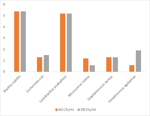
Footnote: AG=Adamawa Gudali, RB=Red Bororo, Cfu=Colony forming unit, ml=millilitre. Figure 1: Colony forming unit of Bacterial Species from the Milk Samples of Badejo and Kusulwa.
| Bacterial Species | AG No. of Occurrence | % | RB No. of Occurrence | % |
|---|---|---|---|---|
| Bacillus subtilis | 6 | 30 | 6 | 30 |
| Escherichia coli | 2 | 10 | 3 | 15 |
| Lactobacillus acidophilus | 5 | 25 | 5 | 25 |
| Micrococcus luteus | 2 | 10 | 1 | 5 |
| Staphylococcus aureus | 4 | 20 | 2 | 10 |
| Streptococcus agalactiae | 1 | 5 | 3 | 15 |
| Total | 20 | 100 | 20 | 100 |
Table 4: Percentage distribution and occurrence of Bacteria identified from the milk samples of Adamawa Gudali and Red Bororo.
| Bacterial Species | AG No. of Occurrence | % | RB No. of Occurrence | % |
|---|---|---|---|---|
| Bacillus subtilis | 6 | 30 | 6 | 30 |
| Escherichia coli | 2 | 10 | 3 | 15 |
| Lactobacillus acidophilus | 5 | 25 | 5 | 25 |
| Micrococcus luteus | 2 | 10 | 1 | 5 |
| Staphylococcus aureus | 4 | 20 | 2 | 10 |
| Streptococcus agalactiae | 1 | 5 | 3 | 15 |
| Total | 20 | 100 | 20 | 100 |
Table 5: Percentage distribution and occurrence of Bacteria identified from the milk samples of Adamawa Gudali and Red Bororo.
No. = Number, %=Percent, AG= Adamawa Gudali and RB=Red Bororo. Table 4: Percentage distribution and occurrence of Bacteria identified from the milk samples of Adamawa Gudali and Red Bororo.
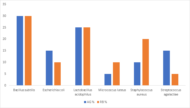
Footnote: AG=Adamawa Gudali, RB=Red Bororo, %=Percent. Figure 2: Percentage Occurrence and Distribution of Bacterial Species from the Milk Samples of Badejo and Kusulwa.
Results’ Interpretations
In this study research, the bacterial load in Red Bororo was significantly higher represented in total viable count of 159 (15.9×10⁻¹⁰) than Adamawa Gudali, which had 153 (15.3×10⁻¹⁰ Cfu/ml) as shown in Table 2, whereas, the gram stains indicates that, the Bacillus subtilis and Lactobacillus acidophilus are rod-shaped gram-negative bacteria, whereas Staphylococcus aureus and Streptococcus agalactiae are gram-positive bacteria that possess cocci-shaped, while Micrococcus aureus is a gram-negative bacterium with a cocci shape and Escherichia coli is a gram-negative rod-shaped bacterium, as shown in Table 1.
Therefore, in this work, the number of colonies and colonies forming unit in respect of individual bacterium such as Bacillus subtilis 54(5.4×10⁻¹⁰Cfu/ml) in Adamawa Gudali and Red Bororo were the same and did not significantly varied (p>0.05), followed by Lactobacillus acidophilus with 52 (5.2 × 10⁻¹⁰ Cf-u/ml) found in both milk samples which did not significantly varies (p>0.05), whereas Streptococcus agalactiae greater 19 (1.9 ×10⁻¹⁰ Cfu/ml) in Red Bororo significantly different (p<0.05) from Adamawa Gudali which had lesser 9 (0.9 × 10⁻¹⁰ Cfu/ml) value. The Escherichia coli was significantly higher (p < 0.05) at 15 (1.5 × 10⁻¹⁰ Cfu/ml) in Red Bororo than 13 (1.3 × 10⁻¹⁰ Cfu/ml) that had lower value, while Staphylococcus aureus had 13 (1.3 × 10⁻¹⁰ Cfu/ ml) in both milk samples, showing that significantly were not different (p > 0.05) and Micrococcus luteus was significantly greater (p > 0.05) with the value of 12 (1.2 × 10⁻¹⁰ Cfu/ml) in Adamawa Gudali, which had significant value than Red Bororo with 6 (0.6 × 10⁻¹⁰ Cfu/ml), as shown in Table 3.
However, in this study, the number of occurrences and percentages of bacteria isolated in the milk samples of Adamawa Gudali and Red Bororo are shown in Figure 3 & Table 4. The Bacillus subtilis possesses 6 (30%) in both samples, whereas Lactobacillus acidophilus had 5 (25%) in two different samples, while Staphylococcus aureus have 4 (20%) in the samples of Adamawa Gudali than Red Bororo with 2 (10%). Escherichia coli was higher with 3 (15%) in the Red Bororo sample than Adamawa Gudali with 22 (10%), but Streptococcus agalactiae followed the same pattern with 3 (15%) in sample of Red Bororo than the Adamawa Gudali with 1 (5%) and finally, Micrococcus luteus had a greater value of 2 (10%) in the sample of Adamawa Gudali than Red Bororo with 1 (5%).
Discussion
Fresh Dairy milk is the most available source of lactic acid bacteria, often, commonly been found from cattle breeds kept or raised. In this study, the isolated and identified bacteria are of non-fish source; however, they are of lactic acid origin that are employed in fish culture as probiotics. This is commensurate with Muniyandi, et al. [10]; Yeshambel [24]. The application of probiotics in fish production is of cognisance attainment due to its potentiality in enhancing growth, quality of water and bolstering disease resistance. This is corroborated with Yaslikan, et al. [25]. Apart from these attributes, it improves flesh and carcass quality. Nonetheless, other probiotics utilised in fish production are originated from terrestrial environment, rarely be from their immediate media on which host fish species dwells. This is in agreement with Doan, et al. [26].
In this current study research, however, the overall bacterial load between milk samples revealed that the Red Bororo was significantly represented higher (p < 0.05) total bacterial count as compared with Adamawa Gudali’s samples had shown in Table 2, similar research also revealed higher TBC in the Gudali breed [12]. Contrarily, the findings of Luka, et al. [27] was reported lower bacterial count in the same Bokolooji breeds in Nigeria. At this juncture, these milk samples exhibit higher contamination with bacterial load when compared with the several studies that isolated bacteria from cattle milk. Thus, the sources of microbial contamination in milk might be due to infected animals, milk handling processes, equipment, handlers and storage. This is corroborated with the findings of Luka, et al. [27].
Therefore, this work had reported the total bacteria counts and colonies forming unit per millilitre in respect of breeds differed significantly (p < 0.05), whereas individual bacterium revealed that Bacillus subtilis did not vary significantly (p > 0.05). Likewise, Lactobacillus acidophilus reported similar in both milk samples, which did not significantly differed (p > 0.05) had showed in Table 3 and disagreed with the findings of Frunza_, et al. [28]. In accordance with Aldohail, et al. that isolated the same bacteria from yoghurt in Malaysia. This is contrary to the work of Ogunshe, et al. [29] that isolated _L. fermentum from fermented food known as Fufu while L. plantarum from fermented beverage called Ogi. Consequently, LAB might be used as probiotics due to their symbiotic relationship with the host. This is also agreed with Ishthiaq, et al. [30]. Although this could be attributed to their antagonistic behaviour towards fish pathogens. This is in line with Khidirova, et al. [31], though this work is juxtaposed with the study of Folorunsho, et al. [32], which isolated four Lactobacillus species from fish and similar to reports of Jimoh Di, et al. [33].
Furthermore, the probiotic bacteria: acidophilus can improve the health condition of aquatic organisms and counterbalance microbes. This was matched with views of Adesina, et al. [34]. In addition, LAB: B. subtilis that constituted in such as commercial probioticsYuge® and AQUA PHOTOS [18, 35], probiotic starter contains and B. subtilis and Lactobacillus species [36] enhances growth and survival of C. gariepinus They can be used to control streptococcosis in fish [37, 38]. The intestinal microflora of fish determined by the environmental factors. Thus, temperature encourages fermentations to proliferate LAB. This is in conformity with Updhyayi.
Moreover, in these findings, Streptococcus agalactiae significantly greater (p < 0.05) in Red Bororo in comparison had shown in Table 3. This was contrasted with Kelany, et al. [39] that isolated higher CFU/ml from farmed tilapia. This could be due to different substrates and hygienic nature of the rearing facilities. Hence, B. subtilis is resistant against S. agalactiae infection that causes mortality in cichlids [40, 41, 42, 43]. This disease is known as streptococcosis [44]. This might cause economic losses, and this is agreed with Kelany, et al.[39].
In this study, thus, Staphylococcus aureus consisted of the same CFU/mL in both milk samples, and not significantly differed (p > 0.05) is depicted in Table 3. This disagreed with Ayuba, et al. [45]. According to Abdel-Gawad, et al. [46]; Adegunleye, et al. [47] who isolated staphylococcus species from fresh fish and water samples. It also from contaminated fish [48]. It causes pathogenicity and mortality in man, livestock, and fish [49]. Althugh, S. aureus and S. agalactiae are gram-positive bacteria that possessed cocci shape and weheras, E. coli is a gram-negative rod shape had shown in Table 1, and this was in line with Emily_, et al. [50]_.
Nevertheless, this work reported the number of occurrences and percentages of bacteria isolated in the milk samples of Adamawa Gudali and Red Bororo have shown that the B. subtilis constituted higher percentage had showing in Figure 3 and Table 4. Correspondingly, this was contrasted with the commercial probiotic “Yuge®” that contains 200 million CFU/g of B. subtilis reported by Abubakar, et al. [18]. Also, in these findings, L. acidophilus and S. aureus have followed the same pattern and this is in line with O’Gara [51] and Matuszewska_, et al. [49]. Although, _E. coli was significantly higher (p < 0.05) in Red Bororo than Adamawa had shown in Table 3, and this is in agreement with Ayuba, et al. [45]. Although, this is relatively lower and contrasted to the findings of Luka, et al. [23]. Thus, higher bacterial load might be due to the poor hygienic nature of the water used to clean utensils by the milkers and dusty particulate faecal matters surrounded the premises. Defiantly, Kato, et al. [52] who reported that E. coli were also isolated from the guts of C. gariepinus and O. niloticus. This might be due to a specific function in their gastrointestinal tract. This is agreed with the study of Burtseva, et al. [53]. In accordance with Dinara, et al. [54] who reported that 64G strain of E. coli has no toxicity and is non-infectious.
Notwithstanding, beneficial bacteria are also proven to improve immunity, intestinal health, tolerates infections, prevent and resist diseases in fish species [55, 56, 57]. Probiotics enhance growth performance and survival in fish species [18, 40, 41, 58]. Inaddition, minimise stresses, it also improves reproduction, engulfs and removes heavy metals in fish [59]. In this research work, Micrococcus luteus was significantly greater (p < 0.05) in the milk sample of Adamawa Gudali comparatively had shown in Table 3. M. luteus is a gram- negative bacterium with Cocci shape had depicted in Table 1, and this was confirmed by Ayuba, et al. [45]. Contrarily, Kato et al. [60] isolated from fish’s parts. According to Azza, et al. [61], M. luteus inhibits Vibrio vulnificus. It’s found naturally in aqueous environments [62]. It also emerges as pathogenic bacterium [63]. Henceforth, Micrococcosis is similar to Staphylococcosis and Streptococcosis in terms of economic loses in aquaculture. This was corroborated with Pekalaa, et al. [64]; Matuszewska, et al. [49]. Thus, lactic acid bacteria are used to treat S. agalactiae [65, 66].
Conclusion and Recommendations
This present study revealed that the Lactic Acid Bacteria (LAB) that were isolated from the milk of Red Bororo and Adamawa Gudali as an alternative to those found in fish source samples, fermented substrates, etc., play an important role in the survival, growth performance, and physiological functions of the cultured fish species as a result of the mutual relationship between the host and these microbial candidates that are subsequently used in lieu of chemotherapy. In this study, the lactic acid bacteria (LAB) detected from dairy milk suggested using them as probiotics in fish culture, though previously, bacteria determined from other similar breeds were never recommended for this purpose [67, 68, 69, 70]. Therefore, the total bacterial counts were significantly higher in Red Bororo than Adamawa Gudali, and these entail bacteria from non-fish sources were isolated, characterised, identified, counted, and colonies forming units were reported higher in this research as compared to the similar work from milk and milk products, unlike those that were reported from different fish organs and parts as well. Lactic acid bacteria from other available sources at any given locality should be analysed to determine their presence, total bacterial count, individual viable count, and molecular level.
Moreover, most of the probiotics used in aquaculture were sourced from fish body parts reported by several authors; thus, bacillus species such as Bacillus subtilis and Lactobacillus acidophilus were previously tested by many researchers to challenge the pathogenicity of E. coli as well as Lactococcus bacteria: Micrococcus luteus, Staphylococcus aureus, and Streptococcus agalactiae are known to cause economic losses; thus, the Lactococcus bacteria could be used to fight infectious bacteria in intensively managed fish species. In a nutshell, probiotics are significant in growth performance, survivability, defence mechanisms against foreigners in the body systems, as well as pathogenic microorganisms and disease resistance due to their antagonistic effects.
References
-
Omorwohwovie EE, Taiwo IO, Olapade OA (2019) Levels of Some Metals in Some Frozen Fish Species from Cold Rooms in Ijebu-Ode and Sagamu, Ogun State, Nigeria pp: 396-404
-
Ike-Obasi JJ, Ogubunka SO (2019) The roles of women in fish processing activities in some local government areas of Rivers State, Nigeria. Journal of Agricultural Extension 3(2): 73-77.
-
Jamabo NA, Dienye HE (2016) Effect of Different Commercial Feed on Water Quality and Survival of _Clarias gariepinus_ (Burchell, 1822). International Journal of Innnovative Studies in Aquatic Biology and Fisheries 2(4): 8-11.
-
Adah PM, Adeyemi SO, Shuaibu S, Sogbesan OA (2019) Studies on Dried Moringa Oleifera as Floating Promoter in Pelleting Feed Diet pp: 59-69.
-
Abdullahi AI, Hussaini U, Haruna MY, Saidu M, Geidam MB, et al. (2023) Effects of Sun-Dried Sicklepod (_Senna_ _obtusifolia_) as a Replacement for Soybean (_Glycine_ _max_) in the Diets of African Catfish (_Clarias gariepinus_) Juveniles. International Journal of Fisheries Science Research 5: 8.
-
Ayo-olalusi CI, Eze NC, Jibrin H, Chidume I, Edah B, et al. (2019) Body Composition of _Clarias gariepinus_ Juveniles Fed Fish Meal Supplemented with Maggot Meal. In Conference Proceedings of Fisheries Society of Nigeria.
-
Olajuyigbe OO, Adejonwo OA, Olusola AO, Ezekiel MO, Afolabi AA (2019) Evaluation of Microbial Quality of Some Feed Ingredients from Lagos Market, Lagos State, Nigeria, pp: 134-135.
-
NNLRS/Blench R (1999) Nigerian National Livestock Resource Survey. V L’homme et l’animal dans le bassin du lac Tchad. pp: 627-649.
-
Pandey GS, Voskuil GCS (2011) Manual on Milk safety, quality and hygiene. Golden Valley agricultural Research Trust, Zambia, pp: 52.
-
Muniyandi K, Punamalai G, Chandrasekaran N, Kamaraj Y (2021) Raw Cow Milk as a Potential Source of Beneficial Lactic Acid Bacteria (LAB). Journal of Bacterial Empire 4(1): 37-39.
-
Chizhayeva A, Amangeldi A, Oleinikova Y, Alybaeva A, Sadanov A (2022) Lactic Acid Bacteria as Probiotics in Sustainable Development of Aquaculture. Journal of Aquatic Living Resources 35(10): 1-17.
-
Oladapo AF, Ogunekun TO (2015) Quality Assessment of Fresh Milk from Traditionally Managed Nigerian Bunaji and Bokolooji Breeds of Cattle. The Pacific Journal of Science and Technology 16(1): 279-285.
-
Kouadri BN, Belabbas M, Bekenniche N, Monnoye M, Gerard P, et al. (2023) Probiotic Properties of Lactic Acid Bacteria Newly Isolated from Algerian Raw Cow’s Milk. Journal of Microorganisms 11: 209.
-
Gorg SK (2015) Effect of dietary probiotics mix (SPILAC) on growth performance and nutritive physiology of Nile tilapia (_Oreochromis niloticus_) (Linn) under laboratory condition. ITFAS 3(2): 440- 446.
-
Doan HV, Soltani M, Ring E (2021) _In vitro_ Antagonistic Effect and _In vivo_ Protective Efficacy of Gram Positive Probiotics Versus Gram Negative Bacterial Pathogens in Finfish and Shellfish. Journal of Aquaculture pp: 540.
-
Marco ML, Sanders ME, Ganzle E, Arrieta M, Cotter PD, et al. (2021) The International Scientific Association for Probiotics and Prebiotics (ISAPP) Consensus Statement on Fermented Food. Journal of Natural Review: Gastroenterology and Hepatology 18(1): 196-208.
-
De Marco G, Cappello T, Maisano M (2023) Histomorphological Changes in Fish Gut in Response to Prebiotics and Probiotics Treatment to Improve their Health Status: A Review. Journals of Animals 13: 286.
-
Abubakar MY, Geidam MB (2017) Survival and growth performance of _Clarias gariepinus_ (Burchhell, 1822) Fry Fed Probiotic (Yuge) at different Inclusion levels. In _Proceedings of the 32__nd_ _Annual conference of Fisheries_ _Society of Nigeria._ Nnamdi Azikwe University Awka Anambra State (28): 115-123.
-
Okocha RC, Olatoye IO, Adedeji OB (2018) Food Safety and Impacts of Antimicrobial Use and their Residues in Aquaculture. Journal of Public Health Review 39(21): 1-22.
-
Dauda AB, Folorunso LA, Dasuki A (2013) Use of Probiotics for Sustainable Aquaculture Production in Nigeria. Journal of Agriculture and Social Research 13(2): 35-45.
-
Mirzabekyan S, Harutyunyan N, Manvelyan A, Malkhalyan L, Balayan M, et al. (2023) Fish Probiotics: Cell Surface Properties of Fish Intestinal Lactobacilli and Escherichia coli. Journal of microorganisms 11: 595.
-
HDM (2020) Hardy Diagnostic Manufacturing facility and Quality Management System. West McCoy Lane Santa Maria, CA 93455, USA, pp: 1439.
-
Janet CC, Gayle EB (1979) Selective Medium for Distinguishing _Micrococci_ from _Staphylococci_ in the Clinical Laboratory. Journal OF Clinical Microbiology. American Society for Microbiology 4(5): 455-457.
-
Yeshambel T, Tadesse D, Haben F, Mesfin M (2021) Isolation and Identification of Lactic Acid Bacteria from Cow Milk Products. The Scientific World Journal 1: 1-6.
-
Yaslikan NM, Yaminudin J, Rasdi NW, Karim M (2023) Microfeed Incorporated with Probiotic for Aquaculture: A Review. World Veterinary Journal 13(4): 595-605.
-
Doan H, Ringo E, Esteban AE, Dadar MR, Dawood M, et al. (2019) Host Associated Probiotics: A key Factor in Sustainable Aquaculture. A Journal of Review in Fisheries Science and Aquaculture pp: 1-69.
-
Luka JS, Akinsola OM, Oshibanjo DO, Suleiman UA, Dakatut RA (2023) Evaluation of Raw Milk Quality from Multipurpose Cow in Jos, Nigeria_._ International Journal of Science and Research Achieves 09(01): 257-261.
-
Frunza OA, Khuda LV, lazerenko LM, Khudyi OI, Karpenko OV, et al. (2023) The Usage of Probiotic Microorganisms in Production Technology of European Grayling Fish Stock. Journal of Earth Science 1254: 1-10.
-
Ogunshe AAO, Olabode PO (2009) Antimicrobial potentials of indigenous _lactobacillus_ strains on gram – negative indicator bacterial species from _Clarias_ _gariepinus_ (Burchell) microbial inhibition of fish-borne pathogens. Afr J Microbiol Res 3(12): 870-876.
-
Ishthiaq IB, Ahmed J, Ramalingam K (2021) Probiotics in Brackish Water Fish Farming. Special Focus on Encapsulated Probiotics. Biointerface Research in Applied Chemistry. Platinun Open Access Journal 11(6): 14697-1470.
-
Khidirova M, Khushvaktov E, Mamatraimova M, Tuychiyev K, Chistyakov V, et al. (2022) Bio-encapsulation of Probiotic Bacteria in Black Soldier Larvae (_Hermetia_ _illucens_). In Proceedings of the International Scientific, Practical Conference Development and Modern Problems of Aquaculture. Rostov-on-Don, Russia. Held 27th September to 2nd October, 2022. pp: 160-165.
-
Folorunsho SB (2012) Use of Probiotics as Growth Promoter and Antibacterial for Nile Tilapia (_Oreochromis_ _niloticus_) and African Catfish (_Clarias gariepinus_), Ph.D. Thesis. Department of Aquaculture and Fisheries Management. University of Ibadan, Ibadan, Nigeria. Unpublished. pp: 1-47.
-
Di J, Chub Z, Zhang S, Huang J, Du H, et al. (2019) Evaluation of the Potential Probiotic _Bacillus subtilis_ Isolated from Two Ancient Sturgeons on Growth Performance, Serum Immunity and Disease Resistance of _Acipensada brycinus_. Journal of Shellfish Immunology 93: 711-719_._
-
Adesina I, Adewale YA, Tiamiyu LO, Ajibola MM, Dauda AB (2019) Gut Microbiota and Innate Nmmune Response of _Macrobrachium vollenhovenii_ Infected with _Pseudomonas aerugina_ and _Aeromonas hydrophilia_ Fed Diet Supplemented with _Lactobacillus acidophilus._ Journal of Aquaculture: Plant and Vertebrate 5: 93-105.
-
Guimaraes MC, Cerazo IM, Fernandez-Alarcon MF, Natori MM, Sato LY, et al. (2022) Oral administration of probiotics (_Bacillus subtilis_ and _Lactobacillus plantanun_) in Nile Tilapia (_Oreochromis_ _niloticus_) vaccinated and challenged with _Streptococcus agalactiae._ MDPT Fishes (7): 211.
-
Fakhri M, Ekawati AW, Arifin NB, Yuniarti A, Hariati AM (2019) Effect of Probiotics on Survival Rate and Growth Performance of _Clarias gariepinus._ Journal of Nature and Pollution Technology 1(18): 313-316_._
-
Kamgar M, Pourgholam R, Ghiasi M, Ghane Mm(2013) Studies on _Bacillus subtilis_ as Potential Probiotics, on the Biochemical Parameters of Rainbow trout, _Oncorhynchus_ _mykiss_ (Walbaum) to Challenge Infections. _Advanced_ _Studies in Biology._ Hikari Ltd 5(1): 37-50.
-
Bazari-Moghaddam S, Pourjaafari M (2021) The Effects of Four Types of Specific Probiotic on Growth Performance, liver enzymes and immune indices of juvenile Persian sturgeon (_Acipenser persicus_). Iranian Journal of Fisheries Sciences 20(4): 1179-1191.
-
Kelany N, Kotb S, Abdel-Mohsein H, Ismail AEM (2023) Multiple Antibiotic Resistant _Streptococcus agalactiae_ and _Streptococcus iniae_ in Nile Tilapia Aquaculture. Journal of Advanced Veterinary Research 14(1): 180-186.
-
Samson J, Quiazon KM, Choresca C (2020) Application of probiotic _Bacillus_ spp. isolated from African nightcrawler (Eudrilus eugeniae) on Nile Tilapia (_Oreochromis_ _niloticus_ L.). _BioRxi._ Archieves of microbiology pp: 1-13.
-
Zabidi A, Yusoff FM, Amin N, Yaminudin NJM, Puvanasundram P, et al. (2021) Effects of Probiotics on Growth, Survival, Water Quality and Disease Resistance of Red Hybrid Tilapia (_Oreochromis_ spp.)Fingerlings in a Biofloc System. Journal of Animal 11: 3514.
-
Zhang Z (2021) Research Advances on Tilapia _Streptococcosis_. Journal of Pathogens 10: 558.
-
Vijayaram S, Chou CC, Razafindralambo H, Ghafarifarsani H, Divsalar E, et al. (2024) _Bacillus_ sp. as potential probiotics for use in Tilapia Fish Farming Aquaculture: A Review. Annals of Animal Science 2024: 1-31.
-
Taukhid T, Lusiastuti AM, Andriyanto S, Sugiani D, Sumiati T, et al. (2021) Efficacy of inactive bivalent and trivalent *Streptococcus agalactiae* bacteria (Bio Type 1 and 2) Vaccines on Tilapia, *Oreochromis niloticus*. E-3S Web of Conferences 322: 1-1.
-
Ayuba VO, Onah E (2011) Microbial Load on the African Catfish (*Clarias gariepinus*) Sold in Makurdi Metropolis: A Case Study of Selected Markets. Proceedings of Annual Conference of FISON Held between 28th Nov and 2nd Dec, 2011 Held at FUT Minna, Niger, Nigeria.
-
Abdel-Gawad FK, Eweda WE, El-Taweel GE, Shehata SF, Abdel Tawab MI (2015) Detection of *Staphylococcus aureus* from fish and water samples collected from Lake Qarun. International Journal of Scientific and Engineering Research 6(8): 366-372.
-
Adegunleye DV, Sanusi I (2023) Microbiota of Catfish Tissues Harvested from Vials Polluted with Soil from E-Waste Dumpsite. Journal of Fisheries and Aquaculture 11(5): 104-111.
-
Bujjamma P, Padmavathi P (2015) Prevalence of *Staphylococcus aureus* in Fish Samples of Local Domestic Fish Market. International Journal of Current Microbiology and Applied Sciences 4(5): 427-443.
-
Matuszewska M, Dabrowska A, Murray GGR, Kett SM, Vick AJA, et al. (2023) Absence of *Staphylococcus aureus* in Wild Populations of Fish Supports a Spillover Hypothesis. Journal of Microbiology Spectrum 11(4): 1-11.
-
Emily EJ, Edibe SE, Neeraj M, Thomas EG, John WL, et al. (2012) Detection and Enumeration of Four Food Borne Pathogens in Raw Comingled Silo Milk in the United State. Journal of Food Protection 75(8): 1382-1393.
-
O'Gara JP (2017) Into the storm: Chasing the opportunistic pathogen *Staphylococcus aureus* from skin colonization to life-threatening infections. Journal of Environmental Microbiology 19(10): 3823-3833.
-
Kato CD, Mugaanyi MB, Majalija S, Tamale A, Musisi NL, et al. (2018) Isolation and Identification of Potential Probiotics Bacteria from the Gut of *Oreochromis niloticus* and *Clarias gariepinus* in Uganda. British Microbiology Research Journal 17(5): 1-8.
-
Burtseva O, Baulina O, Zaytseva A, Fedorenko T, Chekanov K, et al. (2021) In vitro Biofilm Formation by Bioluminescent Bacteria Isolated from the Marine Fish Gut. Journal of Ecology 81: 932-940.
-
Dinara M, Nurzhan S, Yasin U, Bozhena L (2024) Probiotics in the Creation of Fish-Based Heterodietic Half-Finished Products. Potravinarstro Journal of Food Sciences 18(2024):174-184.
-
Zhu C, Yu L, Liu W, Jiang M, He S, et al. (2019) Dietary Supplementation with *Bacillus subtilis* LT3-1 enhance the growth, immunity and disease resistance against *Streptococcus agalactiae* infection in genetically improved farmed tilapia: *Oreochromis niloticus*. Journal of Aquaculture Nutrition 25(6): 1241-1249.
-
Kasmani FB, Karimi-Torshizi MAK, Mehri M (2018) Effects of *Brevi-bacillus laterosporus* Probiotics on haematology, internal organs, meat peroxidation ileat microflora in Japanese Quails fed Aflatoxin-B1. (*Cyprinus capio*). Journal of Agriculture ScienceTechnology 20: 459-468.
-
Aydin F, Sehriban CY (2019) Effects of Probiotics on Reproductive Performance of Fish. Journal of Natural and Engineering Sciences 4(2): 153-162.
-
Umaru J, Agbugui MO (2022) Influence of Antox® Probiotic, as Water Additive on Growth Performance, Nutrient Utilization and Body Composition of the African Catfish, *Clarias gariepinus* (Burchel,1822) Fingerlings. Bayero Journal of Pure and Applied Sciences 14(2):178-183.
-
Zhai Q, Yu L, Li T, Zhu J, Zhang C, et al. (2017) Effects of Dietary Probiotic Supplementation on Intestinal Microbiota and Physiological Conditions of Nile Tilapia (*Oreochromis niloticus*) under Water Cadmium Exposure. Journal of Antonie Van Leeuwen Hoek 110: 501-513.
-
Kato CD, Kahuma CE, Namulawa VT, Kasozi N (2016) Antibacterial Activity of *Lactobacillus spp* and *Lactobacillus spp* Isolated from Various Parts of Pebbly Fish, Alestes baremoze British Microbiology Research Journal 17(2): 1-7.
-
Azza MAE, Yassir AE, Khal tab Abdul MES (2009) Micrococcus luteus and Pseudomonas species as probiotics for promoting the growth performance and health of Nile Tilapia (*Oreochromis niloticus*).
-
Akayli T, Urku C, Yardimci RE, Çanak O (2019) Bacterial Infection in Cultured Common Dentex (*Dentex dentex*, L.1758). Journal of Marine Science and Fisheries 2(1): 132-138.
-
Saleh OH, Mohamed MH, Abd El Galil MAA, Elkamel AA, Sayed HH (2021) Isolation and Characterization of *Micrococcus luteus* from *Oreochromis niloticus* in Egypt. Journal of Current Veterinary Research 3(2): 16-23.
-
Pekalaa A, Pazdziora E, Antychowiczb J, Bernadc A, Głowackad H, et al. (2018) _Kocuria rhizophila_ and _Micrococcus luteus_ as emerging opportunist pathogens in brown trout (_Salmo trutta_ Linnaeus, 1758) and rainbow trout (_Oncorhynchus mykiss_ Walbaum, 1792). Journal of Aquaculture. 486: 285-289.
-
Dawood MAO, Koshio S, Abdel-Daim MM, Van Doan H (2019) Probiotic Application for Sustainable Aquaculture. Reviews in Aquaculture 11(3): 907-924.
-
Kelany N, Abdel-Mohsein H, Kotb S, Ismail AEM (2024) Inhibitory Activity of Lactic Acid Bacteria Isolated from Tilapia Gut Against _Streptococcus Agalactiae._ Assuit Veterinary Medical Journal 70(181): 133-145.
-
Chauhan K, Andhare P, Marchawala F, Bhattacharya I, Updhyayi D (2021) Microbiology Study of Milk: A Review. International Journal of Biology, Pharmacy and Applied Sciences 10(4): 322-332.
-
Dawood MAO, Koshio S, Esteban MÁ (2018) Beneficial Roles of Feed Additives as Immunostimulants in Aquaculture: A Review. Reviews in Aquaculture 10: 950- 974.
-
Jimoh WA, Iyanda SA, Bello BK, Iderolu LA (2011) Microbial Occurrence of Preserved Tilapia Fish in Abeokuta North Local Government, Nigeria. In _Proceedings of the_ 26nTh _Annual conference of Fisheries Society of Nigeria._ Held at Federal University of Technology Minna, Niger State 26: 178.
-
Musa HM, Abeer EMS, Rashed MA (2021) Using _Lactobacillus acidophilus_ in Fish Feed to Improve Disease Resistance and Immune Status of Cultured Nile Tilapia. Alexandria Journal of Veterinary Sciences 70(2): 15-28.
- Genetic Improvement of Nile Tilapia (Oreochromis niloticus): Advances in Selective Breeding and Genomic Approaches for Sustainable Aquaculture
- Microplastics, Contaminants, and Waste Hotspots: Divergences and Faults in Prioritizing Control Efforts
- Creating a Healthier, More Vibrant Open and Closed Aquatic Environment. A Submersible, Centrifugal Magnetically Affixed Current Changing Aquarium Pump
- An Attempt to Assess Alpha Diversity and Sample Size: Using the Ostracod Assemblages off Kumamoto Port, Japan
- Assessment of the Efficiency of Common Fishing Gears and Crafts Used at Mohananda River of Chapai Nawabganj, Bangladesh
- Fish Productivity and Biodiversity Status of Sundarban Mangrove in Bangladesh