Biosynthesis of Pyramidical Nanosilver Using Andrographis paniculata (Burm.f.) and its Efficacy against Anopheles stephensi (Liston) Larvae in Laboratory
We have tested the larvicidal efficacy of AgNPs (Silver nanoparticles) synthesized by using stem extract of Andrographis paniculata (Burm.f.). The AgNPs synthesized using green bio-reduction methods which is economical as well as ecofriendly. The AgNPs formation was confirmed by UV-VIS spectrophotometer, XRD (X-Ray Diffraction) and SEM (Scanning Electron Microscopy). It reveals the geometry including size, shape and morphology of synthesized AgNPs. The biosynthesized AgNPs were spherical, L shaped and Pyramidical shaped, also the average sizes of synthesized silver nanoparticles were found to be 9.59 nm. The larvicidal bioassay further reveals LC50 and LC90 values against I, II and III instars of Anopheles stephensi (Liston) after 24 hr and 48 hr The AgNPs synthesized using stem extract of Andrographis paniculata have potent effect on larvae of Anopheles stephensi. Silver nanoparticles synthesized by stem extract of Andrographis paniculata are effective and economical with its eco-friendly characteristics. For controlling Anopheles stephensi in its larval stage this can be useful in tropical countries having malarial dominance. Further the Pyramidical structure observed in our study can be used in drug delivery for disease like malaria, cancer, dengue, etc.
Introduction
Mosquitoes are commonly known vector for diseases such as Dengue, Malaria, Chikungunya, Japanese Encephalitis, Yellow fever, Filariasis, Zika fever and many others. Mosquitoes are ectoparasite and are responsible for different pathogenic diseases such as Anopheles (Liston) is responsible for Malaria; Aedes (Linnaeus) for Chikungunya, Dengue fever etc.; Culex (Say) for Japanese Encephalitis, West Nile fever etc. Female mosquitoes are serious problem to mankind and animals. Malaria is reported to be high, it infects more than 216 million people per year and kill more than one million [1]. In Africa this is responsible for very high child mortality.
Hence there is a need to control these vectors. Various methods have been used to control these mosquitoes. Plants, fungus, bacteria and many chemicals have been used for the killing mosquitoes in different developmental stages. Andrographis paniculata known as ‘King of bitters’ has been used for years for many properties like antibacterial, antiprotozoal, fungicidal, larvicidal etc. [2]. For larvicidal and insecticidal properties active compound found in A. paniculata are andrographolides, homoandrographolide and Andrographis [3, 4]. Nanoparticles formed by plant extracts have environmental application to dye degradation [5].
Therefore, we assumed that Nanotechnology could play a tremendous role in the field of mosquito control. Nanoparticles have been synthesized using various chemical and natural methods. The chemical methods are not target specific. Exposing them several times causes resistant problem in the target organism. CuNPs have been synthesized using the A. paniculata leaf extract and their antimicrobial properties have been studied [6]. Also, the geometry of Nanoparticles plays a role with different wavelengths. The specific geometrical shapes give distinct spectra responses. In addition including subtle changes in the particle’s morphology by heating causes a shift in the individual particle spectrum and provides a simple means of turning the spectral response to a desired optical wavelength [7].
In present study we have attempted and have synthesized and characterized silver nanoparticles. Also, we have tested the larvicidal property of silver nanoparticles synthesized with the help of stem extract of A. paniculata against malarial vector Anopheles stephensi and recorded the results.
Materials and Methods
Collection of Plant
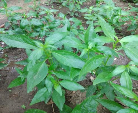
The fully grown plant of Andrographis paniculata (Kalmegh) was collected from the field of Dayalbagh as Figure 1.
Collection and Maintenance of Larvae
An. stephensi larvae were collected from various localities of Dayalbagh. They were maintained at 30 ± 2°C in distilled water in laboratory.
Preparation of Plant Extract
The plant was washed and dried, after that the stems and twigs are separated. They were crushed using mortar and pestle. 20 gm of crushed stem were boiled in 100 ml of de ionized water at 160°C for 20 minutes. The extract was cooled and filtered by using cotton and then by Whatman filter paper no.1. The extract was stored at 4°C for a week.
Green Synthesis of Silver Nanoparticles
1 mM AgNO3 solution was prepared than 5 ml of prepared aqueous plant extract was added to 45 ml of prepared 1mM AgNO3 solution. This was incubated in a dark chamber for 48 hr. Change in colour is the preliminary test for the synthesis of nanoparticles which were further subjected to UV-VIS Spectrophotometer, XRD and SEM analysis.
Characterization of Silver Nanoparticles
The colour change was observed visually. UV-VIS spectrophotometer analysis was performed by using UV- VIS spectrometer. (Shimadzu UV spectrophotometer, UV- 1800). The wavelength was recorded under range of 200 to 800 nm. For AgNPs and aqueous extract of A. paniculata. To perform scanning electron microscopy and XRD the AgNPs which are settled at the bottom were scratched out together with the solutions through micropipette and few drops of AgNPs are dropped on a slide and are dried using oven. After drying one more layer was coated in a similar manner. Then the prepared slide was subjected for the analysis. The size of synthesized silver nanoparticles was determined through XRD (Bruker, D8 Advance) by using Scherrer’s equation [8]. Scherrer’s equation: D = (0.9 λ) / (β cos θ) Where, D = diameter; λ = 0.1541 (wavelength of X- rays); β = Full Width Half Maxima (radians) and θ = Half of 2θ (degree). The surface morphology of AgNPs was determined by using scanning electron microscope (Sec e-beam pioneer, SNE 3200M).
Larvicidal Bioassay
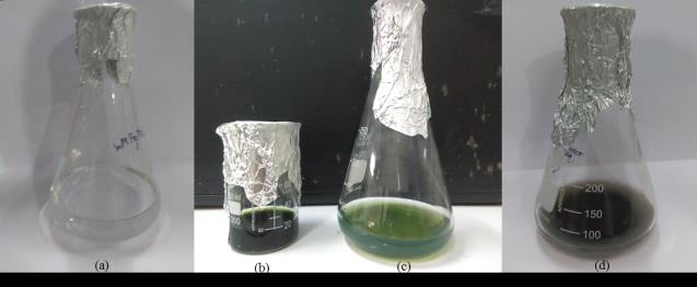
The larvicidal bioassays of AgNPs synthesized by A. paniculata were performed against I, II and III instars of An. Stephensi in distilled water. The mortality was recorded after 24 hr and 48 hr of exposure. WHO, 2005 protocols were followed [9]. Larvae were taken in the batches of 20 in 100 ml of distilled water and the experiment was performed in replicates. The temperature was 30+2°C and the larvae were provided a photoperiod of 12hrD: 12hrL (Dark: Light). No food was given during the time of bioassay. The ranges of concentration used were 15.2, 30.42, 45.63, 60.84, 76.05, 91.26 ppm. The positive and negative controls were setup. The mortality in controls (5% to 20%) was corrected using Abbott’s formula [10]. Abbott’s formula: C%M = (T- C) / (100 – C) *100 Where, C%M = Corrected % Mortality, T = % Mortality in treatment and C = % Mortality in control The colour of 1mM AgNO3 as shown in Figure 2a changes immediately from transparent to colloidal green as shown in Figures 2b & c and after the incubation of 48 hr is becomes colloidal brown as shown in Figure 2d which indicates the synthesis of AgNPs.
The experiments having greater mortality were discarded. The LC50 and LC90 values were calculated using Finney’s table [11] and probit analyzer [12].
Results
UV-VIS Spectrophotometer Analysis
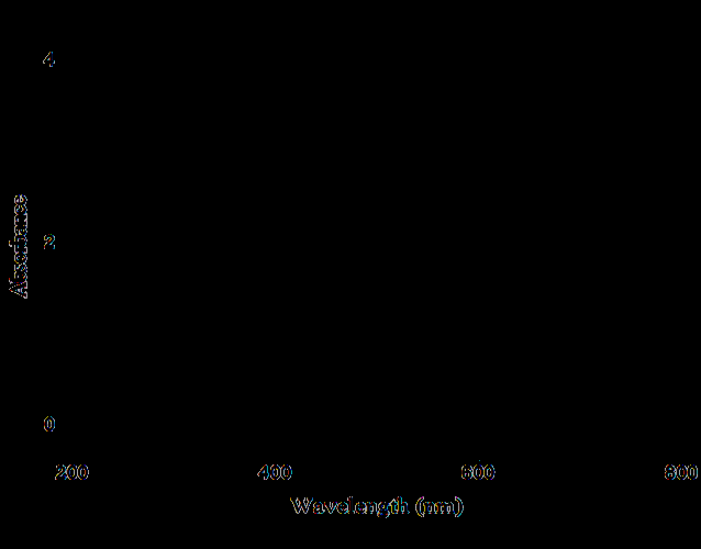
The peaks of UV spectra for both the synthesized AgNPs and aqueous extract of A. paniculata was recorded on 363.53 nm and 645.99 nm respectively as shown in Figures 3a & b.
The synthesized AgNPs were characterized using UV spectrophotometer after the incubation of 48 hr.
XRD Analysis
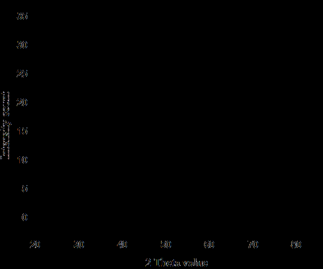
The peaks recorded as in Figure 4. For synthesized Silver nanoparticles are compared with the standard JCPDS card for silver file No. 04-0783 and the average size of AgNPs were found to be 9.59 nm.
SEM Analysis
The morphology revealed that the particles were well dispersed and were of different shapes – spherical, L shaped and Pyramidical shaped as in Figure 5.
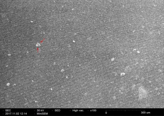
The Geometry of the Pyramid
Nanoparticles having specific geometry are of great significance in research. In our study some of the particles are found to be Pyramidical shaped as the pyramid has special binding property which may able to bind with the drugs and they can easily targeted to cancerous cell or any other disease.
The possibilities of curing non-curable diseases would be increased. According to the image there are two possibilities: the pyramid having square as base and the pyramid having triangle as base. a) Area of pyramid having square as a base: area of square + 4(area of triangle) which was found to be 1792 nm2. b) Area of pyramid having triangle as a base: area of triangle + 3(area of triangle) which was found to be 1008 nm2.
Efficacy Test of AgNPs Synthesized by A. Paniculata against An. Stephensi Larvae
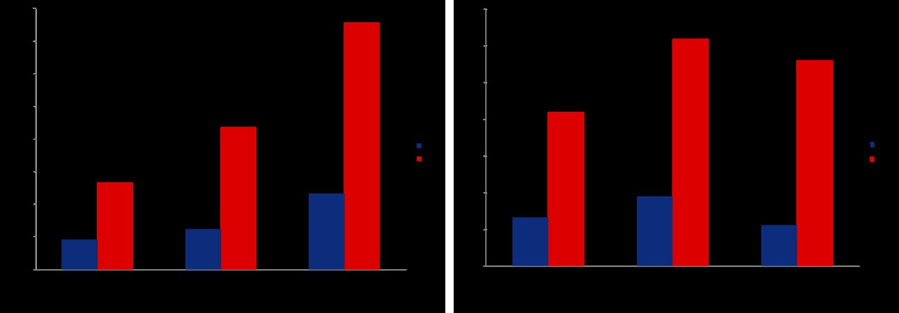
AgNPs synthesized by A. paniculata shows potent effect against I, II, III instars of An. stephensi after 24hr and 48hr of exposure. The instar shows 100% mortality after 24 hr on 91.26 ppm concentration. II instar shows 100% after 48hr on 91.26 ppm concentration. III instar shows 100% mortality on 76.05 ppm after 48hr.
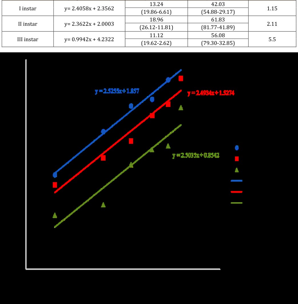
The LC50 and LC90 values after 24 hr depict that the AgNPs synthesized using the stem of A. paniculata are more effective on I instar as compare to III instars. Effectiveness decreases from I > II > III instars. Similarly, after 48 hr the effectiveness was I > III > II instars as represented in Tables 1 & 2 and are shown in Figures 6a & b.
- Probit equation
- LC50 (ppm) LC90 (ppm)
- Instar χ2
- (Regression line)
- (UFL-LFL)
- (UFL-LFL)
- 18.15
- 53.19
- I instar y= 2.5255x +
- 0.67
- (24.65-
- 1.857
- 11.65)
- (68.2-38.14)
- 24.72
- 87.11
- II instar y= 2.4934x +
- 0.89
- (32.86-
- 1.5274
- 16.57)
- (121.18-53.04)
- 46.47
- 151.16
- III instar y= 2.5035x +
- 2.97
- (56.86-
- 0.8542
- 36.08)
- (229.38-72.94)
- Instar
- Probit equation
- LC50 (ppm)
- LC90 (ppm) χ2 (Regression line)
- (UFL-LFL)
- (UFL-LFL)
- I instar y= 2.4058x + 2.3562
- 13.24
- 42.03
- 1.15
- (19.86-6.61)
- (54.88-29.17)
- II instar y= 2.3622x + 2.0003
- 18.96
- 61.83
- 2.11
- (26.12-11.81)
- (81.77-41.89)
- III instar y= 0.9942x + 4.2322
- 11.12
- 56.08
- 5.5
- (19.62-2.62)
- (79.30-32.85)
Table 1: Depicting probit equation, LC50 and LC90 values
Discussion
In this study we successfully synthesized silver nanoparticles of Pyramidical shaped and different size using aqueous stem extract of A. paniculata. These nanoparticles were tested as a larvicide of mosquito vector against I, II and III instars of An. stephensi. These silver nanoparticles were also not synthesized previously using the aqueous stem extract of A. paniculata. Rather, silver nanoparticles synthesized by whole plant extract of A. paniculata have shown antiplasmodial activities [13]. The larvicidal effect is also very good at low concentration and the results were different at different larval stages due to metamorphosis [14], change in chitin and other environmental factor. The tested LC50 and LC90 values for I, II and III instars after 24 hr and 48 hr are represented in Figures 7a & b. The bar diagram showing high mortality on low concentration. The oviposition deterrent, Ovicidal and gravid mortality of An. stephensi have been previously observed using ethanolic extract of A. paniculata [15]. The silver nanoparticle have been synthesized using leaves of Catharanthus roesus and their antiplasmodial activity have been studied previously [16]. Previous synthesis of nanoparticles has been done by using Eclipta prostrata leaf against filarial and malarial vector [17]. Also the nanoparticles have been synthesized using fungus Aspergillus niger and tested against the mosquito larvae [18] showed potent effect. Also, these nanoparticles are the possibility and can be used for malarial and filarial vector [14]. Their study showed that the larval stage of An. stephensi was more susceptible to AgNPs than pupae and adults. Recently, the green synthesis of AgNPs using Azadirachta indica have been performed [19] and their antimicrobial properties were studied, they successfully synthesized AgNPs and result revealed that lower ratio of plant extract is optimum for synthesis of AgNPs. Now, the Pyramidical shape of AgNPs was observed due to certain properties of A. paniculata. Its extract was able to synthesized Pyramidical shaped AgNPs under certain condition of temperature and pH [20]. Also, CuNPs synthesized were having certain properties were discussed previously revealed that synthesized CuNPs have antimicrobial properties [6]. The role of Pyramidical AgNPs can be used as drug interfacing device in various chronic diseases such as cancer, tumor, malaria, etc. which is significantly been found in our study.
Conclusion
It is therefore, concluded from the investigation performed that the AgNPs synthesized by stem extract of A. paniculata have potent effect against I, II and III instars of An. stephensi. However, SEM image further confirmed that some of the AgNPs have formed Pyramidical shape which can be studied in future as the geometry have greater role to effect the efficacies at nano level. The Pyramidical shape is useful in drug delivering for malaria, filarial, dengue, chikungunya and disease like cancer and tumor as it has significant binding properties. Further, there are some peaks observed in XRD other than those of silver which may because of the binding of plant constituent on other peak this can be studied further. Some of the peaks are due to noise which was because of the denaturing nature of plant extract. Further research is warranted to identify all the active elements of this plant and could be treated directly in-vitro study on different Plasmodium species to find out the most economical option available for removing malaria vector in tropical countries like Africa and Asia having endemic population of malaria as well as An. Stephensi.
Acknowledgment
We are sincerely grateful to Prof. P. S. Satsangi Sahab, Chairman of Advisory Committee on Education, Dayalbagh Educational Institute. We thank Prof. P.K. Kalra, Director, Dayalbagh Educational Institute for providing support and encouragements for the work. We profusely thank Mr. Anil Oberoi for providing Kalmegh. We wish to thank Prof. Sahab Das, Dept. of Chemistry, Dayalbagh Educational Institute, for providing UV-VIS Spectrophotometry, XRD and SEM facilities. We also thank Dr. Richa for her help in editing the manuscript.
References
-
WHO (2017)http://www.who.int/mediacentre/facts heets/fs094/en/
-
Akbar S (2011) _Andrographis paniculata_: a review of pharmacological activities and clinical effects. Altern Med Rev 16(1): 66-77.
-
Elango G, Rahuman AA, Kamaraj C, Zahir AA, Bagavan A (2010) Studies on effects of indigenous plant extracts on filarial vector _Culex tritaeniorhynchus_ Giles. Parasitol res 107(1): 167-176.
-
Chatterjee N, Biswas S, Saha NC, Biswas SJ (2014) _Andrographis paniculata_ a traditional herb with pharmacological properties: a review. Global J Res Med Plants & Indigen Med 3(5): 206-214.
-
Francis S, Joseph S, Koshy EP, Mathew B (2017) Green synthesis and characterization of gold and silver nanoparticles using _Mussaenda glabrata_ leaf extract and their environmental applications to dye degradation. Environmental Science and Pollution Research, 24(21): 17347-17357.
-
Devasenan S, Beevi NH, Jayanthi SS (2016) Synthesis and characterization of Copper Nanoparticles using Leaf Extract of _Andrographis paniculata_ and their Antimicrobial Activities. International Journal of ChemTech Research 9(4): 725-730.
-
Mock JJ, Barbic M, Smith DR, Schultz DA, Schultz S (2002) Shape effects in plasmon resonance of individual colloidal silver nanoparticles. The Journal of Chemical Physics 116(15): 6755-6759.
-
Goudarzi M, Mir N, Mousavi KM, Bagheri S, Salavati NM (2016) Biosynthesis and characterization of silver nanoparticles prepared from two novel natural precursors by facile thermal decomposition methods. Scientific reports.
-
WHO (2005) World Health Organisation Guidelines for laboratory and field testing of mosquito larvicides, pp: 139.
-
Abbott W S (1925) Formula for correction of observed mortality due to natural mortality/our econ Ent, 18(2): 265-267.
-
Finney DJ (1971) Probit Analysis: 3rd (Edn) Cambridge University Press, pp: 333.
-
Reddy PJ, Krishna D, Murty US, Jamil K (1992) A microcomputer FORTRAN program for rapid determination of lethal concentrations of biocides in mosquito control. Comput Appl Biosci 8(3): 209-213.
-
Rajasekar P, Priyadharshini S, Rajarajeshwari T, Shivashri C (2013) Bio-inspired synthesis of silver nanoparticles using _Andrographis paniculata_ whole plant extract and their antimicrobial activity over pathogenic microbes. Int J Res Biomed Biotech 3(3): 47-52.
-
Soni N, Prakash S (2014) Silver nanoparticles: a possibility for malarial and filarial vector control technology. Parasitol res 113(11): 4015-4022.
-
Chenniappan K, Kadarkarai M (2008) Oviposition deterrent, ovicidal and gravid mortality effects of ethanolic extract of _Andrographis paniculata_ Nees against the malarial vector _Anopheles stephensi_ Liston (Diptera: Culicidae). Entomological Research 38(2): 119-125.
-
Ponarulselvam S, Panneerselvam C, Murugan K, Aarthi N, Kalimuthu K, et al. (2012) Synthesis of silver nanoparticles using leaves of _Catharanthus roseus_ Linn. G. Don and their antiplasmodial activities. Asian Pacific journal of tropical biomedicine 2(7): 574-580.
-
Rajakumar G, Rahuman AA (2011) Larvicidal activity of synthesized silver nanoparticles using _Eclipta_ _prostrata_ leaf extract against filariasis and malaria vectors. Acta tropica 118(3): 196-203.
-
Soni N, Prakash S (2012) Synthesis of gold nanoparticles by the fungus _Aspergillus niger_ and its efficacy against mosquito larvae. Rep Parasitol 2: 1-7.
-
Roy P, Das B, Mohanty A, Mohapatra S (2017) Green synthesis of silver nanoparticles using _Azadirachta_ _indica_ leaf extract and its antimicrobial study. Applied Nanoscience 7(8): 843-850.
-
Gupta P (2017) Efficacy of _Andrographis paniculata_ (Burm.f.) Integrated with Silver Nanoparticles against _Anopheles_ _stephensi_ (Liston) in Laboratory. Dissertation, Dayalbagh Educational Institute.
- California Red-Legged Frog and Non-Listed Amphibians Response to Non-Native Fish Removal
- Industrial Standardization of the Bio-OS: Algorithmic Codification of Resilience Engineering Guidelines and Version V8 Architecture
- Climate Variability and the Sustainability of Snail Farming in Nigeria: Past Trends, Present Challenges and Potential Outlook
- The Evaluation of the Surveillance System of Anthrax in Gilgit-Baltistan, Pakistan, 2018
- Natural Decline to Extinction of A New Zealand Rabbit Population
- Mitochondrial Bio-Logistics: Steering Co-Enzyme Q10 and Lycopene Synergies within the Science 4.0 Bio-OS Framework