Identification of Bacterial Species from Injured Cuticle of Mud Crab, Scylla serrata
Crustacean aquaculture production is one of the major economic activities worldwide with prime importance in the developing countries. Infectious diseases from viral, bacterial, and in some cases, fungal origin affecting the crustacean aquaculture production due to intensification of farming activities. The microbial population of 11 x 10-5 to 215 x 10-3 CFU were observed in the scraps of naturally injured/wounded cuticle of mud crab, Scylla serrata. Three predominant bacterial species were identified including Bacillus sp., Escherichia coli and Pseudomonas aeruginosa in the scraps based on the series of biochemical analysis as well 16s rDNA sequence analysis. These findings may provide the baseline to reveal the pathogenecity of host specific and invading pathogenic bacteria in the cultivable crustaceans.
Introduction
Crustaceans are the second largest group, next to insects, in the phylum arthropoda comprising approximately more than 30,000 known species including lobsters, crabs, shrimps, prawns, krill, copepods etc., and have a predominant role in aquatic food chain, especially as primary and secondary consumers as well as food for human [1, 2]. The semi and intensive culture of edible crustaceans was started in 1970s as an industrial activity, developed rapidly with a huge increase in the number of hatcheries as well as aqua farms [3]. Even though, crustacean aquaculture sector is still highly dependent upon marine capture fisheries for sourcing key dietary nutrient inputs, including fish meal and fish oil. This dependency is particularly strong within compound aqua feeds and farmed marine shrimps [4].
Diseases in Cultivable Crustaceans
The aquaculture industries witnessed major growth till 1980s, followed by a sudden decline in its production due to environmental degradation and infectious as well as non-infectious diseases caused by many microbes including bacteria, viruses and fungi [3].
A much larger proportion of bacterial diseases are actually external infections, where a variety of microorganisms attack the exoskeleton and affect crustacean aquaculture production [5]. These chitinolytic microorganisms do not penetrate into the soft tissue underlying the exoskeleton [6] but may provide a portal entry to epidermal tissue for secondary invaders. The shell disease, which is most often reported among brachyuran crabs, appears to be associated with the microbes such as Vibrio sp. and Pseudomonas sp. [7]. Vibriosis, caused by bacteria belongs to family Vibrionaceae, is one of the predominant diseases in shellfish and finfish aquaculture, often responsible for mass mortality in aqua farms with high economic loss worldwide [8, 9, 10, 11, 12].
Materials and Methods
The scraps obtained from naturally injured/wounded cuticle site on the dorsal side of crabs were diluted in sterile sea water, plated on nutrient agar medium. The colonies grown were counted after 24-48 hours by colony counter wherein pigmentation, relative size and shape of the colonies were analyzed. The bacterial isolates detected were subjected to staining procedures, biochemical and other tests to study the characteristic features by culturing in various media for their identification.
Staining Methods
Gram’s stain was used to study the morphology and staining properties of the bacterial isolates obtained from the mud crab. The endospores of bacterial isolates were stained with malachite green and then counter stained with safranine [13].
Biochemical Analysis of Bacterial Isolates
Motility test was carried out to determine the differences, if any, among the bacterial isolates, in their motility in a liquid medium [13]. The basic biochemical test for the identification of bacterial isolates from crab were analyzed by following tests including, Oxidase test, Catalase test, Citrate utilization test, Methyl red and Voges Proskauer (MR & VP) test, Indole production test, Urease test, Starch hydrolysis test, Nitrate and nitrite reduction test Hugh-Leif son oxidative fermentative test [14]. Bacterial isolated colonies were grown in nutrient agar and some of the selective media (EMB agar, Cetrimide agar base medium and TCBS agar) for further identification based on colony morphology, size and pigmentation variability.
Molecular Characterization of Isolated Bacterial Species
Total DNA was extracted from bacterial cells using phenol chloroform method [15]. DNA concentration was measured spectrophotometrically at 260 nm in a spectrophotometer. Aliquots of the DNA (100 ng) were then used for Polymerase Chain Reaction (PCR). The 16S rDNA was amplified from the total genomic DNA by PCR using a universal primer. The forward and reverse primer sequences used were 27F 5’-AGAGTTTGATCCTGGCTCAG-3’ and 1492r 5’-TACGGTTACCTTGTTACGACTT-3’ [15]. The PCR products were gel purified and sequenced in one direction only with forward primer using Ocimum Bio solutions DNA Sequencing Services, Hyderabad, India. The gene sequences were aligned in FASTA format and then analyzed with ClustalW using MEGA 4 software. The program was then used to calculate the best match for selected sequences, similarities and differences were identified, through viewing the phylograms based on N-J phylogeny. Finally, based on the morphological, biochemical and other characteristic features, the colonies were identified by using Bergey’s Manual of Determinative Bacteriology [16].
Results
Detection and Identification of Bacterial Species
In order to detect and isolate the microbes from the scraps of injured/wounded cuticle of the mud crab, S. serrata, and total microbial count was analysed for 24 to 48 hours, based on the extent of bacterial colonies grown in nutrient agar plates. The data showed the presence of a strong microbial population ranging from 11 x 10-5 to 215 x 10-3 CFU (10-5 to 10-3 dilutions) in the scraps obtained from naturally injured/wounded cuticle. In addition, a dilution of 10-5 appears conducive to get a maximum 23 types of colonies which could be detected under hand held lens or colony counter (Table 1).
| Dilution factor | Number of colonies (CFU/ml) |
|---|---|
| 3-Oct | 215 X 10^{-3}$ |
| 4-Oct | 48 X 10^{-4}$ |
| 5-Oct | 11 X 10^{-5}$ |
Table 1: Total aerobic microbial count (bacterial load as CFU) grown in nutrient agar from scraps of naturally injured/ wounde
CFU: Colony Forming Units. Table 1: Total aerobic microbial count (bacterial load as CFU) grown in nutrient agar from scraps of naturally injured/ wounded cuticle of the mud crab, Scylla serrate.
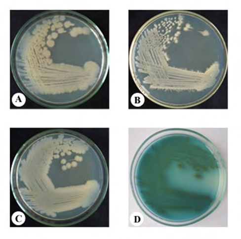
Of the twenty three bacterial colonies detected from the injured/wounded cuticle of the mud crab, S. serrata, at least four rich and morphologically (colour, growing pattern and size of the colony) distinct colonies were isolated. Morphological Phenotypic Characteristics of Bacteria Isolates of these four distinct colonies were sub cultured in nutrient agar medium showed the distinct pigmentation, size and a pattern of growth. As shown in Figure 1, colony 1, 2 and 3 (A, B and C) appeared large with white colouration ranging from cloudy to bright white. Among these three, colony 1 and 2, although appeared similar in overall colouration, dry surface and the size of colonies varied distinctly (Table 2). Further, while both these colonies (colony 1 & 2) grew very well and appeared large, raised with irregular margin, colony 3, which was also white but with wet surface did not grew as raised but with distinct surface appeared with a smooth margin. On the other hand, colony 4, appeared green in colour but were relatively small sized compared to colony 1,
2 and 3, although all the four colonies appeared to be bacilli under light microscope.
| Bacterial colony# | Morphological characteristics | ||
|---|---|---|---|
| Pigmentation | Relative size | Shape of the cell | |
| Colony 1 | cloudy white | large raised, irregular margin | short bacilli |
| Colony 2 | white | large raised, irregular margin | short bacilli |
| Colony 3 | milky white | large | bacilli |
| Colony 4 | green | small | short bacilli |
Table 2: Morphological characteristics of bacterial colonies isolated from the scraps of naturally injured/wounded cuticle of the
# Colonies grown in nutrient agar medium. Table 2: Morphological characteristics of bacterial colonies isolated from the scraps of naturally injured/wounded cuticle of the mud crab, Scylla serrate.
Primary Staining and Biochemical Characteristics of Isolated Bacterial Colonies
In primary test, all the 4 colonies appeared motile and aerobic. Among them, while colony 1, 2 and 3 grew well at anaerobic condition, colony 4 did not show any significant growth. Gram’s and endospore staining showed that colony 1 and 2 were Gram-positive and endospore forming, while colony 3 and 4 were negative for Gram’s and endospore staining (Table 3).
| Primary tests | Bacterial isolates | |||
|---|---|---|---|---|
| Colony 1 | Colony 2 | Colony 3 | Colony 4 | |
| Gram’s reaction (young culture) | + | + | - | - |
| Aerobic growth | + | + | + | + |
| Anaerobic growth | + | + | + | - |
| Endospore | + | + | - | - |
| Motility | + | + | + | + |
| Biochemical tests | Bacterial isolates | |||
| Colony 1 | Colony 2 | Colony 3 | Colony 4 | |
| Oxidase reaction | + | + | - | + |
| Catalase test | + | + | + | + |
| Citrate utilization | + | - | - | + |
| Methyl red | - | - | + | + |
| Voges-Proskauer | - | - | - | + |
| Indole production | - | - | + | + |
| Urease | - | + | - | + |
| Starch hydrolysis | - | + | + | - |
| TSI | ND | ND | - | + |
| Nitrate reduction | ND | ND | + | + |
Table 3: Primary tests for identification of bacterial colonies isolated from the scraps of naturally injured/wounded cuticle
+ Positive - Negative Table 3: Primary tests for identification of bacterial colonies isolated from the scraps of naturally injured/wounded cuticle of the mud crab, Scylla serrate.
Biochemical characteristics, as presented in Table 4, showed that, while all the four colonies are positive for catalase, colony 1, 2 and 4 are positive to oxidase. Tests with methyl red, indole production or nitrate reduction indicated positivity for colonies 3 and 4 only.
ND: not determined; + Positive; - Negative; TSI: triple sugar iron test. Table 4: Biochemical characteristics of bacterial colonies isolated from the scraps of naturally injured/wounded cuticle of the mud crab, Scylla serrate.
As shown in Table 5, the carbohydrate fermentation tests showed that all the four bacterial isolates invariably reduced or fermented all the 15 sugars tested. The colonies 1, 2, 3 and 4 reduced six sugars namely D-xylose, sucrose, D-glucose, D-sorbitol, L-arabinose and L-rhamnose while, melibiose was reduced by colony 1, 2 and 3. Further, colony
1, 2 and 4 did reduce the carbohydrates such as D-fructose, inositol, trehalose, D-mannose. D-galactose was reduced by colonies 1 and 4, while colony 3 and 4 reduced the sugar D-mannitol. Furthermore the colonies 1 and 3 reduced the sugars maltose and lactose respectively.
| Biochemical test | Bacterial isolates | |||
|---|---|---|---|---|
| Colony 1 | Colony 2 | Colony 3 | Colony 4 | |
| Carbohydrate fermentation | ||||
| D-Xylose | + | + | + | + |
| Sucrose | ± | ± | + | + |
| Lactose | - | - | + | - |
| D-Glucose | + | ± | + | + |
| D-Fructose | ± | ± | - | + |
| Maltose | + | - | - | - |
| D-Galactose | + | - | - | + |
| D-Sorbitol | ± | ± | + | + |
| Inositol | ± | ± | - | + |
| L-Arabinose | + | + | + | + |
| D-Mannitol | - | - | + | + |
| Melibiose | ± | ± | + | - |
| Trehalose | ± | ± | - | + |
| L-Rhamnose | ± | ± | + | + |
| D-Mannose | ± | ± | - | + |
Table 4: Growth of bacterial colonies isolated from scraps of the naturally injured/wounded cuticle of the mud crab, _Scylla_
Growth in Selective Media
The isolated colonies were grown in certain specific selective media, including Eosin Methylene Blue (EMB), Cetrimide agar and Thiosulphate citrate bile salt agar (TCBS) showed that colony 1 and 2 did not grow at any of the three selective media tested, while colony 3 and 4 grew well in EMB agar with metallic green colour and in cetrimide agar with green colour respectively. Interestingly, none of these 4 colonies grew in TCBS agar, showing that the isolated colonies did not belong to Vibrio species (Table 6).
| Media | Growth of bacterial isolates | |||
|---|---|---|---|---|
| Colony 1 | Colony 2 | Colony 3 | Colony 4 | |
| EMB agar | - | - | + | - |
| (metallic green) | ||||
| Cetrimide agar base | - | - | - | + |
| (green) | ||||
| TCBS agar | - | - | - | - |
Table 5: Growth of bacterial colonies isolated from scraps of the naturally injured/wounded cuticle of the mud crab, _Scylla_
EMB agar: Eosin Methylene Blue agar TCBS agar: Thiosulphate Citrate Bile Salt agar + Growth; - No growth Table 6: Growth of bacterial colonies isolated from scraps of the naturally injured/wounded cuticle of the mud crab, Scylla serrata in selective media.
Based on these results, it may be safely concluded that colony 3 and 4, bacterial isolates from the scraps of injured/wounded cuticle of the mud crab S. serrata belongs to enterobacteriaciae and colony 3 could be identified as Escherichia coli as it grew well in EMB agar media with metallic green coloured colonies. The possible growth of colony 4 in cetrimide agar with green colour colonies along with its ability to reduce 12 of the 15 sugars tested (Table 5) indicates that the colony 4 could be identified as Pseudomonas aeruginosa. The ability to produce endospores and positivity for Gram’s staining, biochemical, phenotyphic characteristics and inability to grow in any of the three selective media tested indicate that colony 1 and 2 may belong to genus Bacillus.
Molecular Characterization of Bacterial Isolates
In order to identify the species of bacterial colonies 1 and 2 (Bacillus spp.), an attempt was made to characterize the bacterial isolates by amplifying 16S rDNA of the colonies using specific primers. The DNA was isolated from colony 1 and 2 by phenol choloroform method and subjected to electrophoresis on 0.8% agarose gel. The quantity of genomic DNA obtained was approximately 200 ng/µl (Figure 2A). The 16S rDNA amplified from the bacterial genomic DNA by PCR using specific primers produced a single band at approximately 1500 bp in agarose gel (Figure 2B). The purified PCR product was then sequenced and the nucleotide sequences were analysed using BLAST search. The results revealed the sequence similarities with Bacillus sp. and Bacillus flexus in the NCBI database_. The sequence data obtained in the present study was deposited in the GenBank database and accession numbers were obtained, _Bacillus sp. N1 (HM997150) and Bacillus flexus N3 (HM997151).
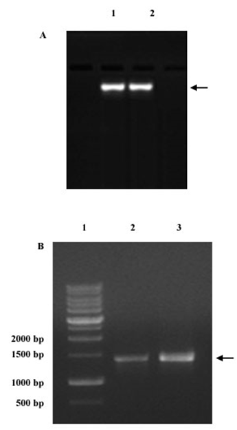
Figure 2A: Genomic DNA isolated from the bacterial isolates 1 and 2 were analyzed on agarose gel electrophoresis. Lane 1 and 2 represents the genomic DNA isolated form the colony 1 and 2 respectively. Figure 2B: PCR amplification products targeting the 16S rDNA gene from colony 1 and colony 2 using specific primers. Lane 1: Molecular weight marker; Lane 2: PCR product of approximately 1500 bp of 16S rDNA gene from DNA of colony 1; Lane 3: PCR product of approximately 1500 bp 16S rDNA gene from DNA of colony 2.
The sequence data of Bacillus sp. N1 and B. flexus N3 obtained for our sample were aligned against similar such Bacillus nucleotide sequences available in the sequence data from NCBI Gen Bank and evolutionary relationships were analysed using MEGA software version 4 (Figures 3&4).
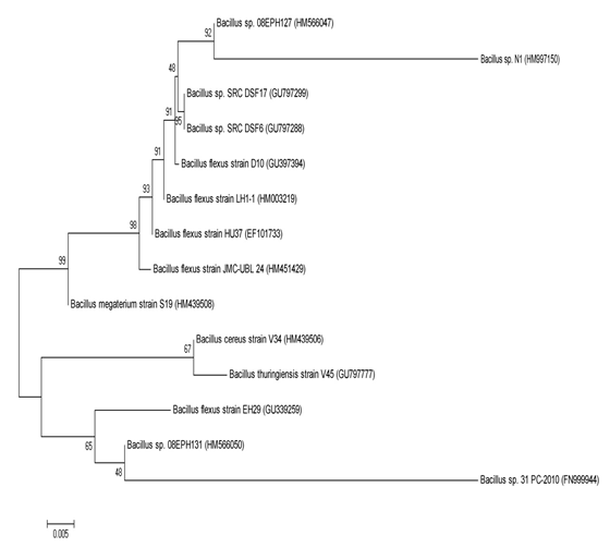
Figure 3: Phylogenetic relationships of colony 1 (Bacillus sp. N1) derived from 16S rDNA gene sequence homology. The sequences have been retrieved from NCBI databases and the tree was drawn using neighbour joining algorithm with kimura 2 parameter distances in MEGA 4 software. The bar represents distance values calculated in MEGA and values at nod represent percentage of 1000 bootstrap replicates. Numbers in bracket represent GenBank accession numbers.
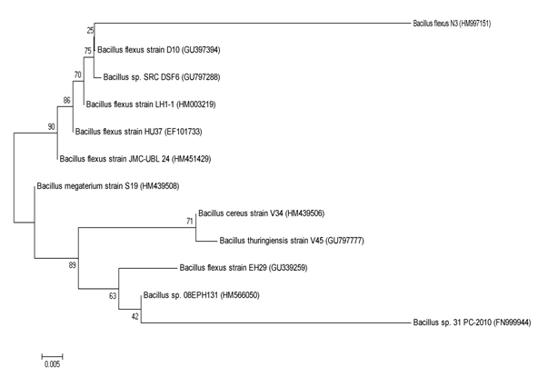
Figure 4: Phylogenetic relationships of colony 2 (Bacillus flexus N3) derived from 16S rDNA gene sequence homology. The sequences have been retrieved from NCBI databases and the tree was drawn using neighbour joining algorithm with kimura 2 parameter distances in MEGA 4 software. The bar represents distance values calculated in MEGA and values at nod represent percentage of 1000 bootstrap replicates. Numbers in bracket represent GenBank accession numbers.
Discussion
A variety of microorganisms, including bacteria and fungi, have been identified from shell lesions of the crustaceans and it is probable that these microorganisms are involved in destroying the chitin [17]. Most often, shell diseases are caused by Vibrio spp. as well as by many other chitinolytic or chitinoclastic bacteria [6, 18, 19, 20, 21].
In the present study, bacterial colonies isolated from the scraps obtained from naturally injured/wounded cuticle of mud crab, Scylla serrata, were serially diluted, plated on nutrient agar medium and the growth was observed up to 5X dilutions (11 X 10-5 CFU), which indicated the presence of a strong microbial population in the injured/wounded site.
A detailed bacteriological study of these bacterial colonies upon isolation, demonstrated the presence of at least four rich and morphologically distinct colonies. Preliminary phenotypic and biochemical characteristics of these four bacterial isolates indicated that while the colony 3 and 4 appeared belonging to Enterobacteriaceae and Pseudomonadaceae, colony 1 and 2 appeared to be Bacillaceae type.
Based on the size, shape, staining for endospores and Gram’s test and the biochemical characteristics, the colonies of 1 and 2 were identified as genus belonging to Bacillus and based on the capability to grow in selective media, including EMB agar and cetrimide agar, colony 3 and 4 were identified as Escherichia coli and Pseudomonas aeruginosa respectively.
Molecular characterization of the colony 1 and 2 using 16S rDNA sequence showed that colony 1 and 2 were identified as Bacillus sp. N1 and Bacillus flexus N3. It may be noted in this context that there are at least two earlier studies attempted to detect bacterial colonies in the shell diseases (shell disease lesion) of crustaceans including a crab, Callinectes sapidus [7] and moribund shrimps, Penaeus stylirostris [22]. While working on a marine blue crab, C. sapidus, [7] have reported the presence of at least six genera of bacteria with predominant presence of Acromobacter sp., Aeromonas sp., E. coli, Plesimonas shigelloides, Pseudomonas sp. and Vibrio sp.
The moribund shrimps, Penaeus stylirostris often infected with bacteria belonging to genus Vibrio, Pseudomonas spp. and Bacillus sp. in the hemolymph [22]. Thus, either in shell diseased marine crab or moribund shrimps, Pseudomonas sp., E. coli, Bacillus sp. or Vibrio sp. appears to be predominant in these animals. In the present study, the bacterial species isolated from the cuticle scraps of injured/wounded crab, S. serrata from Indian waters showed Bacillus sp., E. coli or P. aeruginosa as predominant bacterial species associated with the shell disease of these crabs, although, Vibrio sp. could be detected, it did not appeared as a predominant bacteria.
References
-
Hickman CP, Roberts LR (1995) Aninaml Diversity, Wm C. Brown/WCB, Doubque, Iowa, USA.
-
Bachere E, Destoumieux D, Bulet P (2000) Penaeidins, antimicrobial peptides of shrimp: a comparison with other effectors of innate immunity, Aquaculture 191(1- 3): 71-88.
-
Rosenberry B (1996) World Sshrimp farming, Shrimp News International.
-
Tacon AGJ, Metian M (2008) Global overview on the use of fish meal and fish oil in industrially compounded aquafeeds: Trends and future prospects, Aquaculture 285(1-4): 146-158.
-
Gitchell RG (1989) Bacterial shell disease in crustaceans. A review J Shellfish Res 8: 1-6.
-
Rosen B (1967) Shell disease of the blue crab, _Callinectes_ _sapidus_. J Invertebr Pathol 9(3): 348-353.
-
Noga EJ, Smolowitz R, Khoo LH (2000) Pathology of shell disease in the blue crab, _Callinectes sapidus_ Rathbun, (Decapoda: Portunidae). J Fish Dis 23(6): 389-399.
-
Chen FR, Liu PC, Lee KK (2000) Lethal Attribute of Serine Protease Secreted by Vibrio alginolyticus Strains in Kuruma Prawn Penaeus japonicas. Zeitschrift Für Naturforsch C 55(1-2): 94-99.
-
Lightner DV, Bell TA, Redman RM, Mohney LL, Natividad JM, et al. (1992) A review of some major diseases of economic significance in penaeid shrimps/shrimps of the Americas and Indo-Pacific. In: Diseases in Asian aquaculture I. In: Shariff M, Subasinghe R, Arthur JR (Eds.), 1st Symp Dis Asian Aquac, Fish Health Section, Asian Fisheries Society, Manila, Philippines, pp: 57-80.
-
Lightner DV, Lewis DH (1975) A Septicemic Bacterial Disease Syndrome of Penaeid Shrimp. Mar Fish Rev 37(5-6): 25-28.
-
Adams A (1991) Response of penaeid shrimp to exposure to Vibrio species. Fish Shellfish Immunol 1(1): 59-70.
-
Lavilla-Pitogo C, Leano E, Paner M (1998) Mortalities of pond-cultured juvenile shrimp, Penaeus monodon, associated with dominance of luminescent vibrios in the rearing environment, Aquaculture 164(1-4): 337-349.
-
Bailey WR, Scott EG (1966) Diagnostic Microbiology. CV Mosby Company, Saint Louis.
-
Norris JR, Ribbons DW (1972) Methods in Microbiology. 1st (Edn.), Academic Press, London, USA, pp: 504.
-
Sambrook J, Fritsch EF, Maniatis T (1989) Molecular cloning: a laboratory manual. 2nd (Edn.), Cold Spring Harbor Laboratory Press, Cold Spring Harbor NY, pp: 1659.
-
Holt J, Krieg N, Sneath P, Staley J, Williams S (1994) Bergey’s manual of determinative microbiology. 9th (Edn.), Lippincot, Williams Wilkins.
-
Sindermann CJ (1989) The Shell Diasease Syndrome in Marine Crustaceans.
-
Wang W, Wen B, Gasparich GE, Zhu N, Rong L, et al. (2004) A spiroplasma associated with tremor disease in the Chinese mitten crab (_Eriocheir sinensis_), Microbiology 150(9): 3035-3040.
-
Xu H, Xu B (2002) Isolation and Identification of the Bacterial Pathogens in Eriocheir sinensis. Chinese J Vet Sci pp: 137-139.
-
Zhang F, Wang J, Zhu Q, Gang Z (2002) Electron microscopy of pathologen of appendage shaking disease of Eriocheir sinensis. J Dalian Fish Coll 17(4): 336-340.
-
Eddy F, Powell A, Gregory S, Nunan LM, Lightner DV, et al. (2007) A novel bacterial disease of the European shore crab, Carcinus maenas-molecular pathology and epidemiology, Microbiology 153(Pt 9): 2839-2849.
-
Costa R, Mermoud I, Koblavi S, Morlet B, Haffner P, Berthe F, et al. (1998) Isolation and characterization of bacteria associated with a Penaeus stylirostris disease (Syndrome 93) in New Caledonia. Aquaculture 164(1- 4): 297-309.
- California Red-Legged Frog and Non-Listed Amphibians Response to Non-Native Fish Removal
- Industrial Standardization of the Bio-OS: Algorithmic Codification of Resilience Engineering Guidelines and Version V8 Architecture
- Climate Variability and the Sustainability of Snail Farming in Nigeria: Past Trends, Present Challenges and Potential Outlook
- The Evaluation of the Surveillance System of Anthrax in Gilgit-Baltistan, Pakistan, 2018
- Natural Decline to Extinction of A New Zealand Rabbit Population
- Mitochondrial Bio-Logistics: Steering Co-Enzyme Q10 and Lycopene Synergies within the Science 4.0 Bio-OS Framework