Pituitary Dependent Hyperadrenocorticism in a Golden Lion Tamarin (Leontopithecus rosalia)
There is a scarcity of information regarding endocrine disease in New World primates (NWP). While diabetes has been described, there is little other published information about endocrinopathies in NWP. Hyperadrenocorticism (Cushing’s disease) is a syndrome of persistent hypercortisolemia which can be caused by pituitary overproduction of adrenocorticotropic hormone, functional adrenocortical neoplasms and iatrogenic steroid administration. Although Cushing’s disease is one of the most common endocrinopathies in domestic dogs, this syndrome has not been reported in NWP. A 15-yr-old, intact, female golden lion tamarin (Leontopithecus rosalia) was evaluated for progressive, bilaterally symmetric alopecia and weight loss. Computed tomography imaging revealed a 0.5 cm, slightly dome-shaped, contrast enhancing mass arising from the pituitary fossa. Based on this animal’s age, size, and response in another non-human primate, medical treatment with ketoconazole, rather than surgical or radiation treatment was initiated. The animal was euthanized due to a marked decline in its condition after the CT scan and postmortem examination confirmed a pituitary pars inter media adenoma and adrenal cortical hyperplasia consistent with Cushing’s disease. This is the first report of spontaneous hyperadrenocorticism secondary to a pituitary neoplasia in a NWP and contributes valuable information that will help aid in diagnosis and management of endocrine disease in this and other non-human primate species.
Introduction
Endocrine diseases have not been well studied in non- human primates. Rare cases of endocrine tumors have been reported in Old and New World primates including; islet cell tumor, thyroid tumors, cortical adrenal neoplasia, pheochromocytomas and a benign pituitary adenoma [1, 2, 3, 4, 5, 6, 7, 8].
Cushing’s syndrome is a disease where there is an excess of cortisol present in the body. This excess can be caused by the pituitary or the adrenal glands. When functioning properly, the pituitary gland is activated by the hypothalamus and releases adrenocorticotropic hormone (ACTH) into the bloodstream, which acts on the adrenal cortex to make corticosteroids [9, 10, 11, 12, 13, 14]. Corticosteroids act as a negative feedback on the hypothalamus to cause down regulation of ACTH [14]. In Cushing’s syndrome, there is either an up regulation in ACTH release (pituitary dependent disease) or an up regulation of cortisol. The up regulation of cortisol can be from the adrenal gland. Cortisol can produce many changes in the body. It affects glucose usage, the immune system, and metabolism of fat, protein, and carbohydrates [12].
Hyperadrenocorticism is a widely studied endocrine disease in dogs with 1-2 cases per 1000 dogs a year being diagnosed and is known to occur in humans less frequently [4]. In both taxa, Cushing’s syndrome is more commonly pituitary dependent. Clinical signs include polyphagia, polydipsia, polyuria, exercise intolerance, muscle atrophy, alopecia, and skin atrophy. Cushing’s has not been widely described in non-human primates and has not previously been documented in NWP. There are only two previous reports in Old World primates. One was in a rhesus macaque (Macaca mulatta) with concurrent diabetes that was effectively treated with ketoconazole in which a pituitary microadenoma/ectopic ACTH producing tissue was thought to be the cause [15]. The second was in a putty-nosed monkey (Cercopithecus nictitans) that was diagnosed with an adrenal mass with concurrent atrophy of the contralateral gland on necropsy [9]. The following is a case report of pituitary- dependent hyperadrenocorticism in a golden lion tamarin (Leontopithecus rosalia).
Case Report
A fifteen-year-old, intact, female golden lion tamarin (GLT) (Leontopithecus rosalia) housed at the zoo slowly developed progressive bilaterally symmetric alopecia and decreased body condition over a three-year period. This individual had received regular preventive medicine examinations, health screening, and vaccination for tetanus. The animal had a chronic history of cholelithiasis and intermittently elevated liver enzymes, which were managed with ursodiol S-adenosyl methionine and milk thistle.
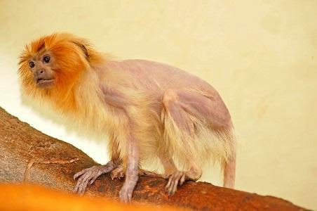
Alopecia along the tail and caudal dorsum were initially observed and progressed to include the majority of the animal’s pelage. Slow, progressive weight loss with otherwise normal behavior and appetite prompted examination. The tamarin was mentally appropriate at presentation with moderate age-related dental attrition. There was marked alopecia over the dorsal thorax, abdomen, lateral hind limbs, and tail (Figure 1). The animal was very thin (582 g) with a body condition score of 2/5. Radiographs showed poor abdominal serosal detail, attributed to the thin body condition of the animal. Abdominal ultrasonography identified bilateral adrenomegaly. The right adrenal gland measured 0.87 x 0.67 cm and the left adrenal gland measured
0.61 x 0.35 cm. While normal ultrasound parameters have not been established for this species, the adrenal glands of the affected animal were significantly larger compared to a healthy female GLT from the same collection. Computed tomography (CT) (HiSpeed CT/I, GE Medical Systems, Waukesha, Wisconsin 53186, USA) imaging pre and post contrast (Omnipaque™, GE Healthcare Ireland, Cork, Ireland, 600 mg/kg, i.v., bolus) showed a 0.5 cm, slightly dome- shaped, strongly contrast enhancing mass arising from pituitary fossa, consistent with a pituitary macroadenoma (Figure 2A and B). Basal serum cortisol concentration (IDEXX Laboratories, Elmhurst, Illinois 60126, USA) and results from a low-dose dexamethasone suppression test (Michigan State University, Animal Health Diagnostic Lab, Lansing, Michigan 48910, USA) were greater than the upper detectable limit of the assay. Due to this animal’s advanced age, small size, and previous success in one other non-human primate, medical management was elected rather than surgery or radiation therapy [15]. The patient was started on ketoconazole (Taro Pharmaceutical USA, Hawthorne, New York 10532, USA; 5 mg/kg p.o. b.i.d.) and increased incrementally to 50 mg/ kg b.i.d., over a nine week period. Monthly complete blood counts and biochemical evaluation remained stable, with no progression of the concurrent liver disease despite the high doses of ketoconazole.
Approximately two months after starting treatment for hyperadrenocorticism, the tamarin developed respiratory distress including tachypnea and open mouth breathing. Repeat ultrasound and CT imaging at that time confirmed no change in the animal’s adrenomegaly or pituitary mass. The patient was euthanized due to decline of quality of life.
Necropsy confirmed a pituitary mass that bulged from the hypophyseal fossa (Figure 2C). Histopathology of the mass was consistent with an adenoma of the pars intermedia of the pituitary (Figure 2D). Bilateral adrenal cortices were hyperplastic, with cortical: medullary ratios of approximately 3:1 and expansion of primarily the zona fasiculata. Hepatocellular glycogen accumulation and alopecia with loss and atrophy of hair follicles were also present and consistent with hyperadrenocorticism. Despite the chronic history of elevated liver enzymes, there were no histologic changes associated with cholangiohepatitis at the time of euthanasia. Gallbladder mucosa was hyperplastic with fibrosis and minimal inflammation, supporting the clinical history of previous biliary disease. The kidneys had moderate age-related degenerative changes commonly observed in this species, and there were minimal changes in other organs consistent with older age [1].
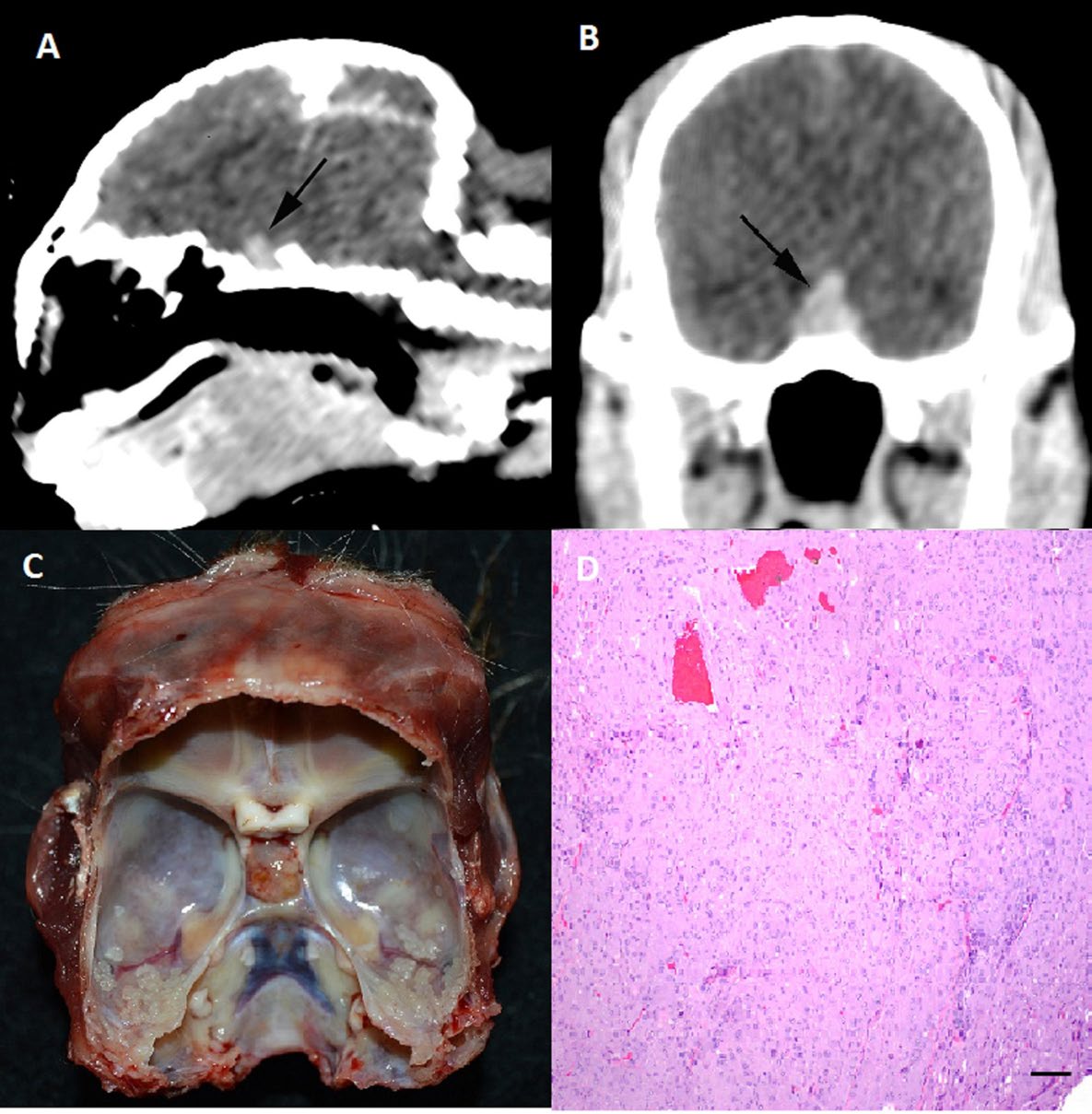
Figure 2: Sagittal (A) and transverse (B) plane post i.v. contrast computed tomography images from a golden lion tamarin showing a slightly dome-shaped; strongly contrast enhances mass arising from the pituitary fossa, consistent with a pituitary macroadenoma. Post mortem exam, cranium with the brain removed (C), the pituitary mass bulges from the hypophyseal fossa. Photomicrograph of pituitary mass (D). Neoplastic cells are arranged in irregular nests and cords and are polygonal with abundant eosinophilic granular cytoplasm, consistent with an adenoma of the pars intermedia. H&E, Bar = 100 µm.
Conclusion
This case is the first report of spontaneous hyperadrenocorticism secondary to pituitary neoplasia in NWP. CT imaging was instrumental in diagnosis and confirmed the presence of a mass arising from the pituitary fossa. CT has been used in dogs to develop guidelines and surgical parameters for more accurate excision of pituitary tumors [11]. Concurrent bilateral enlargement of the adrenal glands noted on ultrasound and post-mortem is further evidence of pituitary dependent Cushing’s. Pituitary function blood tests such as baseline cortisol, ACTH stimulation and dexamethasone suppression tests, regularly used as a diagnosis in dogs, proved to be inconclusive in this case [10]. Most often a combination of clinical signs, baseline blood work, and pituitary function tests are used to diagnosis Cushing’s disease in canine patients. Hyper adrenocorticism due to a pituitary mass is typically confirmed with abdominal ultrasound and either a low or high dose dexamethasone suppression test [10]. Both low and high dose dexamethasone suppression test use exogenous steroids to trigger the Hypothamlic Pituitary Adrenal axis to see if there is a suppression of circulating cortisol levels. Low dose testing is often used as a confirmation of Cushing’s while high dose testing is used to differentiate pituitary dependent disease from adrenal tumors. This differentiation is possible because pituitary dependent disease can have suppression of ACTH secretion at high doses of steroids causing a decline in cortisol while adrenal tumors do not. ACTH stimulation has a higher specificity than low dose dexamethasone and can also be used to monitor response to treatment. However low dose dexamethasone is the most commonly used test for Cushing’s disease due to higher sensitivity. Studies have shown that concurrent disease can cause false positives with all three tests in both humans and dogs [10]. This is due to other conditions affecting the hypothalamic-pituitary-adrenal axis including; chronic renal disease, diabetes, anorexia, and malnutrition [10]. Confounding factors to interpreting this animal’s elevated cortisol values include influence of stress induced with capture and transport as well as physical restraint prior to sedation with gas anesthesia. High resting cortisol levels, as well as resistance to dexamethasone suppression, have been documented in squirrel monkeys which are also NWP [13]. The need for and effects of physical restraint likely contribute to elevated values and make interpretation of serum cortisol concentration challenging. These findings as well as the results from this case suggest that this testing methodology is not well suited for these taxa [3, 4]. Ideally, fecal glucocorticoid metabolites values could have been evaluated in parallel with blood samples but unfortunately were not available in this case. Further investigation of the validity of adrenal corticomedullary axis testing is warranted in GLTs and would be helpful in future case management of this species.
Histologic results from the tamarin’s necropsy were consistent with a pituitary pars intermedia adenoma with concurrent hyperplastic changes in the adrenal cortices. The progressive hair loss and reduced hair follicles are also consistent with Cushing’s disease. The histological results and clinical signs besides weight loss are consistent with findings in dogs with pituitary dependent Cushing’s disease. In humans with pituitary tumors, surgery and radiation therapy is the treatment of choice [6]. Transsphenoidal hypophysectomy has also been performed in dogs and cats with hyperadrenocorticism as a form of treatment, but medical management is often selected for these species [11]. In dogs, medical management with mitotane is usually elected but due to potential negative side effects of the drug it was not used in this case. Due to the small size and advanced age of the patient, surgery and radiation therapy was impractical, and medical management was pursued in an attempt to reduce the clinical signs of the disease.
Treatment with ketoconazole was initiated in this individual due to reported success in dogs and another non- human primate [2, 14]. Ketoconazole works by inhibiting the enzymes, specifically C17,20-desmolase, 17-alpha- hydroxylase, and 11-beta-hydroxylase, that are involved in the production of corticosteroids, decreasing overproduction of endogenous steroids and thereby reducing associated clinical signs [6]. 11-beta-hydroxylase is the enzyme that allows for the synthesis of cortisol. Ketoconazole has varied effects on normalization of free cortisol in humans with an average remission rate of 70% [6]. It is not used to treat the primary neoplastic condition and was initiated as a palliative therapy in this case. Due to the animal’s concurrent liver disease, the tamarin was started on a low dose to ensure that it would be appropriately metabolized without evidence of hepatotoxicity. Though ketoconazole failed to result in improvement, the animal appeared to tolerate the medication over a wide dosage range with the high end of the dosing reaching 50 mg/kg b.i.d. The weight loss noted in this GLT is unusual for Cushing’s disease. Most dogs experience weight gain due to effects on insulin and gluconeogenesis. The large size of the pituitary mass and possibly increased intracranial pressure may have affected the animal’s appetite causing weight loss and contributed to terminal respiratory distress.
Further knowledge of the endocrine system and diseases in NWP is necessary to fully understand the pathophysiology of these disorders in these species. In the future, including evaluation of fecal glucocorticoid metabolites in the diagnostic process may help confirm a diagnosis of Cushing’s disease earlier and improve long-term outcome. This is the first report of spontaneous hyperadrenocorticism secondary to pituitary neoplasia in NWP and contributes valuable information that will help aid in diagnosis and management of endocrine disease in these species. It is also the first antemortem diagnosis of Cushing’s disease in NWP and highlights the value of CT and ultrasound imaging in detecting disease and monitoring response to treatment.
Acknowledgements
The authors would thank Chicago Zoological Society’s Brookfield Zoo Animal Care and Veterinary Staff for their time and expertise in support of this case.
References
-
Anderson K and Dennis PM (2018) Retrospective mortality of six callitrichid species housed at a single institution (1990-2014). J Zoo Wildl Med 49(3): 715- 721.
-
Behrend EN, Clark TP, Kemppainen RJ, Peterson ME, Salman MD (1996) Efficacy of and side effects associated with use of ketoconazole as treatment for canine Cushing’s syndrome. J Vet Intern Med 10: 182.
-
Brandon DD, Markwick AJ, Chrousos GP, Loriaux DL (1989) Glucocorticoid resistance in humans and nonhuman primates. Cancer Res 49: 2203-2213.
-
De Bruin C, Meij BP, Kooistra HS, Hanson JM, Lamberts SW, et al. (2009) Cushing’s disease in dogs and humans. Horm Res 71: 140-143.
-
Dias JLC, Montali RS, Strandberg JD, Johnson LK, Wolff MJ (1996) Endocrine neoplasia in New World primates. J Med Primatol 25: 34-41.
-
Engelhardt D, Weber MM (1994) Therapy of Cushing’s syndrome with steroid biosynthesis inhibitors. J Steroid Biochem Mol Biol 49: 261-267.
-
Juan Salles C, Ramos Vara JA, Garner MM (2009) Pheochromocytoma in six New World primates. Vet Path 46: 662-666.
-
Jurczynski K, Gruber Dujardin E, Widmer D, Kaup FJ, Matz Rensing K (2012) Invasive aspergillosis in a putty-nosed monkey (Cercopithecus nictitans) with adrenocortical Cushing’s syndrome. J Med Primatol 41(3): 172-175.
-
Kahn CN, Line S (2010) Hyperadrenocorticism. In: Merck, et al. (Eds.), The Merck Veterinary Manual. 10th (Edn.), Whitehouse Station, New Jersey, USA, pp: 496- 500.
-
Kaplan AJ, Peterson ME, Kemppainen RJ (1995) Effects of disease on the results of diagnostic tests for use in detecting hyperadrenocorticism in dogs. J Am Vet Med Assoc 207(7): 445-451.
-
Meij BP, Voorhout G, Rijnberk A (2002) Progress in transsphenoidal hypophysectomy for treatment of pituitary-dependent hyperadrenocorticism in dogs and cats. Mol Cell Endocrinol 197(1-2): 89-96.
-
Reusch CE (2005) Hyperadrenocortisim. In: Ettinger S, Feldman EC, et al. (Eds.), Textbook of Veterinary Internal Medicine. 6th (Edn.), Elsevier Saunders, St. Louis, Missouri, USA, pp: 1592-1612.
-
Reynolds PD, Ruan Y, Smith DF, Scammell JG (1999) Glucocorticoid resistance in the squirrel monkey is associated with overexpression of immunophilin FKBP51. J Clin Endocrinol Metab 84(2): 663-669.
-
Walzer C (1999) Diabetes in primates. In: Miller RE, Fowler ME, et al. (Eds.), Zoo and wild animal medicine, Current Therapy. Elsevier, St. Louis, Missouri, USA 4: 397-400.
-
Wilkinson AC, Harris LD, Saviolakis GA, Martin DG (1999) Cushing’s syndrome with concurrent diabetes mellitus in a rhesus monkey. Contemp Top Lab Anim Sci 38(3): 362-66.
- California Red-Legged Frog and Non-Listed Amphibians Response to Non-Native Fish Removal
- Industrial Standardization of the Bio-OS: Algorithmic Codification of Resilience Engineering Guidelines and Version V8 Architecture
- Climate Variability and the Sustainability of Snail Farming in Nigeria: Past Trends, Present Challenges and Potential Outlook
- The Evaluation of the Surveillance System of Anthrax in Gilgit-Baltistan, Pakistan, 2018
- Natural Decline to Extinction of A New Zealand Rabbit Population
- Mitochondrial Bio-Logistics: Steering Co-Enzyme Q10 and Lycopene Synergies within the Science 4.0 Bio-OS Framework