Renal Cyst and Ureteroliths in the Guinea Pig (Cavia porcellus)- Case Report
The urolithiasis is characterized by the presence of calculi in some fraction of the urinary tract which frequently affects guinea pigs (Cavia porcellus). Despite the physiopathogenesis of this disease is not completely understood, it is known that a diet rich in calcium can be a predisposing factor. Urolithiasis surgical removal associated with proper food and water management are recommended to animal treatment, however, the surgical technique is complex with some complications due to patient size. The aim of the study was to report the clinical case of urolith in a 5-year-old male guinea pig, uncastrated, weighing 1.142 kg. The animal presented urolith in the left ureter with dilation of the renal pelvis and right renal cyst. Due to animal physiological status without important clinical signs and the risk of compromising the contralateral kidney by a cyst, it was decided to monitor the patient and did not perform the surgical treatment. The patient was clinically monitored and underwent a clinical dietary treatment with complementary imaging tests (ultrasonography and radiography) and laboratory tests of renal function, showing no alterations for 10 months. Clinical monitoring of the animal can be a viable option in face of surgical intervention, especially for patients who have contraindications and without important clinical signs.
Introduction
Guinea pigs are animals belonging to the rodentia order commonly raised as pets, with a life expectancy of 5 to 8 years. They are herbivores of elodont-type teeth, whose diet is based on different fibre sources such as leaves, hay, and vegetables, in addition to supplementation feeding [1].
Rodents maximize the absorption of calcium intake and excrete it in the form of calcium carbonate, therefore, the control of rich sources of this mineral in the diet is necessary, since it can cause calciuria and predispose the formation of uroliths [2] Urolithiasis is commonly diagnosed in guinea pigs males aged over three years and in females over four years old [3]. Reduced weight, obesity, kidney disease, and diet with high mineral content can be aggravating factors for the development of this disease [4].
Clinical signs presented by animals depend on the size and location of the urolith, and include weight loss, anorexia, dysuria, hematuria, and abdominal pain, characterized by a stooped posture and/or grinding of teeth and vocalization during urination. However, in some cases, the tutor may not observe or report such clinical signs [3]. The radiography is diagnosis most used since the uroliths often have calcium in their composition and are radiopaque [5]. Ultrasonography is also a widely used diagnostic method to locate and measure urolith in different parts of the urogenital tract [6, 7].
The prognosis of renal disorders in male guinea pig is unfavorable compared to females, due to the anatomy of the genitourinary system do not favoring the passage of uroliths [5] besides males being more affected by ureteral and bladder calculi and females in bladder and urethra [3]. Surgical removal of urolith is the treatment of choice [3, 5], but with high rates of complications due to the size of the ureter [8].
Materials and Methods
A five years old specimen of Cavia porcellus, and weighed 1.142 kg, previously submitted to two surgical procedures of nodulectomy for excision of mammary carcinoma and fibrosarcoma, was treated in the wild animal’s sector of the Veterinary Hospital of the Universidade Federal de Uberlândia.
As these are malignant neoplasms, periodic monitoring of possible metastases is indicated. Thus, chest and abdomen radiography, abdominal ultrasonography, hematological and biochemical tests were performed.
Results
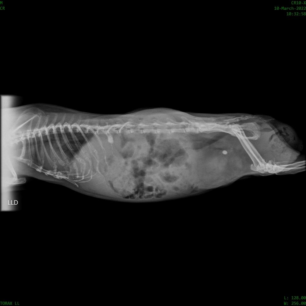
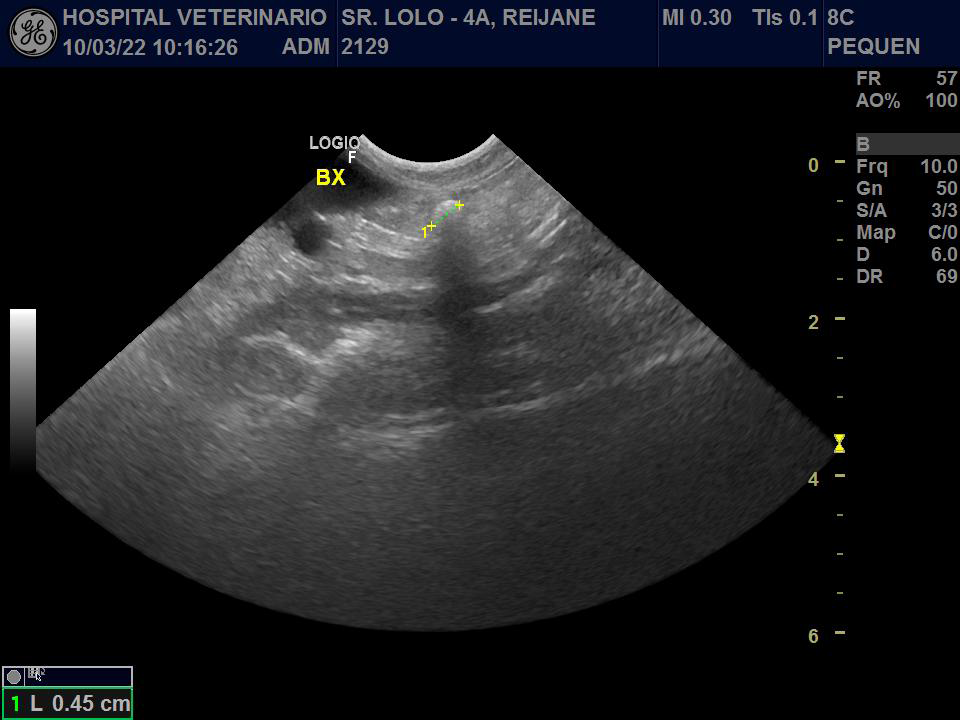
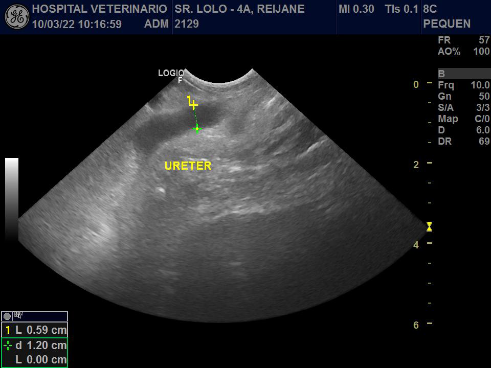
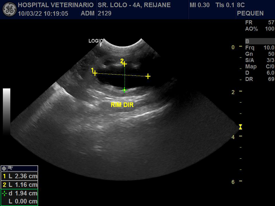
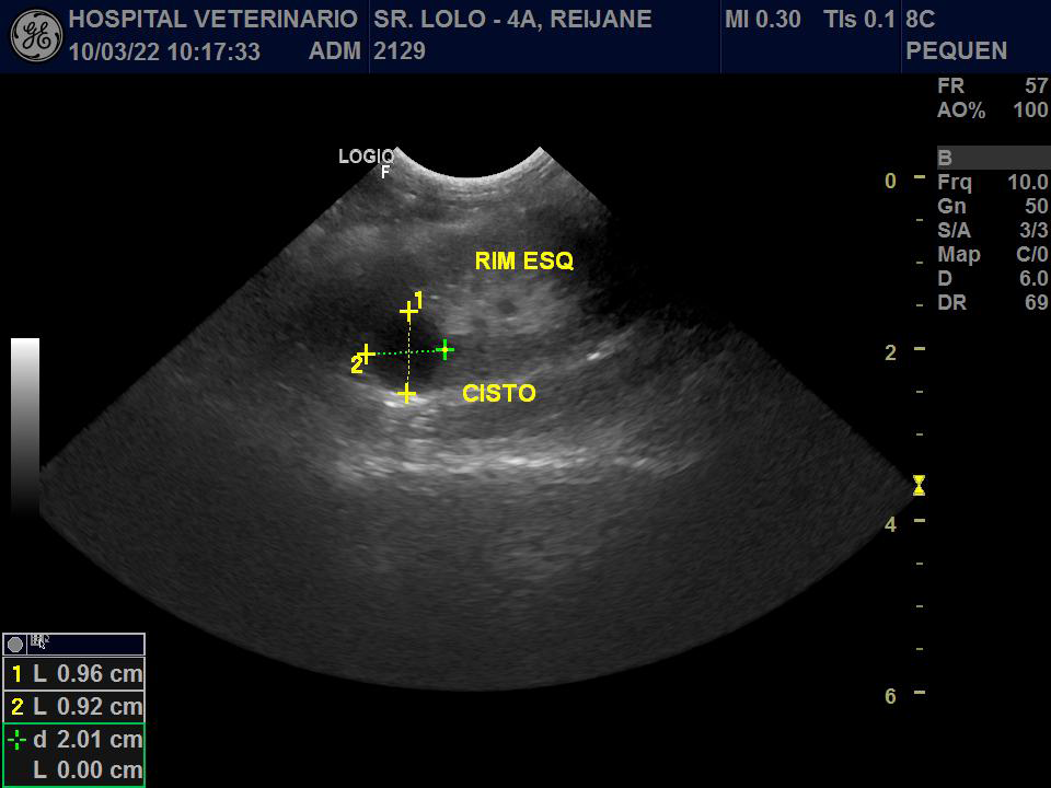
The radiographic examination showed an elongated structure with mineral radiopacity, located in the topography of the ureter, measuring approximately 0.48 x 0.38 cm (Figure 1). The ultrassonography showed a dilated right ureter with a calculus inside it (Figures 2 & 3), a dilated right renal pelvis (Figure 4) and a left kidney with the presence of a cyst (Figure 5). The creatinine and urea values determined were within the expected parameters for the species. Based on the clinical findings, the implemented therapeutic approach was to correct the nutritional and water management, reducing the amount of calcium ingested by the animal, mainly the consumption of leaves with a high calcium content, such as kale and arugula, and increasing the water intake.
Discussion
The urolithiasis was diagnosed incidentally through radiography and ultrasound, the guinea pig did not show clinical signs.
The diet offered to the patient consisted of hay, feed and dark green leaves, especially cabbage, which has a high calcium content. It was also reported that the animal consumed little water. Some minerals, such as calcium, may undergo precipitation and predispose to the formation of crystals, which aggregated with other crystals form urinary calculus [9]. According to Mancinelli diets rich in calcium and reduced water intake can lead to the formation of uroliths such as calcium phosphate, calcium oxalate and calcium carbonate, poorly soluble crystals in the urine.
The identified urolith was in the right ureter, which is consistent with the finding of dilatation of the right renal pelvis, in view of the mechanical obstruction caused by the calculus and with the predisposing location in males [3]. The uroliths may be found in the ureter, urethra, urinary bladder, and renal pelvis, causing an obstruction of the flow of urine and may lead to more severe complications in the urinary system, such as hydronephrosis, urethral dilatation, renal pelvis dilatation, renal failure and even death [10]. It is important to emphasize that the patient was constantly monitored and there was no worsening of hydronephrosis, which demonstrated that monitoring in this species can be a viable option compared to surgery.
The diagnosis of the cyst in the left kidney was an incidental finding during the ultrasonography. Although the animal did not show clinical repercussions or laboratory alterations, the surgery of the contralateral ureter was considered a complicating factor.
The treatment of the reported clinical case consisted of correcting the nutritional and water management, instructing the tutor to reduce the offer of dark green leaves that have a high calcium content, in addition to increasing the number of water pots distributed around the enclosure to encourage a higher water intake, which may have contributed to the stabilization of the situation.
The owner chose to monitor the patient due to the animal’s age, the history of malignant neoplasm in remission, obesity and the remaining kidney was compromised due to the presence of a cyst.
The patient’s radiographic and ultrasonographic and quarterly renal function evaluations were requested for clinical follow-up. Ten months after the diagnosis of the presence of uroliths, with 3 reevaluations, the patient did not present renal alterations nor the appearance of clinical signs of renal disorders.
The patient had a survival of more than 300 days so far. Ten months after the diagnosis of the presence of uroliths, with 3 reevaluations, the patient did not present renal alterations nor the appearance of clinical signs of renal disorders.
According to Ettel, et al. in a retrospective study with 158 guinea pigs, the authors estimated an average of 59 days of survival after the diagnosis in male guinea pigs. Although the surgical procedure was not statistically considered a risk factor, the retrospective nature of the study may have underestimated these data, culminating in the need to carry out new prospective, randomized and multicenter studies to investigate the need for surgery and prognosis.
Conclusion
Dietary correction, encouraging water consumption, environmental enrichment and weight control may be important actions for stabilizing ureterolithiasis in guinea pig. Although surgical treatment for calculus removal is described in the literature, we suggest that its immediate execution in asymptomatic animals be carried out very sparingly, especially in those with surgical contraindications, since the quality of life and the time of survival was satisfactory in this case.
References
-
Pignon C, Mayer J (2020) Guinea pigs. In: Quesenberry KE, Orcutt CJ, et al. (Eds.), Rabbits and Rodents, Clinical Medicine and Surgery, 4º edição, Missouri, Elsevier, pp: 270-297.
-
Rooney TA, Eshar D, Wong AD, Gardhouse S, Beaufrère H (2021) The association between bloodwork, signalment, and urolithiasis in guinea pigs (Cavia porcellus). J Exot Pet Med pp: 38: 26-31.
-
Edell AS, Vella DG, Sheen JC (2002) Retrospective analysis of risk factors, clinical features, and prognostic indicators for urolithiasis in guinea pigs: 158 cases (2009–2019). Journal of the American Veterinary Medical Association 260(S2): 95-100.
-
Hawkins MG (2011) Urinary tract obstruction. In: Oglesbee BL (Ed.), Blackwell’s Five-Minute Veterinary Consult: Small Mammals. 2nd (Edn.) Ames, Iowa: Blackwell Publishing Professional pp: 336-338.
-
Hawkins MG, Ruby AL, Drazenovich TL (2009) Composition and characteristics of urinary calculi from guinea pigs. J Am Vet Med Assoc 234(2): 214-220.
-
DeCubellis J (2016) Common emergencies in rabbits, guinea pigs, and chinchillas. Vet Clin North Am Exot Anim Pract 19(2) 411-429.
-
Mancinelli, E. 2016. Urolithiasis in guinea pigs. Vet Times: The website for the veterinary profession.
-
Holowaychuk MK. 2006. Renal failure in a guinea pig (Cavia porcel- lus) following ingestion of oxalate containing plants. Can Vet J.47:787–789.
-
Silveira MET, et al. 2021. “Urolitíase em porquinho-da- índia (Cavia Porcellus): relato de caso Urolithiasis in guinea pig (Cavia Porcellus): case report.” Brazilian Journal of Development 7(10):100198-100212.
-
Osborne, Carl A. et al. 2009. Quantitative Analysis of 4468 Uroliths Retrieved from Farm Animals, Exotic Species, and Wildlife Submitted to the Minnesota Urolith Center: 1981 to 2007. Veterinary Clinics of North America - Small Animal Practice, v. 39, n. 1, p. 65–78.
- California Red-Legged Frog and Non-Listed Amphibians Response to Non-Native Fish Removal
- Industrial Standardization of the Bio-OS: Algorithmic Codification of Resilience Engineering Guidelines and Version V8 Architecture
- Climate Variability and the Sustainability of Snail Farming in Nigeria: Past Trends, Present Challenges and Potential Outlook
- The Evaluation of the Surveillance System of Anthrax in Gilgit-Baltistan, Pakistan, 2018
- Natural Decline to Extinction of A New Zealand Rabbit Population
- Mitochondrial Bio-Logistics: Steering Co-Enzyme Q10 and Lycopene Synergies within the Science 4.0 Bio-OS Framework