Histopathological Investigation of the Giant Prawn Macrobrachium rosenbergii in the Extensive Culture Ponds of Nellore Region, Andhra Pradesh, India
The present study was carried out in Muthukur area of Nellore region of Andhra Pradesh located at 14.29 19 latitude and 80.10383 longitude), India since this area is actively engaged with aquaculture activities of fish and prawn farming. A recent data indicated that 280 to 984 kg per hectare per crop achieved in Macrobrachium rosenbergii under extensive system in a culture period of 180 to 210 with stocking density of 3 to 5 juveniles per square meter. Since the scampi Macrobrachium rosenbergii was the candidate as well as prominent species in that region, histopathological study was carried out to the see quality of the prawn for the present study. During the investigation of shrimp disease in the culture pond, the prawn Macrobrachium rosenbergii were found to die due to swollen head disease having enlargement of cephalothoracic region containing gill, heart and hepatopancreas, etc. Another kind of symptom of black spots were noticed on the dorsal portion of anterior exoskeleton in the cephalothorax region. Enlargement of pigmentary with black spots formed in the periphery of dorsal side of exoskeleton might be due to the presence of suspected white spot syndrome virus. This kind of disease was found to attack commonly in Macrobrachium rosenbergii in most of the culture ponds. Apart from the study of shrimp disease in culture ponds, attempts were also made to identify the disease in the post larvae of Macrobrachium rosenbergii in the hatchery in and around Nellore region. Mass mortality was found to occur in the post larval stages due to a kind of white muscle disease. It has led to great loss of economy not only to the entrepreneurs involved in the hatchery at Nellore region but also to the country as well. WSSV was characterised by prominent eosinophilic to pale basophilic intra nuclear inclusion bodies in hypertrophied nuclei of most commonly in the epithelial cells and connective tissue cells of the above target tissues. Result of the study showed that multiple infection is quite common in the prawn to be cultured once the water quality has become deteriorated and It is suggested that although it is a extensive culture system, water management and supplement of feeds are prerequisite to maintain the pond.
Introduction
The global aquaculture of penaeid shrimp has grown rapidly during the past two to three decades. In 1998, world shrimp farmers produced an estimated 737 200t of whole shrimp (this includes 530 200 t from the eastern hemisphere and 207 000 t from the western hemisphere a record 12% increase from 660 200 t produced in 1997 [1, 2]. The shrimp sector suffered a serious setback during 1995-96. Lack of technically qualified manpower, improper site selection, defective farm design, rapid intensification, overcrowding of farms in restricted locations and disproportionate development of the industry relative to supply of quality farm inputs paved the way for poor environmental conditions in ponds. The ultimate effects of the above-mentioned problems was serious harm to the industry. However, the recent disease caused by a viral pathogen (white spot viral disease) during 1993-1994 delivered a lethal blow to the global shrimp farming industry, which could not recover immediately. In other words, this specific viral pathogen still plays a key role in controlling the development of the global shrimp farming industry.
Viral diseases are a major problem for shrimp aquaculture all over the world. Although several viral diseases have been reported in Asia wide spread viral diseases have been reported mainly due to Monodon baculovirus (MBV) and white spot syndrome virus (WSSV). WSSV has been shown to affect a wide range of wild crustaceans including crabs, lobsters and shrimps, both penaeid and non-penaeid [3, 4, 5, 6, 7] Diseases of mixed etiologies are more common place in penaeid shrimp than generally documented [8]. Likewise, the presence of multiple disease-causing agents (i.e., infectious pathogens) or factors (i.e., toxicants, nutritional imbalances, environmental extremes, etc). Sometimes result in the misdiagnosis of a particular disease syndrome. Viral infections are typically accompanied by secondary bacterial and epi commensal infestations, which may actually be the ultimate cause of death in a shrimp already severely compromised by the initial viral infection [9, 10].
Although a large portion of the world’s farmed shrimp is produced in Asia, shrimp culture operations do not succeed over the entire area. There have been several problems and impediments that are yet to be resolved. One of these problems is disease. Several shrimp diseases have threatened shrimp production, but the most devastating ones are the viral diseases, namely, yellow-head virus (YHV) and White Spot Syndrome Virus (WSSV). At present, there is no curative procedure for the two viral diseases. As such, preventive measures have been studied and introduced, but the most acceptable one is the improvement of grow out systems. Asian Shrimp culture employs three shrimp culture techniques: the extensive, semi-intensive, and intensive systems. Beginning with the extensive about 3 decades ago, parts of this system were modified into a semi- intensive system in 1980, and later the intensive type was introduced. The extensive type evolved from the hunting and gathering of food by the nearshore communities. Mangrove environments were modified into trapping ponds for livestock during the stormy months. This system uses low technology and requires vast and low-lying areas, such as mangrove forested areas. The intensive system uses higher technology, requires a smaller area and higher elevations. Thus, it does not require mangrove areas, but it is suitable for areas behind the mangroves. The production is quite high when compared with the two other systems. Because of the high stocking density and intensive feeding, however, this type of shrimp culture is faced with several problems, such as coastal pollution caused by the farm effluent and disease problems. The intensive system was later modified to a more bio secure system, that is, a closed recirculating water system, a reduced or zero water exchange system and shrimp culture at inland locations away from coastal influences. These innovations have made shrimp culture more efficient in controlling diseases more sustainable and more environmentally friendly.
Tomoya Kono, et al. [11] studied on the detection of white spot syndrome virus in shrimp by loop mediated isothermal implication and gave a standardized lamp procedure to defect the presence of WSSV in the heart, stomach and lymphoid organ from infected shrimp. Severity of MBV infection means that infection by one virus predisposes shrimp to infection by the other. Macrbrachium elegans showed multiple occlusion bodies in the hepatopancreatic cells. Pond-reared penaeid shrimp typically serve as hosts for a multitude of parasitic and epi commensal organisms [12, 13, 14, 15, 16, 17, 18, 19, 20, 21, 22, 23] and their presence may not necessarily equate to disease. Classical microbiological methods were applied early in the development of shrimp pathology to help determine the identity of the agent of vibriosis, shell disease and certain fungal diseases [24, 25, 26, 27, 28].
Gross and clinical signs, with the most commonly applied laboratory test being direct examination and microscopy using the light microscope, classical microbiology with isolation and culture of the agent, and routine histology and histochemistry. ‘Classic’ diagnostic techniques that are important, but are used less frequently, include techniques such as bioassay and enhancement, which are used for the detection of subclinical or carrier-state infections by certain pathogens [29, 30, 31, 32]. Horizontal transmission through water and feeding of infected shrimps has been suggested by Mohan, et al. [33] as the probable route for the spread of white spot disease virus. Lo, et al. [34] and Mohan, et al. [33] proved that vertical transmission of this viral agent is possible from brooders to offspring. DNA hybridization probes for the white spot disease virus have been developed by several laboratories [35, 36]. The primers for detection of this virus by PCR technology have also been developed [37].
Sahul Hameed, et al. [38] reported the pathogenicity of systemic ectodermal and mesodermal baculovirus and its detection in shrimp by immunological methods. The result of their study showed the presence of SEMBV in all the organs and tissues except in hepatopancreas. Elpidio Cesar, et al. [39] reported detection of yellow head virus and Chinese baculovirus in Penaeid shrimp by the western blot technique and suggested a protocol which was highly specific, rapid and sensitive enough to detect the presence of the viruses before appearance of over symptoms.
Tomoya Kono, et al. [11] studied on the detection on white spot syndrome virus in shrimp by loop mediated isothermal implication and gave a standardized lamp procedure to detect the presence of WSSV in the heart, stomach and lymphoid organs from infected shrimp. Piamsak Menasveta [40] studied on Improved shrimp grow out systems for disease prevention and environmental sustainability in Asia and suggested that the most practical preventive measures is the improvement of grow out systems and showed the intensive system modified to a more bio secure system that is a closed recirculating water system, a reduced or zero water exchange system and shrimp culture at inland locations away from coastal influences.
To know the reason for causing the disease, the present work has been attempted on histopathological investigation of prawn in Nellore region in relation with pond management.
Materials and Methods
For the present study, histopathological study was carried out in Muthukur area of Nellore region, Andhrapradesh which is located in 14.2919 latitude and 80.10383 longitude. Although aquaculture practice is intensively progressing in this area, disease outbreak has devastated the production of prawn Macrobrachium rosenbergii predominantly with the virus of White Spot Syndrome Virus (WSSV), Monodon bacculovirus, (MBV) and Hepatopancreatic Virus (HPV) etc. The details for pond management containing environmental parameters of water, water exchange, feed application, stocking density of the candidate species, mode of water supply etc. were collected and interpreted. To know the reason for causing of this disease in the candidate species of prawn, the present work has been attempted on histopathological investigation of prawn in Nellore region in relation with pond management. During the study period, weekly samples of the giant prawn Macrobrachium rosenbergii was collected from the extensive culture ponds for histopathological investigation, for which the tissues like hepatopancreas, gills, eyestalk, soft tissue and gut were dissected out in live condition and immediately fixed in Davidson’s fixative for 24 hours. Tissues fixed were transferred to 70% ethanol after two days for further study. The target tissues such as hepatopancreas, gills, eyestalk, soft tissue fixed were subjected for histopathological work as given below.
Processing and Sectioning
The tissues of hepatopancreas, gills and eyestalk fixed were washed overnight in running tap water to remove the excess of picric acid. The fixed tissues were dehydrated using and alcohol series (30%-100%) and cleared in xylol. The tissues were further cold impregnated overnight with wax using xylene and wax shavings in 1:1 ratio. Subsequently the sample was evaporated by placing the tissues in an oven at 58 C. The tissues were then transferred through two changes of fresh molten wax (Paraffin wax M.P 58-60 C). The tissues embedded blocks were prepared by using proper orientation of wood or metal. Serial sections of the block were cut at approximately 6-8 m thickness using a rotary microtome. Sections were fixed on clear glass slides using fresh Mayer’s egg albumin and flattered by placing on a slide warmer with a drop of distilled water. Subsequently the water was drained off and the slides were allowed to dry. These slides were then used for histological staining.
Staining
Routine staining for gross morphological observations was carried out using Harris Hematoxylin stain with 1% alcoholic eosin as the counter stain. Sections to be stained were first deparaffinized in two changes of xylene and then hydrate through a descending series of propanol grades. Section was blued using tap water or ammonia solution. Eosin stained sections were repeatedly washed in an ascending series of propanol grades to remove excess eosin and cleared in xylene. Sections were mounted with DPX mountant and examined under microscope.
Results
Bore water containing 10-15 ppt saline was used in extensive culture pond for Macrobrachium rosenbergii. Water was exchanged 10-25% irrespective of growth phase of prawn, the pH was ranged between 7.8-8.5 and turbidity became clear with slightly green in colour. Cp brand fed was routinely given in the culture pond starting from larvae to adult prawn in most of the farms studied. When the water become acidity in condition, the lime was added with quantity of 25kg/acre once in a week. 12,000-15,000 seeds/ acre were stocked in extensive pond. There was no scientific method of aeration provided in the culture ponds. Prawns were harvested at 30 counts /kg after the periods lasted from 110 to 120 days (Figures 1-11).
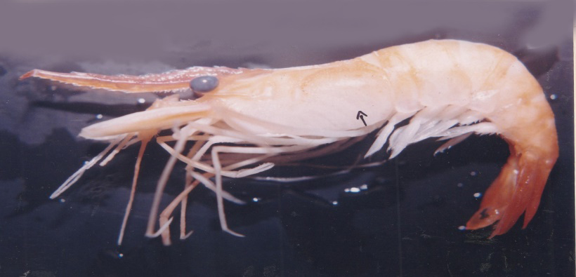
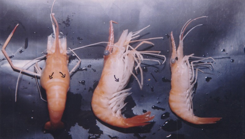

During the investigation of shrimp disease in the culture pond, the prawns Macrobrachium rosenbergii were found to die due to swollen head disease having enlargement of cephalothoracic region containing gill, heart and hepatopancreas etc (1-11). This kind of swollen head disease is mainly caused by bacterial disease. When the animal was seemed to be affected by this swollen head disease, it was found to move towards the bottom of the pond and die within one week of attack. During this time, the animals became moribund and lethargic in condition. In Telugu commonly known as “Dhavadavapu “ disease was found in the giant prawn Macrobrachium rosenbergii in most of the culture pond (muthukuru ) of Nellore region . Dorsal and lateral view of prawns attacked with swollen head disease are shown in plates 3a and 3b.
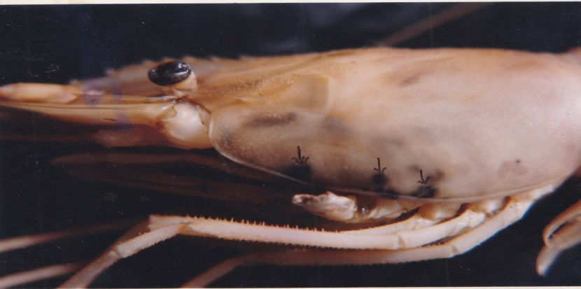
Another kind of symptom of black spots were noticed on the dorsal portion of anterior exoskeleton in cephalothoracic region. Enlargement of pigmentary with black spots formed in the periphery of dorsal side of exoskeleton might be due to the presence of suspected white spot syndrome virus (Figure 7). This kind of disease was found to attack commonly in Macrobrachium rosenbergii in the anterior region are shown in Figures 3 & 4.
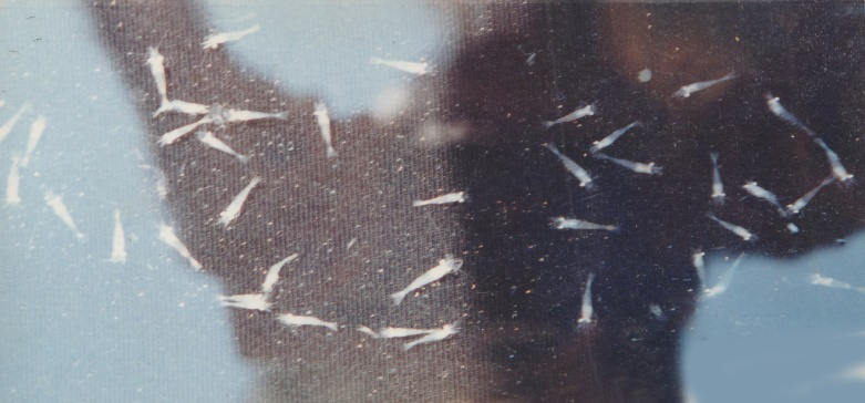
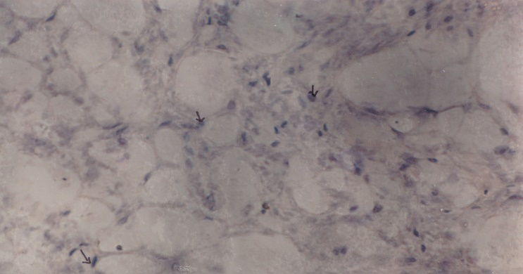
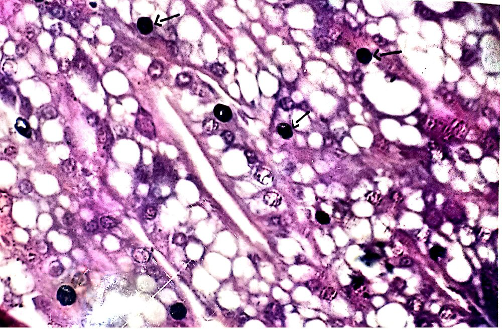
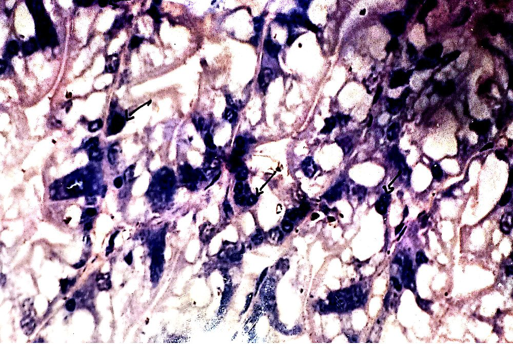
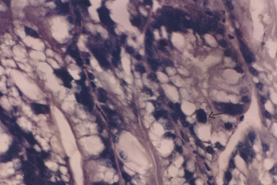
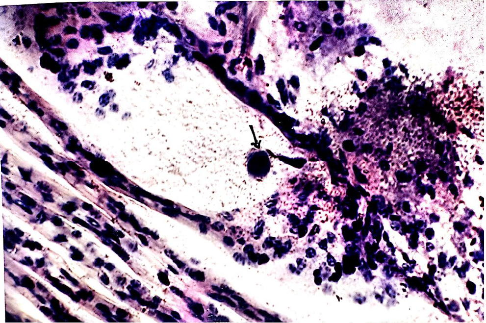
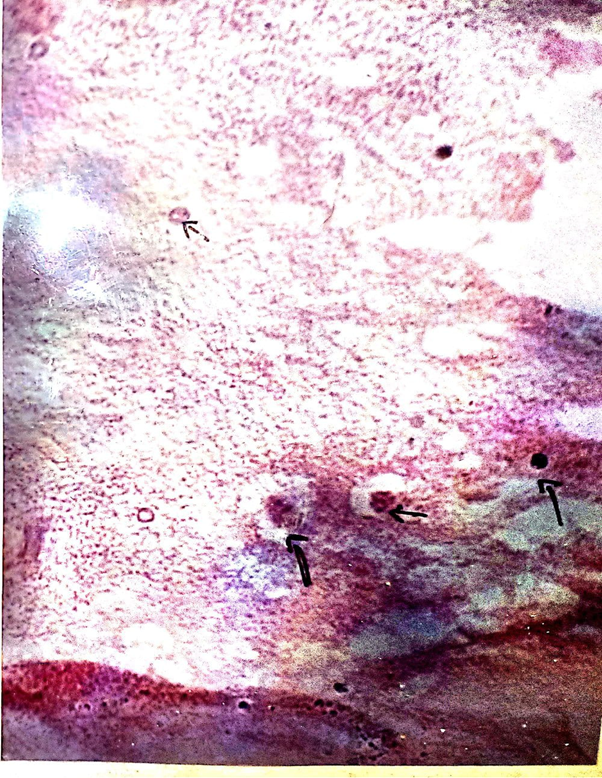
Apart from the study of shrimp disease in culture ponds, attempts were also made to identify the disease in the post larvae of Macrobrachium rosenbergii in the hatchery in and around Nellore region. Mass mortality was found to occur in the post larval stages due to a kind of white muscle disease (Figure 5). It has led to great loss of economy not only to the entrepreneurs involved in the hatchery at Nellore region but also to the country as well. White muscle disease infected in the post larval stages Macrobrachium rosenbergii is clearly shown in Figure 5. Target tissue such as gills, guts, eye tissue and soft tissues of cephalothoracic region were dissected out from the Macrobrachium rosenbergii suspected with the disease of white spot syndrome virus (WSSV). WSSV was characterised by prominent eosinophilic to pale basophilic intranuclear inclusion bodies in hypertrophied nuclei of most commonly in the epithelial cells and connective tissue cells of the above target tissues (Figure 4) .
Discussion
Most of the economy of India from aquaculture sector depends on the euryhaline species and among which, giant freshwater prawn Macrobrachium rosenbergii is one of the important candidate species. Like penaeid prawn, non penaeid prawn have been affected due to improper management of pond, over stocking density and commercial feed containing antibacterial residues etc . Due to various factors, the disease outbreak has become quite common with various levels in the prawns in unfavourable condition, particularly with WSSV, MBV, HPV and SEMBV etc.
In the present study, WSSV and MBV and other bacterial diseases were noticed to cause major damage or necrosis in the target tissues of hepatopancreas, gills, gut and eye stock etc in the species of Macrobrachium rosenbergii. Characterisations made at WSSV and MBV were quite common in the tissues for the present study as revealed by many authors in the same species. When the study was made at Nellore area in extensive shrimp culture pond, Macrobrachium rosenbergii was found with swollen head disease and white spot syndrome disease. When the survey was made at Macrobrachium rosenbergii hatchery at Nellore area, most of the post larval stages were found with white muscle disease (Figure 5). Viral diseases remain a major threat to the shrimp aquaculture industry because some can cause massive disease out breaks. This is the agreement as reported by many authors that MBV disease was caused mainly in the tissues of hepatopancreas by improper management of water and failure of aeration. It accepts the common fact that lipid metabolism can be affected due to the spoilage of secretary cells of hepatopancreas. Piamsak Menasveta [40] and Hung Sung, et al. [41] reported that the presence of a large number of vibrios in the hepatopancreas may be associated with growth retardation in shrimps.
Nilskautsky, et al. [42] studied on ecosystem perspectives on management of disease in shrimp pond farming and discussed that from an ecological perspective, the causes behind the development and spreading of pathogens in shrimp aquaculture [43, 44, 45, 46, 47, 48, 49, 50, 51], the risk of disease shrimp farming often increases with culture intensity and high stocking densities and when polyculture is replaced by monoculture. Kasomchandra, et al. studied on detection of white spot baculovirus (WSBV) in giant freshwater prawn Macrobrachium rosenbergii using polymerase chain reaction and suggested that the amplified product from the DNA of the naturally- infected WSSV Macrobrachium rosenbergii was similar to that of WSBV-infected Penaeus monodon. WSSV and swollen head disease were seen in Macrobrachium rosenbergii infecting hepatopancreas, gill, gut, etc (Figures 1-11) and this may be due to improper management of water, over stocking density, high feed dosage and failing of aeration. White muscle disease was found to attack in the post larvae of Macrobrachium rosenbergii due to improper management of hatchery and imbalanced diet application [51, 52, 53, 54, 55, 56, 57, 58, 59, 60, 61, 62, 63, 64, 65, 66]. It is concluded that the hatchery and farming of Macrobrachium rosenbergii under extensive culture system continued to be encouraged with proper management of water parameters and feed supplements.
Acknowledgement
Authors are thankful to the authority of the Thiruvalluvar University, Vellore, India for providing necessary facilities.
References
-
Rosenberry B (1998) Shrimp hits new high. Fish farming International 25(12): 1.
-
Rosenberry B (1999) Farmed shrimp up by 12 percent. Fish farmer pp: 40-41.
-
Lo CF, Lev JH, Ho CH, Chen CH, Peng SE, et al. (1996) Detection of baculovirus associated with white spot syndrome virus (WSBV) in Penaeid Shrimps using polymerase chain reaction. Dis Aquat Org 25: 133-141.
-
Supamattaya K, Hoftmann RW, Boonyaratpalin S, Kanchanaphum P (1998) Experimental transmission of white spot syndrome virus (WSSV) from bloack tiger Shrimp Penaeus monodon to the sand crab Portunus pelagicus, mud crab Scylla serrata and Krill Acetes sp. Dis Aqua Org 34(1): 79-85.
-
Peng SE, Lo CF, Ho CH, Chang CF, Kou GH (1998) Detection of White spot baculovirus (WSBV) in giant freshwater Prawn, Macrobrachium rosenbergii, using Polymerase Chain Reaction, Aquaculture 164: 253-262.
-
Wang YC, Lo CF, Chang PS and Kou GH (1998) Experimental infection of white spot baculovirus in some cultured and wild decapods in Taiwan. Aquaculture 164: 221-231.
-
Wang CH, Lo CF, Leo JH, Yeh CM PY, Chou HY, et al. (1995) Publication and genomic analysis of baculovirus associated with white spot syndrome virus (WSSV) of Penaeus monodon. Dis Aquat Org 23: 239-242.
-
Brock JA, Lea Master B (1992) A look at the principal bacterial, fungal and parasitic diseases of farmed shrimp. In: Wyban J (Ed.), Proceedings of the special session on shrimp farming. World Aquaculture Society, pp: 212- 226.
-
Anderson IG, Shariff M, Nash G (1987) Mortalities of juvenile shrimp Penaeus monodon associated with Penaeus monodon baculovirus, cytoplasmic reo-like virus, rickettsia and bacterial infections from Malaysian brackish water ponds . Asian Fish Sci 1: 47-64.
-
Lightner DV (1996) A Handbook of shrimp pathology and diagnostic procedures for diseases of cultured Penaeid Shrimp. World Aquaculture society, pp: 305.
-
Kano T, Masahirosakai P, Toshiakiitami (2003) Detection of white spot syndrome virus in shrimp by loop mediated isothermal amplification. Journal of virological methods pp: 59-65.
-
Johnson SK (1975) Handbook of shrimp diseases, Sea Grant Publ. SG-65-603. Texas A&M University, College Station TX, pp: 19.
-
Johnson PT, Lightner DV (1988) Rod shaped nuclear viruses of crustaceans: gut –infecting species. Dis Aqual Org 5: 123 -141.
-
Johnson SK (1990) Handbook of Shrimp diseases, Sea Grant publ,. No. TAMU-SG-90-601. Texas A&M University, College Station, TX, pp: 25.
-
Baticados MCL (1988) Diseases of Prawns in the Philippines. SEAFDEC Asian Aquacult, pp: 10: 1-8.
-
Liu CL (1989) Shrimp Disease, prevention and treatment. In: Akiyama DM (Ed.), Proc. Southeast Asia shrimp Farm Management Workshop. American Soybean Assoc., Singapore, pp: 64-74.
-
Baticados MCL, Cruz ER, Duremdez-Ferandez RC, Gacutar RG, Lavilla-Pitogo, et al. (1990) Diseases of Penaeid Shrimps in the Philippines. Aquaculture Extension Manual 16(46).
-
Lightner DV (1993a) Diseases of Penaeid Shrimp. In: Mcvey JP (ED.), CRC Handbook of Marine culture Crustacean Aquaculture, 2nd (Edn.), CRC Press, Boca Raton, FL, pp: 393-486.
-
Lightner DV, Hedrick RP, Fnjer JL, Chen SN, Liao IC, et al. (1987) A survey of cultured Penaeid shrimp in Taiwan for viral and other important diseases. Fish Pathol 22: 127-140.
-
Lightner DV, Bell TA, Redman RM, Mohney LL, Natividad JM, et al. (1992a) A review of some major diseases of economic significance in Penaeid Prawns Shrimps of the Americas and Indo pacific. In: Shariff IM, Subasinghe RP, et al. (Eds.), Diseases in Asian Aquaculture. Fish Health Section, Asian Fisheries Society, Manila, Phillippines, pp: 57-80.
-
Brock JA, Lightner DV (1990) Diseases of crustacea. Diseases caused by microorganisms. In: Kinne O (Ed.), Diseases OF Marine animals, Vol. III, Biologische Anstalt Helgoland, Hamburg, Germany, pp: 245-424.
-
Brock JA (1991) An overview of diseases cultured crustaceans in the Asia Pacific region. In: Fish Health Management in Asia-Pacific. Report on a regional study and workshop on fish Disease and fish health management. ADB Agriculture Department Report Series No.1. Network of Aquaculture centres in Asia- Pacific, Bangkok, Thailand, pp: 347-395.
-
Brock JA (1992) Current diagnostic methods for agents and diseases of farmed Marine Shrimp. In: Fulks W, Main K (Eds.), Proceedings of the Asian Interchange Program Workshop on the diseases of cultured Penaeid Shrimp. Asian Interchange Program, The Oceanic Institute, pp: 209-231.
-
Kusada R, Watada A (1969) A new pathogenic bacterium, belonging to the genus vibrio, isolated from diseased spiny lobster and prawn. Res Rep Kochi Univ 18: 77-79.
-
Egusa A, Ueda T (1972) A Fusarium sp. associated with black gill disease of the kuruma prawn Penaeus japanicus, Bate. Soc Sci Fish 38: 1253-1260.
-
Cook DW, Lofton SR (1973) Chitino clastic bacteria associated with shell disease in Penaeus shrimp and the blue crab (Callinectes sapidus). J Wildl Dis 9: 154-159.
-
Lewis DH (1973) Response of brown shrimp to infection with sp. Proc. 4th Ann Workshop, World Mariculture Society 4: 335-338.
-
Lightner DV, Lewis H (1975) A Septicemic bacterial disease syndrome of Penaeid Shrimp. In: Diseases of crustaceans Mar. Fish Rev 37: 25-28.
-
Lightner DV, Redman RM, Bell TA, Brock JA (1983b) Dection of 1HHN virus in Penaeus stylirostris and P. vannamei imported into Hawaii. J World Maricult Soc 144: 212-225.
-
Overstreet RM, Stuck KC, Krol RA, and Hawkins WE (1988) Experimental infections with Baculovirus Penaei in the white Shrimp Penaeus vannamei as a bioassay. J World Aquacult Soc 19: 175-187.
-
Brock JA, Lightner DV (1990a) Diseases of crustacea. Diseases caused by microorganisms. In: Kinne O (Ed.), Diseases of Marine animals, Vol. III, Biologische Anstalt Helgoland, Hamburg, Germany, pp: 245-349.
-
Lu Y, Tapay LM, Loh PC, Brock JA (1995a) Infection of the yellow head baculo-like virus in two species of Penaeid Shrimp. P.styrirostris and P.vannamei. J Fish Dis 17: 649- 656.
-
Mohan CV, Sudha PM, Shankar KM, Hegde (1997) Vertical transmission of white spot baculovirus in shrimps – A possibility ?. current science 73(2): 109-110.
-
Lo CF, Ho CH, Chen CH, Liok YL, Yeh PY, et al. (1997) Detection and tissue tropism of white spot syndrome baculovirus (WSBV) captured brooders of Penaeus monodon with a special emphasis on reproductive organs. Dis Aquat Org 30: 53-72.
-
Chang PS, Lo CF, Wang YO and Kou GH (1996) Identification of white spot syndrome associated baculovirus (WSBV) target organs in the shrimp, Penaeus monodon by in situ hybridization. Dis Aquat. Org 27: 131-139.
-
Wongteerasupaya C, Vickers JE, Sriurairatana S, Nash GL, Akarajamora A, et al. (1995) Anon – occluded systemic baculovirus that occurs in the cells of ectodermal and mesodermal origin and caused high mortality in the black tiger Prawn Penaeus monodon. Dis Aqual Org 21: 69-77.
-
Lo CF, Lev JH, Ho CH, Clen CH, Pend SE, et al. (1996b) Detection of baculovirus associated with white spot syndrome (WSBV) in Penaeid Shrimp using Polymerase Chain Reaction. Dis Aquat Org 25: 133-141.
-
Poulos BT, Nunan L, Mari J, Swider J, Lighter DV (1995) Use of the polymerase Chain Reaction to detect viral infections in Penaeid Shrimp. In: Sahul Hameed AS, Anilkumar M, et al. (Eds.), Studies on the pathogenicity of systemic ectodermal and mesodermal baculovirus and its detection in shrimp by immunological methods. Aquaculture 160: 31-45.
-
Elpidocesar B, Nadala JR, Lourdes M, Tapay LM, Shurongeao (1997) Detection of Yellow head virus and Chinese baculovirus in the penaeid Shrimp by the western blot technique. Journal of virological methods 69: 39-44.
-
Menasveta P (2002) Improved shrimp grow out systems for disease prevention and environmental sustain ability in Asia. Reviews in fisheries Science 10: 391-402.
-
Sunga HH, Hsua SF, Kunchena C, Tingb YY, Chao WL (2001) Relationships between disease outbreak in cultured tiger shrimp (Penaeus monodon) and the composition of vibrio communities in the pond water and shrimp hepatopancreas during cultivation. Aquaculture 192(2- 4): 101-110.
-
Nilskautsky, Ronnback P, Teddengreu M, Troell M (2000) Ecosystem perspectives on management of disease in shrimp pond farming. Aquaculture 191: 145-161.
-
Chan PS, Lo CF, Wang YC, Kou GH (1996) Identification of white spot syndrome associated baculovirus (WSBV) target organs in the shrimp, Penaeus monodon by in situ hybridization. Dis Aqual Org 27: 131-139.
-
Flegal TW, Boonyaratpalin S, Wityachummamkul B (1996) Current status of research on Yellow -head virus and white -spot virus in Thailand. World Aquaculture; 96. Book of Abstracts. World Aquaculture society, pp: 126-127.
-
Flegal TW, Nielsen L, Thamavit V, konglim S, Pasharawipas T (2004) Presence of multiple viruses in non-diseased, cultivated shrimp at journal of virological methods 119: 1-5.
-
Johnson SK (1995) Handbook of Shrimp Disease. Sea Grant Publ.No TAMU-SG-95-601(r). Texas A&M University College Station ,TX, pp: 25.
-
Lightner DV, Pouls BT, Redman RM, Bruce L, Nunan L, et al. (1994) Development and application of genomic probes for us as diagnostic and research reagents for the Penaeid shrimp Parvoviruses 1HHNV and HPV, and the Baculoviruses MBV and BP U.S. Marine shrimp farming program 10th anniversary Review, Gulf coast Research laboratory special publication, Gulf Research Reports, No.1,Ocean Springs, Ms, pp: 59-85.
-
Lightner DV, Poulous BT, Redman RM, Mari J, Bonami JR (1992c) New developments in Penaeid virology: Application of biotechnology in research and disease diagnosis for shrimp viruses of concern in the Americas. In: Ful KS, Main WK (Eds.), Proceedings of the Asian Interchange Program Workshop on the Disease of cultured Penaeid Shrimp. Asian Interchange Program, The Oceanic Institute, Oahu, HI, pp: 233-253.
-
Lightner DV, Hasson KW, White BL, Redman RM (1995) Chronic toxicity and histopathological studies with Benlate, a commercial grade of benomyl, in Penaeus vannamei ( Crustacea: Decapoda ). Aquat Toxicol 34: 105-118.
-
Lightner DV (1998) Diseases of cultured Penaeid Shrimp and Prawns. In: Sindermam CJ, Lightner DV (Eds.), Disease Diagnosis and Control in North American Marine Aquaculture, Elsevies, Amsterdam pp: 8-127.
-
Manivannan S, Kennedy B, Karunasagar I, Karunasagar I (2003) Department of fishery prevalence of Monodon baculovirus in wild metapenaeus spp. Along the south west coast of India, Aquaculture 232: 1-4.
-
Mari J, Lightner DV, Poulos BT, Bonami JR (1995) Partial cloning of and unusual shrimp parvovirus (HPV): use of gene probes in diseases diagnosis. Dis Auqat Org 22: 129-134.
-
Mari J, Bonami JR, Poulos B and Lightner D (1993b) Preliminary characterization and partial cloning of the genome of a baculovirus from Penaeus monodon (PMSNPV = MBV). Dis Aquat Org 16: 207-215.
-
Momoyama K, Hiraoka M, Nakano H, Koube H, Inonye K et al. (1994) Mass mortalities of cultured Kuruma Shrimp Penaeus japonicus, In Japan in 1993: Histopathological Study. Fish PathoL 29: 141- 148.
-
Nakanc H, Koube H, Erawa SU, Momoyama K, Hiraoka M, et al. (1994) Mass mortalities of cultured Kuruma Shrimp Penaeus japonicus, in Japan in 1993: epi zoolological survey and infection trails. Fish Pathol 29: 135-130.
-
Naticidad JM, Lightner DV (1992) Prevalence and geographic distribution of MBV and other diseases in cultured giant tiger prawns (Penaeus monodon) in the Philippines. In: Fulks W, Main KL (Eds.), Diseases of cultured Penaeid Shrimp in Asia and the United states (Proceedings of a workshop in Honolulu, Hawaii). The Oceanic Institute, Makapuu point, pp: 139-160.
-
Otta SK, Shubha G, Joseph B, Chakraborty A, Karunasagar I, Karunasagar I (1999) Polymerase Chain Reaction (PCR) detection of white spot syndrome virus (WSSV) in cultured and wild crustaceans in India. Dis Aquat Org 38: 67-70.
-
Poulos BT, Mari J, Bonami JR, Readman R, Lightner DV (1994b) Use of non-radioactively labelled DNA probes for the detection of a baculovirus from Penaeus monodon by in situ hybridization on fixed tissue. J Virol Meth 49: 187-194.
-
Rajan PR, Ramasamy P, Purushothaman V, Brennan GP (2000) White Spot baculovirus syndrome in the Indian shrimp Peneaus monodon and Penaeus indicus. Aquaculture 184: 31-34.
-
Ruby PRS, Murali Manohar B, Sundararaj A (1999) Histopathology of white spot diseases in shrimps. In: Sundararaj V, Santhanam R, et al. (Eds.), Abstracts of National Seminar on Development and Transfer of Fisheries Technology pp: 3-5.
-
Shankar KM, Mohan CV (1998) Epidemiological aspects of shrimp viral diseases in India – a review. J Aqua Trop 13(1): 43-49.
-
Sindermann CJ (1988) Disease problems caused by introduced species. In: Sindermann CJ, Lightner PV (Eds.), Disease diagnosis and control in North American Marine Aquaculture, pp: 394-398.
-
Smith PT (1996) Toxic effects of blooms of marine species of oscillatoriales on farmed prawns (Penaeus monodon, Penaeus japonicus) and brine shrimp (Artemia salina). Toxicon 34: 857-869.
-
Takahashi Y, Itami T, Kondo M, Maeda M, Fuji R, et al. (1994) Electron microscopic evidence of baciliform virus infection in kuruma Shrimp Penaeus japonicas. Fish Pathol 29: 121-125.
-
Tapay LM, Nadala ECD, Loh PC (1999) A Polymerase Chain Reaction Protocol for the detection of various geographical isolates of white spot virus. J Virological methods 82(1): 39-43.
-
Tomoyokono, Ramasavan, Masahiro, Toshikiitami (2003) Detection of white spot syndrome virus in shrimp by loop mediated isothermal amplication. Journal of Virological Methods 115: 59-65.
- California Red-Legged Frog and Non-Listed Amphibians Response to Non-Native Fish Removal
- Industrial Standardization of the Bio-OS: Algorithmic Codification of Resilience Engineering Guidelines and Version V8 Architecture
- Climate Variability and the Sustainability of Snail Farming in Nigeria: Past Trends, Present Challenges and Potential Outlook
- The Evaluation of the Surveillance System of Anthrax in Gilgit-Baltistan, Pakistan, 2018
- Natural Decline to Extinction of A New Zealand Rabbit Population
- Mitochondrial Bio-Logistics: Steering Co-Enzyme Q10 and Lycopene Synergies within the Science 4.0 Bio-OS Framework